4461
Effectivity and correlation of parameters derived from diffusion kurtosis imaging and quantitative dynamic contrast-enhanced MRI in the breast imaging1Shanghai General Hospital, Shanghai, China, 2PhilipsHealthcare, Shanghai, China, 3Fudan University, Shanghai, China
Synopsis
The aim of this study is to evaluate the diagnostic efficacy of 3.0T MRI diffusion kurtosis imaging and quantitative dynamic contrast enhancement in benign and malignant breast lesions, and to explore the differential diagnosis ability of different pathological types and molecular subtype lesions. The results showed that 3.0T MRI diffusion kurtosis imaging and dynamic contrast-enhanced quantitative hemodynamic parameters are of great value in the differential diagnosis of benign and malignant breast lesions. The combination of both can significantly improve the diagnostic efficiency.
Objective
To evaluate the diagnostic performance of diffusion kurtosis imaging (DKI), conventional diffusion-weighted imaging (DWI) and quantitative dynamic contrast-enhanced breast MRI (DCE-MRI) in differentiating malignant from benign breast lesions independently or jointly and to explore whether correlations exist among these parameters.Methods
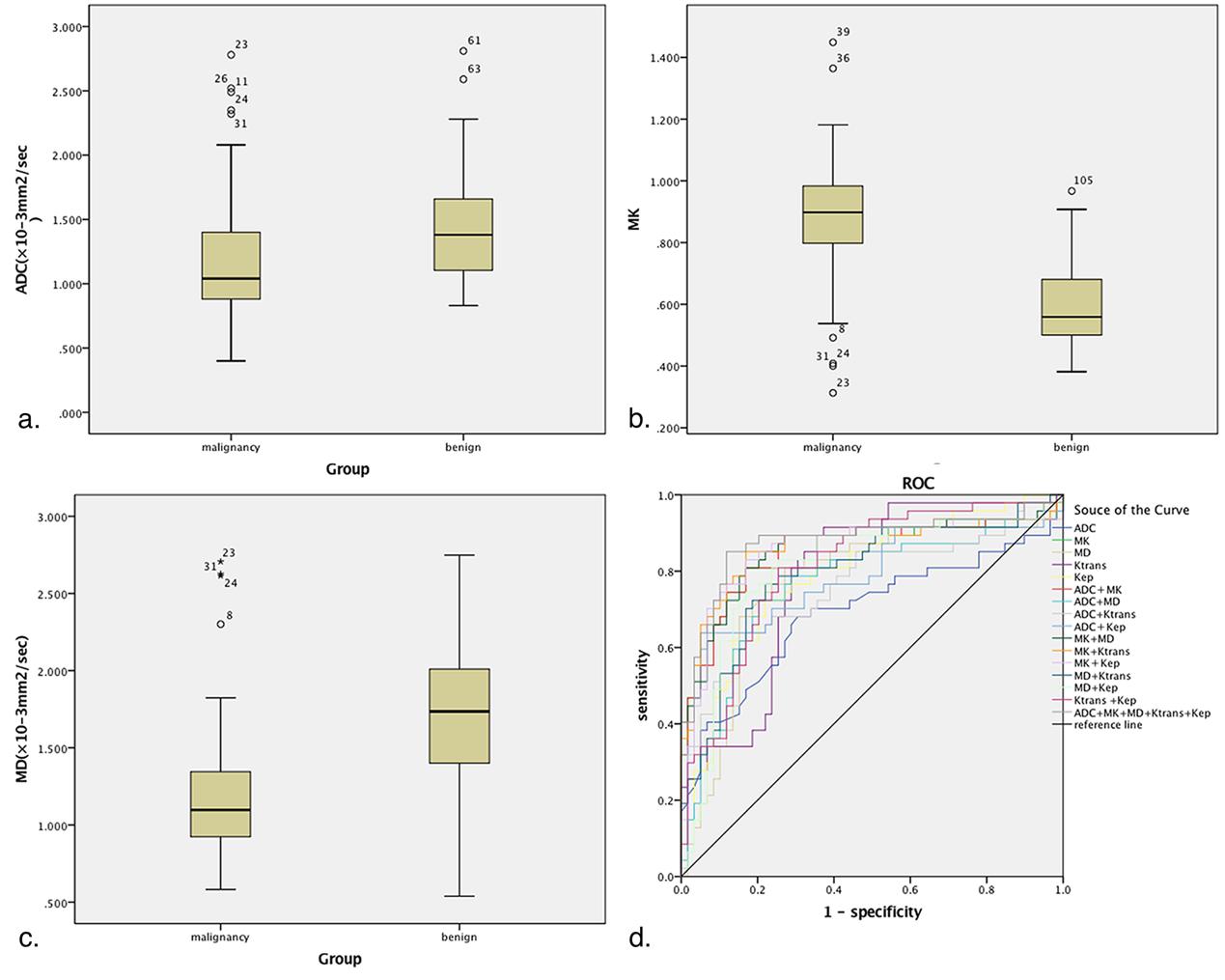
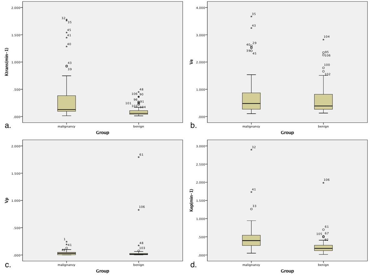
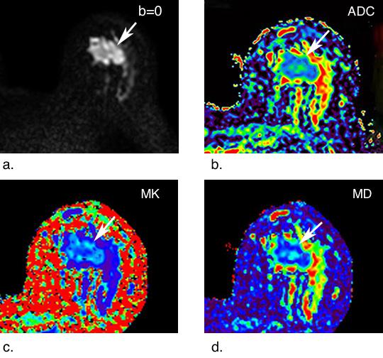
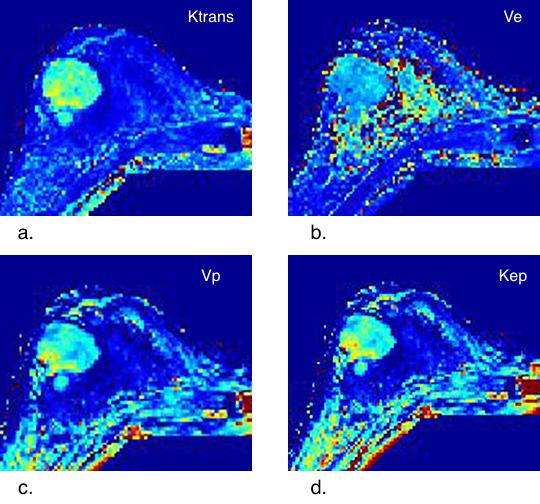
One hundred nine patients were included in the study. Quantitative parameters from DKI (MD, MK), DWI (ADC) and quantitative DCE-MRI (Ktrans, Kep, Ve and Vp) were calculated. Two blinded radiologists evaluated findings in consensus. The diagnostic performances of DKI, DWI and quantitative DCE-MRI, either alone or in combination, were statistically evaluated. The Spearman correlation test was used to evaluate correlations among the DKI-, DWI- and quantitative DCE-MRI-derived parameters.Results
MK and MD from DKI and ADC from DWI and Ktrans and Kep from quantitative DCE-MRI were significantly different between breast cancer and benign lesions (p < 0.05, respectively). MK demonstrated the largest area under the receiver operating characteristic curve (AUC = 0.849) and had the highest specificity (83.1%) and accuracy (82.1%) with a cut-off value of 0.727, while Ktrans showed the highest sensitivity (91.5%) with a cut-off value of 0.067 min-1. The highest diagnostic specificity (93.2%) was obtained when ADC and Kep were combined. The highest accuracy (86.8%) and AUC (0.879) were attained when all of the parameters were combined. Kep was correlated moderately positively with MK (r = 0.516) and moderately negatively with MD (r = -0.527). Ktrans was weakly positively correlated with MK with an r of 0.398 and weakly negatively correlated with MD with an r of -0.450.Conclusions
DKI-derived parameters can significantly improve diagnostic performance compared with the ADC from conventional DWI. DKI exhibits promise as a quantitative technique to augment quantitative DCE-MRI. The diagnosis was improved when the parameters were combined. Diffusion parameters derived from DKI were statistically correlated with parameters from quantitative DCE-MRI.Acknowledgements
No acknowledgement found.References
1. Partridge SC, McDonald ES. Diffusion weighted magnetic resonance imaging of the breast: protocol optimization, interpretation, and clinical applications[J]. Magnetic resonance imaging clinics of North America 2013;21:601-24. PMID: 23928248
2. Kato F, Kudo K, Yamashita H, et al. Differences in morphological features and minimum apparent diffusion coefficient values among breast cancer subtypes using 3-tesla MRI[J]. European journal of radiology 2016;85:96-102. PMID: 26724653
3. Bickelhaupt S, Laun FB, Tesdorff J, et al. Fast and Noninvasive Characterization of Suspicious Lesions Detected at Breast Cancer X-Ray Screening: Capability of Diffusion-weighted MR Imaging with MIPs[J]. Radiology 2016;278:689-97. PMID: 26418516
4. Zaric O, Pinker K, Zbyn S, et al. Quantitative Sodium MR Imaging at 7 T: Initial Results and Comparison with Diffusion-weighted Imaging in Patients with Breast Tumors[J]. Radiology 2016;280:39-48. PMID: 27007803
5. Nogueira L, Brandao S, Matos E, et al. Application of the diffusion kurtosis model for the study of breast lesions[J]. European radiology 2014;24:1197-203. PMID: 24658871
6. Jensen JH, Helpern JA, Ramani A, Lu H, Kaczynski K. Diffusional kurtosis imaging: the quantification of non-gaussian water diffusion by means of magnetic resonance imaging[J]. Magnetic resonance in medicine 2005;53:1432-40. PMID: 15906300
7. Sun K, Chen X, Chai W, et al. Breast Cancer: Diffusion Kurtosis MR Imaging-Diagnostic Accuracy and Correlation with Clinical-Pathologic Factors[J]. Radiology 2015;277:46-55. PMID: 25938679
8. Wu D, Li G, Zhang J, Chang S, Hu J, Dai Y. Characterization of breast tumors using diffusion kurtosis imaging (DKI) [J]. PloS one 2014;9:e113240. PMID: 25406010
9. Cheang MC, Chia SK, Voduc D, et al. Ki67 index, HER2 status, and prognosis of patients with luminal B breast cancer[J]. Journal of the National Cancer Institute 2009;101:736-50. PMID: 19436038
10. Goldhirsch A, Winer EP, Coates AS, et al. Personalizing the treatment of women with early breast cancer: highlights of the St Gallen International Expert Consensus on the Primary Therapy of Early Breast Cancer 2013[J]. Annals of oncology : official journal of the European Society for Medical Oncology / ESMO 2013;24:2206-23. PMID: 23917950
11. An YY, Kim SH, Kang BJ. Differentiation of malignant and benign breast lesions: Added value of the qualitative analysis of breast lesions on diffusion-weighted imaging (DWI) using readout-segmented echo-planar imaging at 3.0 T[J]. PloS one 2017;12:e0174681. PMID: 28358833
12. Zhang L, Tang M, Min Z, Lu J, Lei X, Zhang X. Accuracy of combined dynamic contrast-enhanced magnetic resonance imaging and diffusion-weighted imaging for breast cancer detection: a meta-analysis[J]. Acta radiologica (Stockholm, Sweden : 1987) 2016;57:651-60. PMID: 26275624
Figures