4448
The Value of DKI and IVIM Quantitative Parameters in Differentiating Breast Lesions and Correlations with Histopathologic Factors: A Preliminary Study1National Cancer Center/Cancer Hospital, Chinese Academy of Medical Science and Peking Union Medical College, Beijing, China, 2GE healthcare, Beijing, China
Synopsis
The current study aimed to evaluate the application of diffusion kurtosis imaging (DKI) and intravoxel incoherent motion (IVIM) in the differential diagnosis of breast lesions. The association of DKI and IVIM derived parameters were compared with pathological types, histologic grade, and Ki-67 expression of primary breast cancers. The results indicated that MD, MK, D and f are capable of distinguishing benign from malignant breast lesions. Furthermore, significant correlations were observed between MD and different histologic grade and Ki-67 expression, repsectivley.
Purpose
To assess the clinical application value of DKI and IVIM on differentiating breast lesions and to evaluate the potential associations between the derived quantitative parameters and clinical-pathologic factors of breast cancers.Introduction
Conventional diffusion-weighted imaging(DWI) has been demonstrated as a useful adjunct sequence to breast MRI in order to improve diagnostic accuracy. However, the quantitative parameter of DWI, known as apparent diffusion coefficient (ADC), demonstrated controversial results in the differentiations of breast lesions [1]. On the other hand, DKI and IVIM have both been observed to contribute to higher sensitivity for cancer detection in the corresponding previous studies [2-5]. To be specific, DKI quantifies non-Gaussian diffusion and IVIM was conducted on basis of the perfusion of microcirculation. Therefore, the current study aimed to evaluate diagnostic accuracy of applying DKI and IVIM on patients with breast lesions and to further investigate the potential associations between parameters derived from DKI and IVIM and clinical-pathologic factors of breast cancer.Material and Methods
Sixty-four women (mean age, 44±8.6 years; range age, 15-61years) with a total number of 66 breast lesions(42 malignant and 24 benign) underwent breast MRI including DKI sequence on a 3.0T MR system. The acquisition parameters applied were as follows: 3b-values of 0, 1000, and 2000 s/mm2 and a IVIM sequence with 12 b-values from 0 to 2000 s/mm2 . The status of the breast lesions were all identified through operations or biopsies. DKI related parameters (MD, MK) and IVIM-derived parameters (D, D* and f) were measured. The derived parameters in benign and malignant lesions were compared using Independent-sample t-test. Receiver operating characteristic (ROC) analysis was performed to assess the sensitivity and specificity of MD, MK, D, D* and f in the diagnosis of breast lesion. Additionally, the potential correlation between the Ki-67 expression and clinical-pathologic factors of breast cancer was evaluated by the Spearman’s correlation.Results
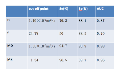
There was statistically significant difference observed between benign and malignant lesions for all the parameters (MD, p<0.001; MK, p<0.001; D, p<0.001; f, p<0.001. MK of malignant lesions (0.86±0.12) was significantly higher than that of benign lesions (0.58±0.12); in contrast, MD of malignant lesions (1.11±0.18(10-3 mm2/s)) was lower than that of benign lesions (1.66±0.23 (10-3 mm2/s)) [Table 1]. As for the IVIM-derived parameters, D of benign lesions (1.03±0.24) was significantly higher than that of malignant lesions (1.17±0.25), and f-value of malignant lesions (38±15) % was significantly higher than that of benign lesions (26±15)% [Figure 1,Table.1]. The area under the curve (AUC) is 0.98 for MD, 0.96 for MK, 0.87 for D, and 0.70 for f, respectively. At a cutoff of 1.35 (10-3 mm2/s), the specificity and sensitivity of MD for identifying malignant lesions were 90.9% and 94.7%, respectively [Table.2]. Meanwhile, in patients with breast cancer, MD was lower in grade 3 tumors than that in grades 1 and 2 tumors (r= -0.44), associated with a negative correlation with Ki-67 expression (r = -0.59), whereas MK, D and f showed no significant correlation.Discussion and Conclusions
DKI and IVIM derived parameters have the capability to assess malignant and benign breast lesions. MD and MK derived from DKI showed higher specificity for the differentiation of malignant from benign lesions than IVIM derived parameters. Furthermore, MD was negatively correlated with histologic grade and Ki-67 expression. Limitations in the present study include limited sample size and potential micro-structural changes after biopsy. Thus, further investigations are required to confirm these findings.Acknowledgements
No acknowledgement found.References
[1] Ei Khouli RH, Jacobs MA, Mezban SD, et al. Diffusion-weighted imaging improves the diagnostic accuracy of conventional 3.0-T breast MR imaging. Radiology. 2010, 256 (1): 64-73.
[2] Nogueira L, Brandão S, Matos E, et al. Application of the diffusion kurtosis model for the study of breast lesions. Our Radiol, 2014, 24(6):1197-1203.
[3] Sun K, Chen X, Chai W, et al. Breast Cancer: Diffusion Kurtosis MR Imaging-Diagnostic Accuracy and Correlation with Clinical-Pathologic Factors. Radiology. 2015, 277(1): 46-55.
[4] Iima M, Yano K, Kataoka M, et al. Quantitative non-Gaussian diffusion and intravoxel incoherent motion magnetic resonance imaging: differentiation of malignant and benign breast lesions. Invest Radiol, 2015, 50(4): 205-211.
[5] Wang Q, Guo Y, Zhang J, et al. Contribution of IVIM to conventional dynamic contrast-enhanced and diffusion-weighted MRI in differentiating benign from malignant breast masses. Breast Care (Basel), 2016, 11(4): 254-258.
Figures
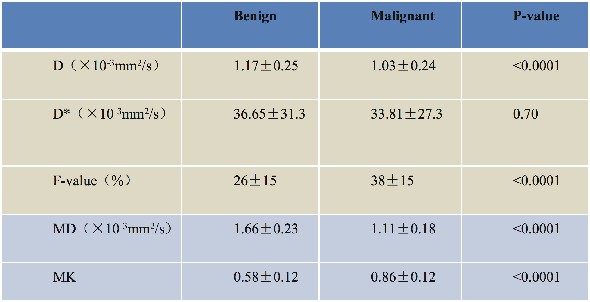
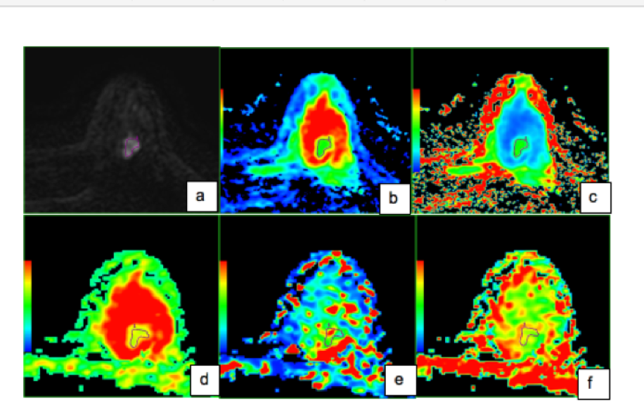
Figure 1. Images of a 37-year-old female showed an invasive ductal carcinoma in the left breast.
(a) Axial b=1000 s/mm2 DKI image; (b) MD=1.29*10-3mm2/s; (c) MK=0.95; (d) D=0.75*10-3mm2/s; (e) D*=0.05*10-3mm2/s; (f) F=23%