4437
On the Properties of Additive Manufacturing Materials for Anatomy Specific 3D-Printed MRI Coils1Electrical and Computer Engineering, University of Wisconsin-Madison, Madison, WI, United States, 2Radiology, University of Wisconsin-Madison, Madison, WI, United States
Synopsis
Additive manufacturing provides a low-cost and rapid means to translate 3D design into the construction of a prototype. For MRI, this type of manufacturing can be used to construct various components including the structure of RF coils. In this abstract, we characterize the material properties (dielectric constant and loss tangent) of several common 3D-printed plastics, and utilize these material properties in full-wave electromagnetic simulations to design and construct a very low-cost subject/anatomy-specific 3D-printed receive-only RF coil that fits close to the body. We show that the anatomy-specific coil exhibits higher signal-to-noise ratio compared to a conventional flat surface coil.
Purpose
Additive manufacturing (or 3D printing) provides a low-cost and rapid means to translate 3D design into the construction of a prototype. For MRI, this type of manufacturing can be used to construct various components including phantom, holders, and the structure of RF coils1-2. In this abstract, we characterize the material properties of several common 3D-printed plastics, and demonstrate a method for design and construction of a very low-cost subject and anatomy-specific 3D-printed receive-only RF coil. .Methods
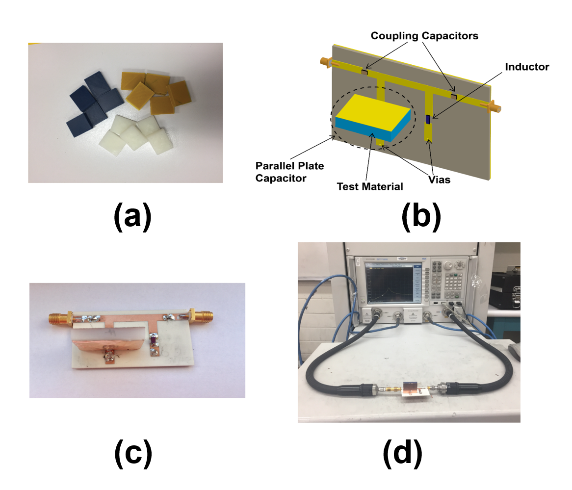
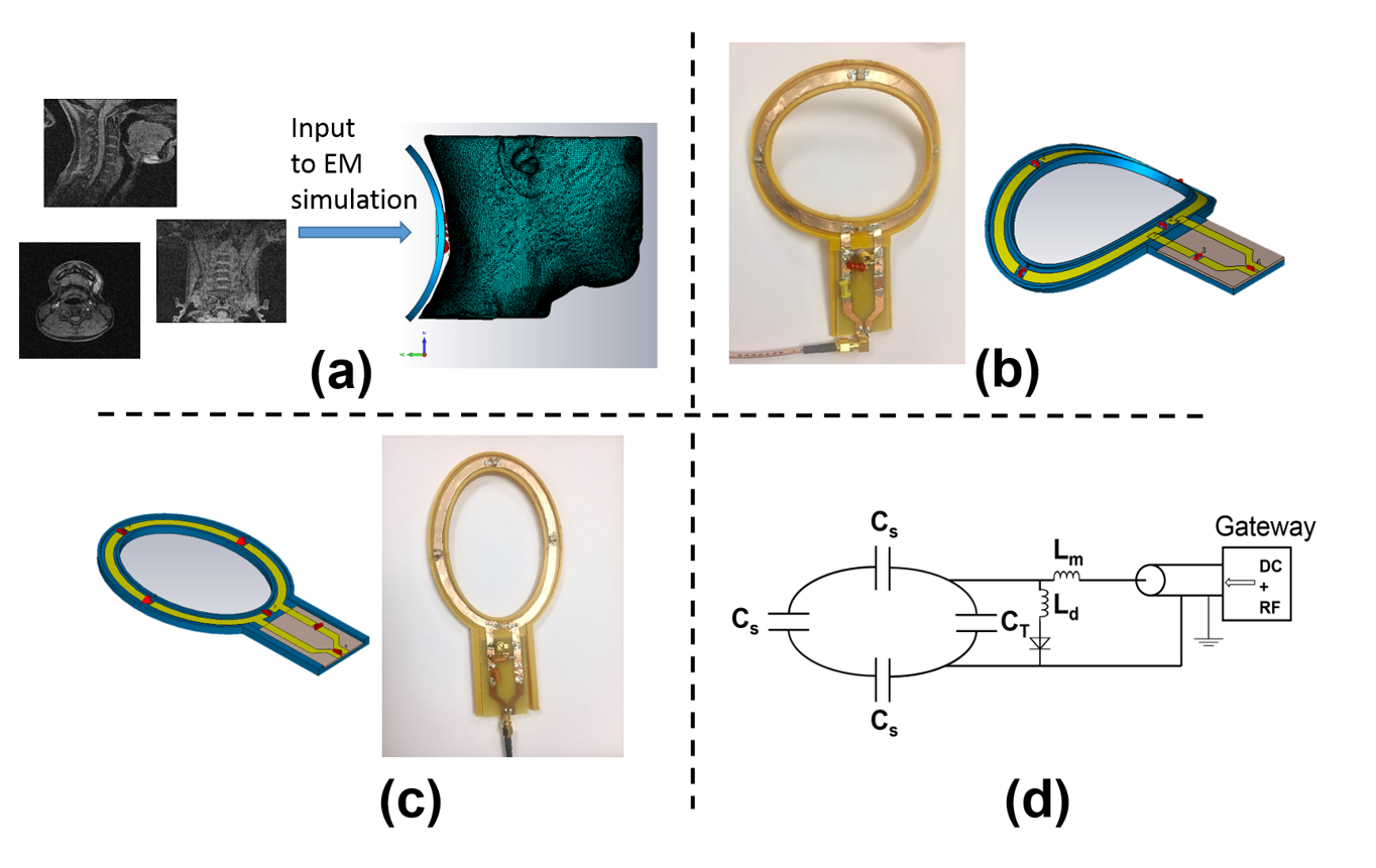
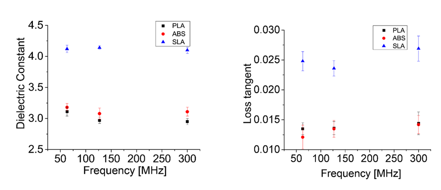
Many materials can be used in additive manufacturing. Although these materials generally have similar properties in terms of mechanical stability, their electrical properties such as dielectric constant and dielectric loss can vary. Choosing a material with a low dissipation factor in RF coil design can improve frequency response and quality factor. In this study, we developed a simple and robust method to characterize the dielectric constant and loss tangent at frequencies relevant to MRI for three common materials used in additive manufacturing (ABS plastic, PLA, and stereolithography photopolymer resin [SLA]). To measure the dielectric properties of the materials, we developed an RLC resonator circuit as a test fixture utilizing the 3D-printed material under test as a parallel plate capacitor in the resonator. Next, full-wave electromagnetic simulations and scattering-parameter measurements were obtained to estimate material properties. Figure 1 shows the test fixture with the material under test (MUT) used to construct the parallel plate capacitor of the parallel RLC circuit. Based on the extracted parameters, two MRI receive-only coils were simulated and constructed for imaging the cervical spine. Both coils were constructed on a FDM 3D printer (Ultimaker Extended 2+) from PLA plastic because of its low loss tangent at 3T compared to other materials (see Figure 3). The first coil is a conventional, flat single-channel surface coil. The other coil is designed to fit against the neck and close to the body. To design the anatomy specific coil, a 3D CAD model of volunteer head and neck was created using 3D MR images of a human volunteer. This 3D model was used as input to electromagnetic (EM) simulation software (CST Studio) to design a specific neck coil for the volunteer. Figure 2-a shows the EM simulation environment. Figure 2-b shows the simulated and constructed anatomy-specific coil. Figure 2-c shows the flat surface coil. Both coils have the same diameter and utilize a path in their support structure printed in negative relief. Conductive elements of both loops are 6mm wide copper tape. Coils were matched to 50 Ω and tuned to 127.8MHz (3T MRI). A detuning circuit were designed for both coils to detune the coil and prevent interactions with the body transmit coil. Figure 2-d shows the circuit diagram of the tuning, matching, and detuning network. Imaging was performed on both MR spherical phantom and the volunteer neck.Results
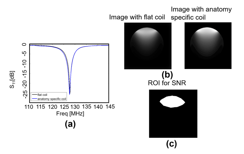
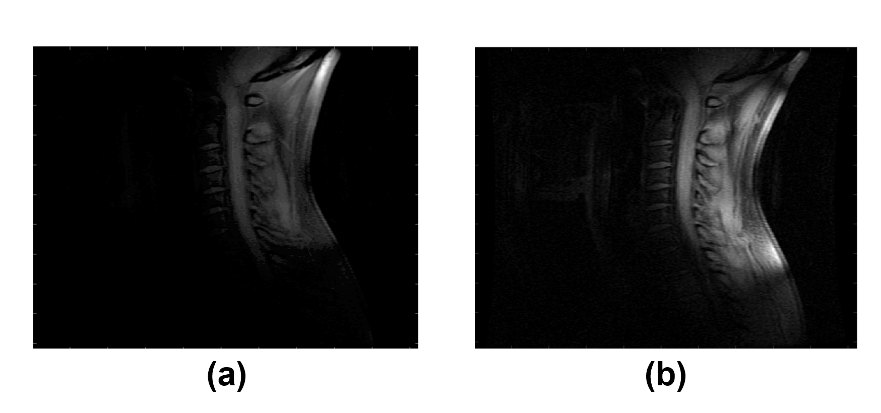
Figure 3 shows the extracted dielectric properties (dielectric constant and loss tangent) for the three 3D printing materials for common MRI RF frequencies using the proposed method. ABS and PLA have similar properties at relevant MR frequencies, whereas SLA has a higher loss. Figure 4-a shows the measured reflection coefficient of the coils. Both coils have almost similar quality factor = 40. Figure 4-b shows the MR images of the phantom scanned by the designed coils. Figure 4-c shows the ROI for calculating the SNR, calculated using NEMA method. The SNR in this region for the anatomy-specific coil was 394 compared to the flat surface coil which was 293. Figure 5 shows the cervical spine images of the volunteer showing low SNR when using a flat coil and high SNR when using the anatomy-specific neck coil placed against the neck.Discussion and Conclusion
The use of additive manufacturing in the development of MRI devices is highly appealing. By characterizing the electrical properties of 3D-printed materials (which to our knowledge has not been conducted at MRI frequencies for these materials), different coil geometries can be quickly designed and printed for evaluation until the design is optimized. This abstract demonstrates the construction of anatomy-specific 3D-printed MR coils that can provide the improved SNR for imaging a specific area of body. These coils can be designed and implemented very quickly with a very low cost for each body region (and potentially for each patient). In particular, this capability likely to be beneficial for the application of MR-based radiation therapy planning and in particular with the increasing availability of combined MR-Linac scanners, where anatomy- and subject-specific coils could be created to perform optimal imaging (and/or treatment) in the therapy position.Acknowledgements
The project described was supported in part by NIH UL1TR000427 and DARPA N66001-17-2-4010. The content is solely the responsibility of the authors.References
1. Herrmann, Karl-Heinz, et al. "3D printing of MRI compatible components: Why every MRI research group should have a low-budget 3D printer." Medical engineering & physics 36.10 (2014): 1373-1380. 2.
2. Wei, Shufeng, et al. "Design and implementation of MRI RF coil based on 3D printing." RF and Wireless Technologies for Biomedical and Healthcare Applications (IMWS-BIO), 2015 IEEE MTT-S 2015 International Microwave Workshop Series on. IEEE, 2015.
Figures