4425
Recording Electrophysiological Signals Through MR Receiver Coils During Concurrent fMRI1Weldon School of Biomedical Engineering, Purdue University, West Lafayette, IN, United States, 2Electrical and Computer Engineering, Purdue University, West Lafayette, IN, United States
Synopsis
Simultaneous recording of fMRI and electrophysiological (EP) signals, e.g. electroencephalography (EEG), electrocorticography (ECoG), and local field potentials (LFP) holds significant potential to evaluate the brain dynamics and its underlying neural circuitry across various spatiotemporal scales. However methodological challenges associated with bio-potential recording within MRI still work as a bottleneck. Here we present a miniaturized, MR-compatible device that can adaptively learn the presence of electromagnetic artifacts and can perform high fidelity EP recording (ECG, LFP, SEP etc.) wirelessly through the MR-receiver coil. The low power device provides a cheap and simple solution for recording various electrophysiological signals during concurrent fMRI.
Background
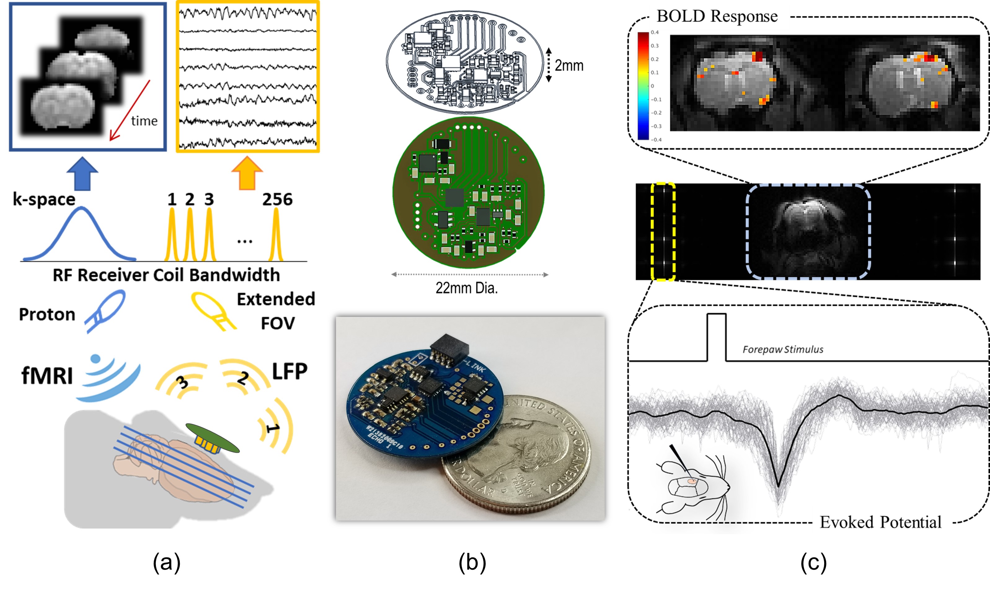
Simultaneous recording of fMRI and electrophysiological signals, e.g. electroencephalography (EEG), electrocorticography (ECoG), and local field potentials (LFP), may help to bridge neural activity across spatial and temporal scales. However, the MRI system is a hostile environment for recording bioelectric signals. Apart from MR-safety and compatibility, major bottlenecks are due to strong electromagnetic artifacts generated from the rapidly changing gradient fields and RF pulses. As a result, the recorded electrophysiological (EP) data is often unusable to researchers without specialized technical expertise. Here, we present a simple solution to address this challenge, by introducing an active and wireless device – MR-Link (Multimodal Recording Link). This device adaptively samples electrophysiological signals to avoid artifacts, and transmits the signal to the RF receiver coil during simultaneous fMRI (Fig.1a).Methods
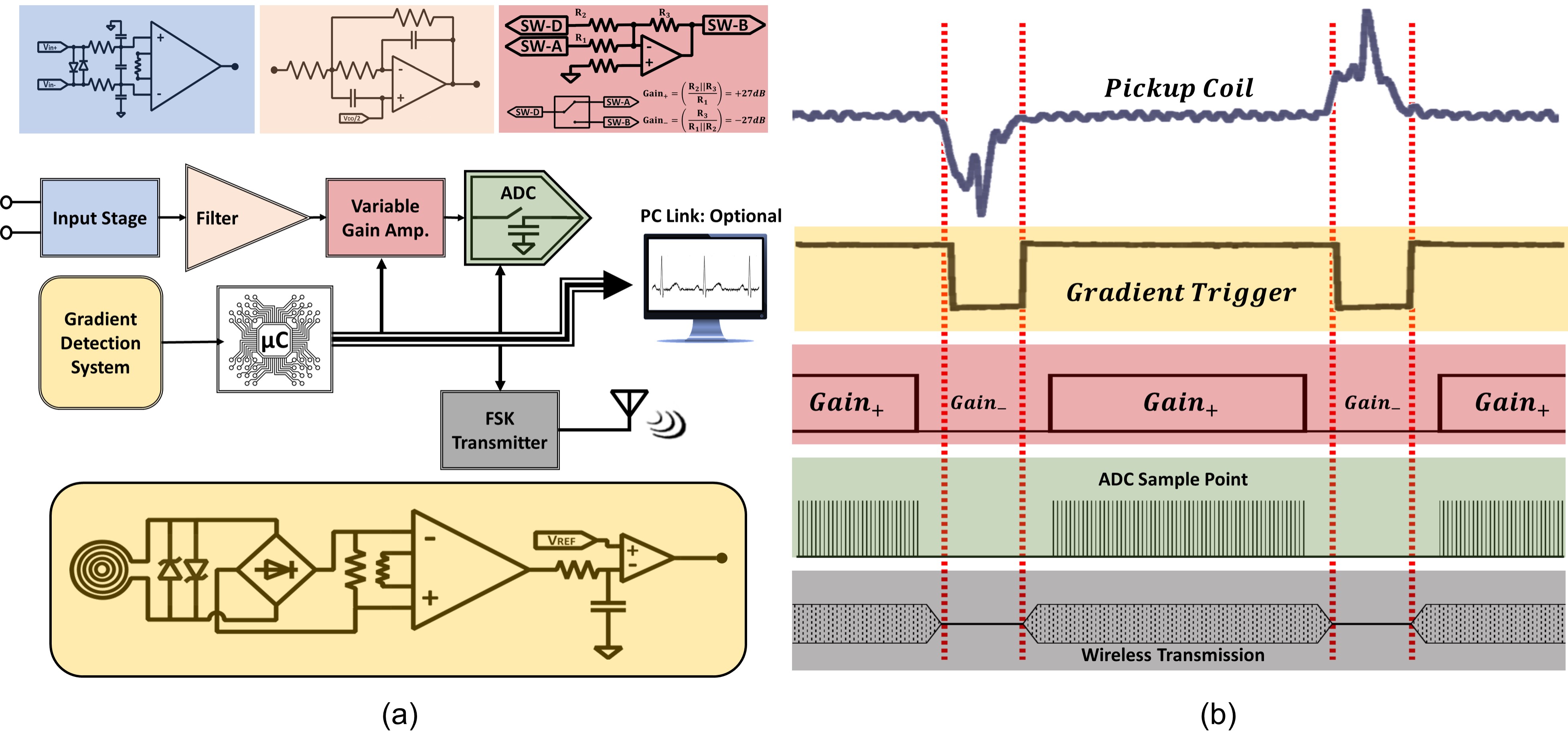
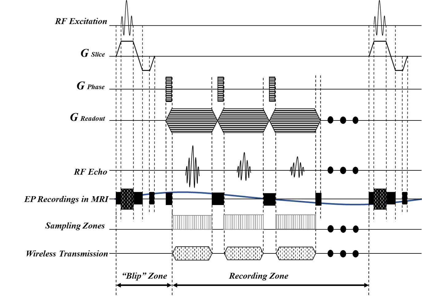
The device utilized a machine-wound copper coil (14mm diameter) to pick up variations in the magnetic field inside the MRI bore. The gradient detection module delineated periods with changing magnetic fields and generated a binary signal (gradient trigger) which provided the onboard microcontroller (μC) with information of when to acquire gradient free EP signals. The gradient trigger also allowed the μC to learn patterns of when gradient noise would corrupt EP data which helped to improve the recorder’s functionality. Based on the state of the gradient trigger, the EP signal was either amplified and digitized, or simply attenuated to assure the analog components did not saturate due to the induced gradient artifacts. The EP signal was primarily processed through an analog front end circuit (voltage limiting circuit and differential low pass filter) before being amplified (+27 dB) or attenuated (-27 dB) by the variable gain amplifiers to provide an extra layer of protection against the strong gradient artifacts (Fig. 2). The analog signal was ultimately digitized with a 0.5 uV resolution during the silent periods of the MRI sequence to provide gradient free EP data. The digitized packets were wirelessly transmitted through a low power ultra-high frequency transmitter using frequency shift keying at discrete non-overlapping RF frequencies. Surplus bandwidth of receiver coil was utilized by increasing the imaging field of view (FOV) in the readout direction to accommodate the non-MR signals (Fig. 1a). The transmitted EP signals and the fMRI images were stored conveniently in the same file following DICOM archiving standards and were later extracted through a custom software based on their respective frequency content. Additionally, the software isolated MR data containing all the imaging information and stored it in the file format compatible with original scanner software for image reconstruction without the presence of the non-MR data lines. All the experiments were carried out with double-shot gradient-echo echo-planar imaging (EPI, 1s repetition time, 16.52ms echo time, 55° flip angle, 1×1×1 mm3 voxel size) in a 7-tesla small-animal MRI system (BioSpec 70/30, Bruker). The pulse sequence, alongside sampling and wireless transmission of data packets can be seen in Fig. 3. The recorder system was evaluated for efficacy and MR-compatibility through a series of in vivo experiments using Sprague Dawley rats and recording evoked potential from the forelimb somatosensory cortex during concurrent fMRI imaging.Results
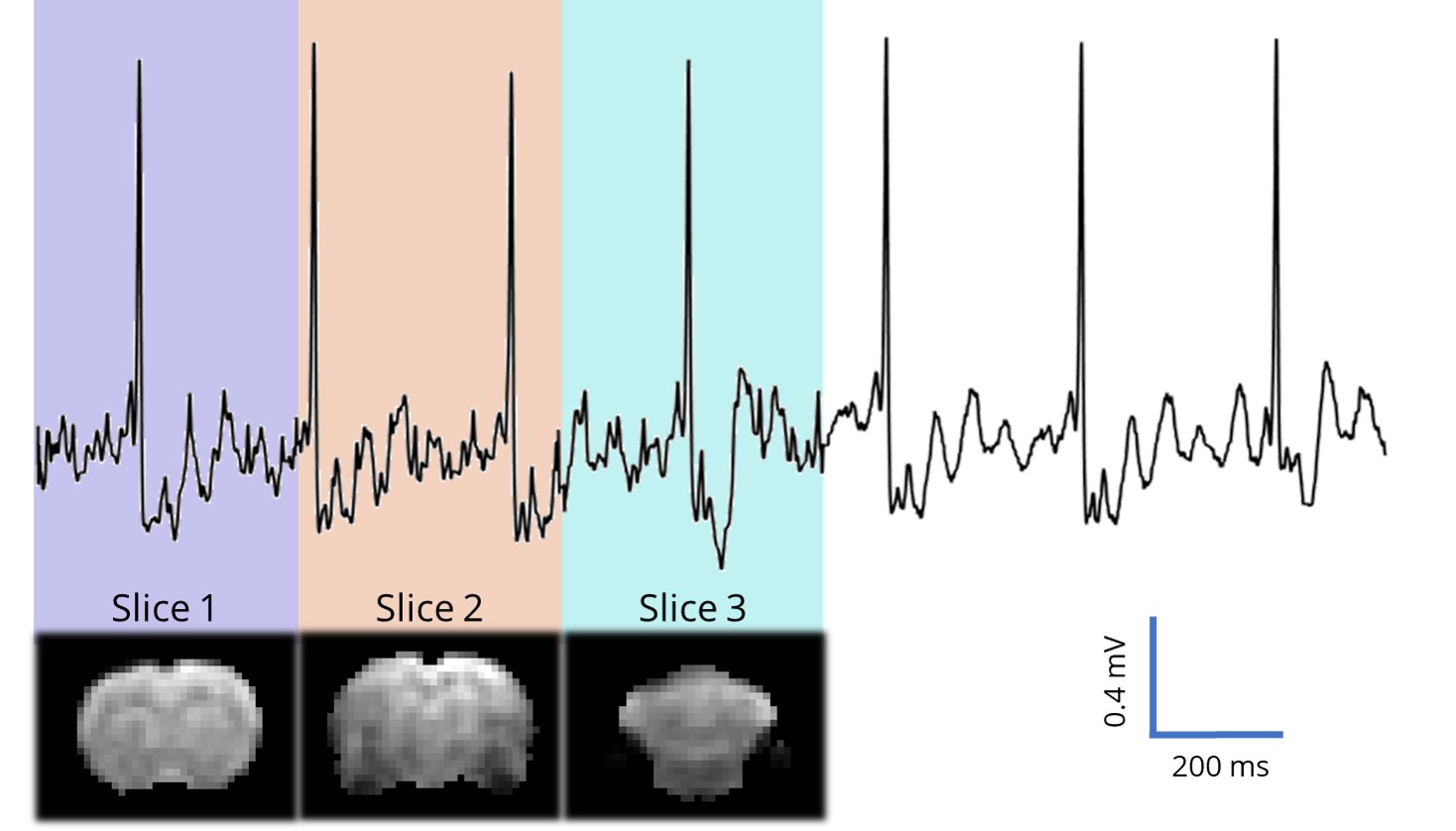
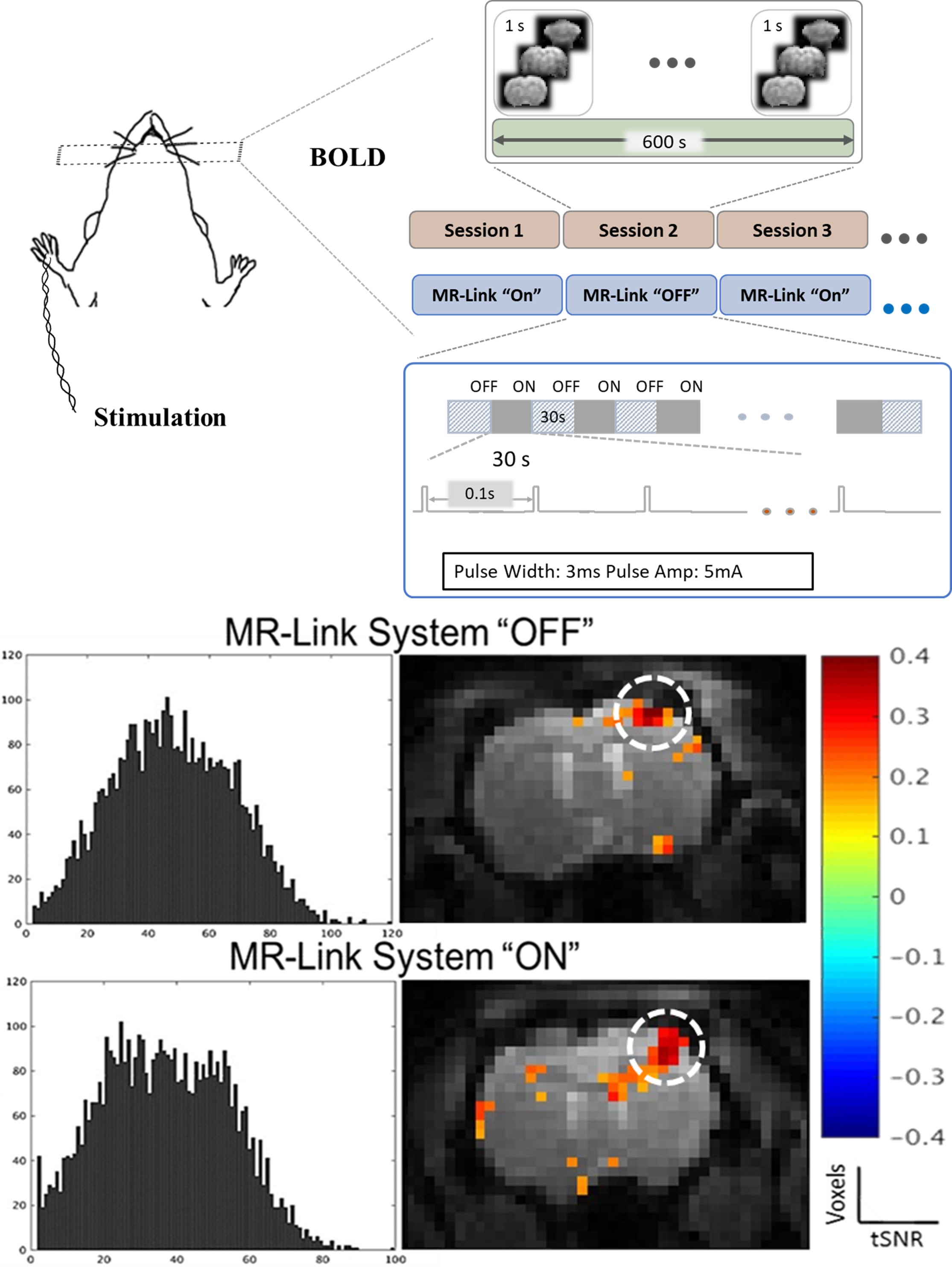
Somatosensory evoked potential (SEP) due to forepaw monophasic current stimulus (3mA, 5ms, 10Hz) was recorded through the device and wirelessly transmitted back to the MRI as the functional images show the non-MR data overlaid as discrete lines in the extended FOV (Fig. 1c). Extraction of MR and EP data demonstrate gradient-artifact free SEP and corresponding bold activation in the R-S1FL region. Co-registration of non-MR data through the fMRI image allowed for accurate synchronization between two datasets (Fig. 4), During each K-space acquisition, EP data were recorded at a sampling rate of 1.32kHz. Additionally, the pickup coil, utilized as a power harvesting tool generated a peak power of 8mW, which was sufficient for the operation of the MR-Link system without the requirement of any additional powering source. Finally, qualitative and quantitative analysis of device’s MR-compatibility showed no identifiable effect on fMRI diagnostic performance as tSNR characteristics and BOLD response (Fig. 5b) were very comparable for the two experimental paradigms shown in Fig. 5a.Conclusions
In this study, we have presented a miniaturized fully wireless platform, suitable for high fidelity neural recording in simultaneous fMRI studies. The cheap and simple solution utilizes MRI electromagnetic interference to adaptively identify various gradient waveforms and modulate analog amplification and remove gradient artifacts. Furthermore, with the miniaturized and low powered design of the recorder, it is suitable to be wirelessly powered using the MRI. The continued development of this technology will pave the path for a fully MRI integrated system for animal and human applications.Acknowledgements
No acknowledgement found.References
1. K. Anami et al., “Stepping stone sampling for retrieving artifact-free electroencephalogram during functional magnetic resonance imaging,”Neuroimage, vol. 19, no. 2, pp. 281–295, 2003.
2. L. G. Hanson, T. E. Lund, and C. G. Hanson, “Encoding of electrophysiology and other signals in MR images,” J. Magn. Reson. Imaging, vol. 25, no. 5, pp.1059–1066, 2007.
Figures