4422
A true comparison of B0 shimming with a very high order spherical harmonic based setup and a multi-coil shim array1Max Planck Institute for Biological Cybernetics, Tuebingen, Germany, 2IMPRS for Cognitive and Systems Neuroscience, University of Tuebingen, Tuebingen, Germany, 3Institute of Physics, Ernst-Moritz-Arndt University, Greifswald, Germany, 4Department of Biomedical Magnetic Resonance, University of Tuebingen, Tuebingen, Germany
Synopsis
In this work, a true experimental comparison between a very high order spherical harmonic based shim setup and a multi-coil shim array is presented. Each technique has its own cons and pros which are studied through several often-used sequences. All evaluations are performed by shimming of the human brain at 9.4T.
INTRODUCTION
Spatial inhomogeneity of the B0 field is a major challenge in many MR sequences with a long echo time. Spatial magnetic field fluctuation degrades data quality in MR spectroscopy and leads to distortion and signal loss in MR imaging1. One remedy is to measure B0 inhomogeneity, decompose it to its fundamental spherical harmonic (SH) components and employ dedicated coils to form SH with the opposite sign. As an alternative, one can decompose the inhomogeneity to predefined basis functions which are generated from a set of small local coils which is known as the multi-coil method2. The aim of this work is to evaluate and make a true experimental comparison between these two common approaches of shimming for human brain applications at 9.4T.METHODS
A 28-channel insert-shim3 (Resonance Research Inc, Billerica, MA) and a 16-channel multi-coil setup4 were used for the comparison (Figure 1). All measurements were performed on a whole-body 9.4T Siemens (Erlangen, Germany) MR scanner. The insert-shim consisted of the full to 4th and partial 5th and 6th order SH terms and the multi-coil consisted of 16 circular coils with a diameter of 100 mm which were equally positioned in two rows. Only up to 4th order insert-shim channels were used for a fair comparison and 16-channel multi-coil setup was used in combined with scanner’s 2nd order shim (25 channels in total for both). Both setups have their own dedicated current amplifiers without any integrated pre-emphasis circuits.
The same subject was measured with both setups in order to have identical brain geometry. The four shimming strategies were static whole brain shimming with the scanner’s 2nd order shim, with the insert-shim and with the multi-coil shim as well as dynamic shimming (only multi-coil). Those strategies were evaluated using B0 maps, EPI and single voxel spectroscopy (SVS). A dual-echo GRE sequence with monopolar readout gradients was used for B0 mapping of the brain with 35 axial slices and a voxel size of 2 mm isotropic. The effectiveness of the shimming was further evaluated with a single-shot EPI scan with 10 axial slices (TE/TR = 25/2000 ms, FOV = 220×220 mm). A STEAM sequence was employed to acquire spectra from a voxel with a volume of 30x30x30 mm3 in the prefrontal cortex. The used sequence parameters were: TE/TR = 11/5000 ms, bandwidth = 8 KHz, 16 averages. The full-width-half-maximum (FWHM) of the water peak was then used to rate shimming efficiency.
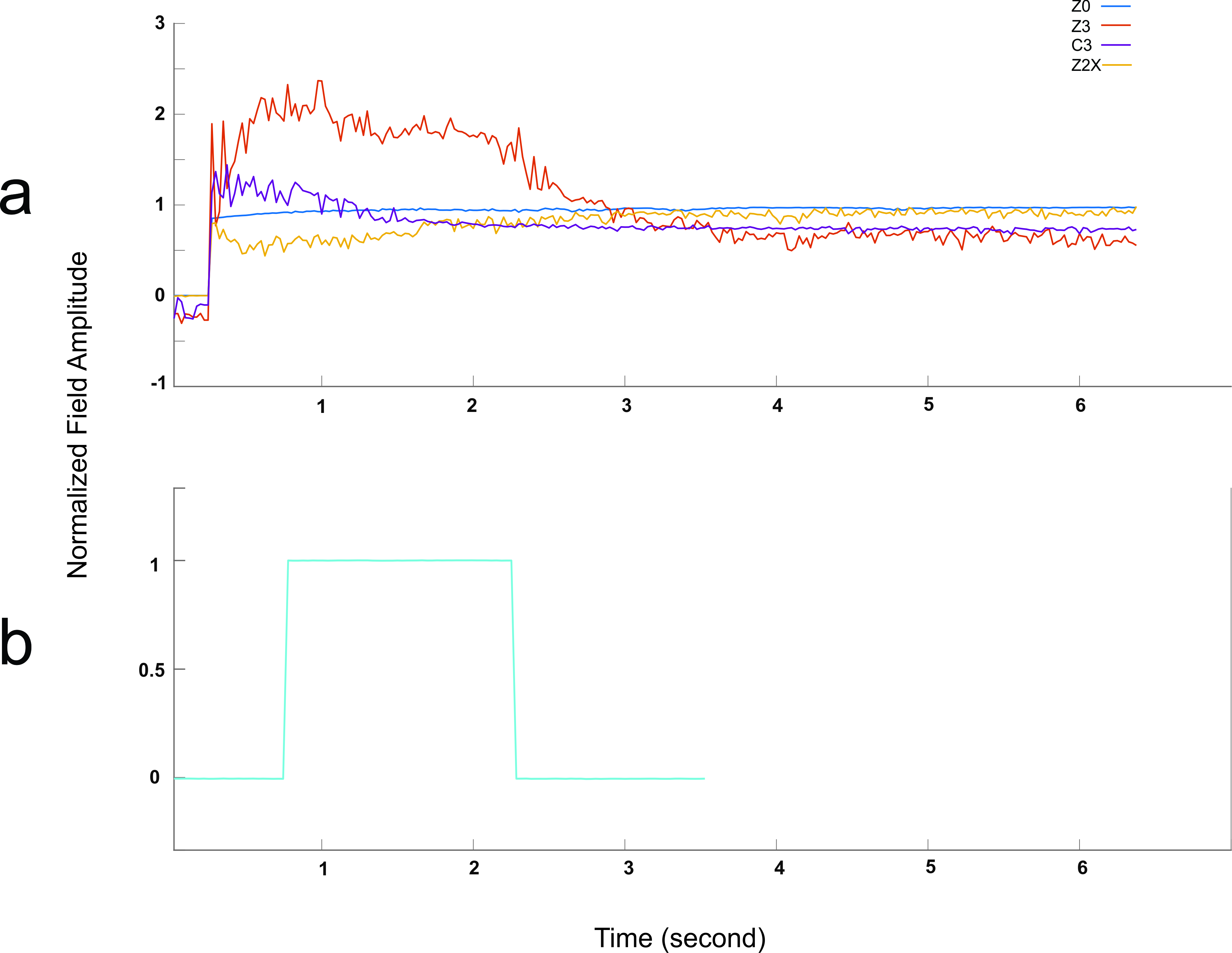
Additionally, eddy current-induced magnetic field perturbations were studied by applying a step input to the individual channels and monitoring spatiotemporal field variation employing NMR field probes. An NMR field camera5 consisting of 12 1H field sensors and 2 19F field sensors6,7 were employed to characterize the behavior of the generated magnetic field8 from the insert-shim and multi-coil setup respectively.
RESULTS AND DISCUSSION
Figure 2 represents a comparison of the B0 maps for the four shimming strategies used in this study. Shimming with the multi-coil setup was performed while the ROI was initially shimmed with the scanner’s 2nd order SH, while the insert shim contains own 2nd order shim coils. Fast switching of the input for dynamic shimming was unapproachable for the insert-shim because the coils cover a large area and have widely overlapping surfaces and high inductance and are accordingly prone to produce severe eddy current artifacts9. Even though the multi-coil was equipped with non-orthogonal basis maps, it performed similar to the insert-shim if driven in dynamic slice-wise mode.
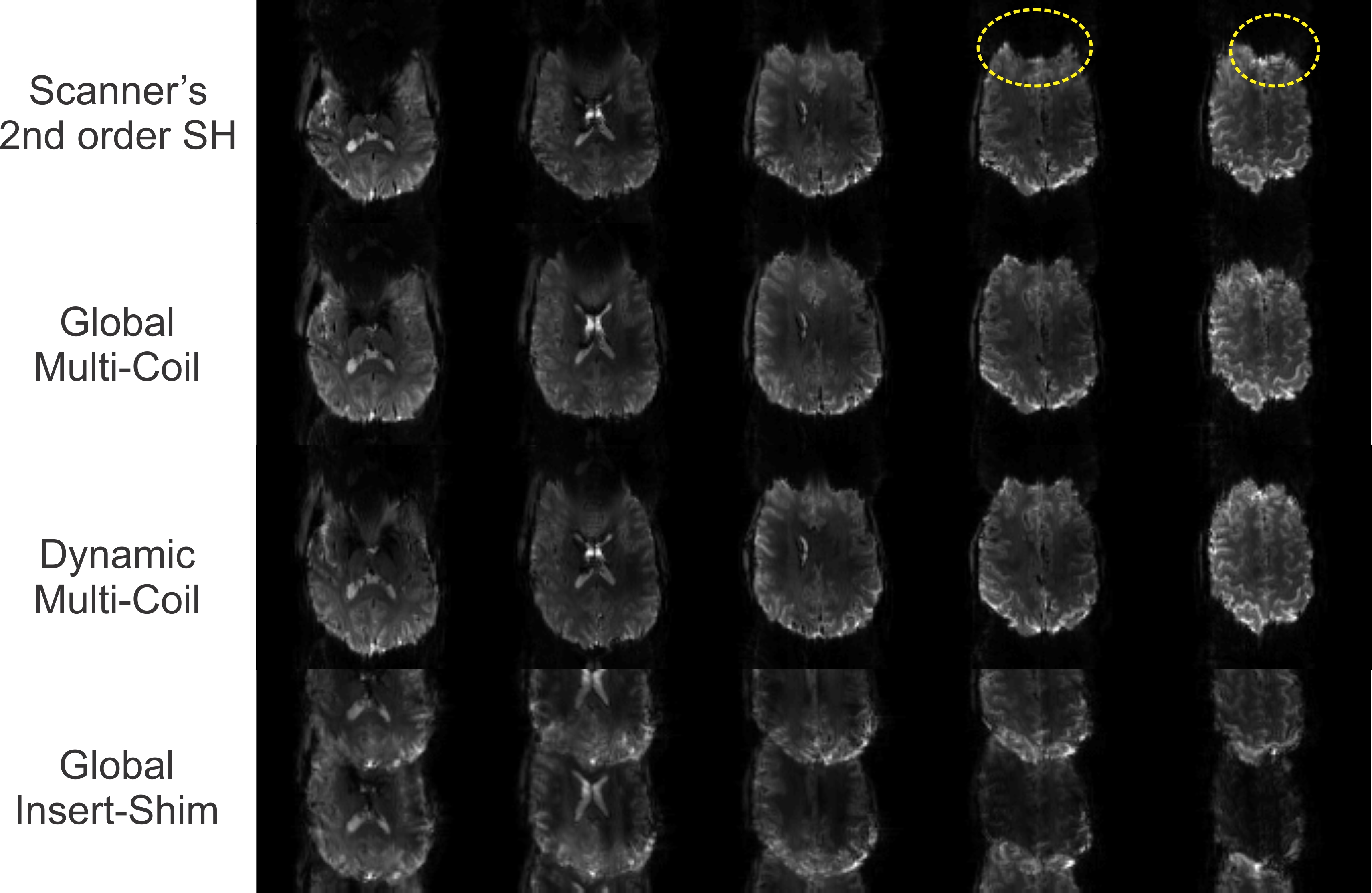
Figure 3 shows the results of the EPI scans acquired after shimming of the brain in the previous step. Although the insert-shim performance was very similar to dynamic multi-coil and better than global multi-coil shimming, very fast gradient switching introduced drastic eddy currents in the insert-shim, and consequently extremely degraded the EPI images. This result was expected due to the characterized eddy current behavior of the insert-shim (Figure 4). Having pre-emphasis can help8,10, but it is difficult to design for so many self- and cross-terms.
As a very sensitive sequence to local field inhomogeneity, SVS was employed. The FWHM of the water peak was 41, 33 and 22 Hz for shimming with the scanner’s 2nd order, multi-coil and insert-shim respectively. The lack of strong rapid gradient switching in spectroscopy avoids the generation of eddy currents, hence the insert-shim can be an ideal solution for SVS.
CONCLUSION
In case of global shimming and SV MRS, the insert-shim setup outperformed the others, particularly when no rapid gradients alterations existed. The multi-coil setup can be readily configured for dynamic slice-wise and real-time shimming to gain the most efficient performance; however, a wide range of sequences are 3D and thus not compatible with slice-wise shimming.Acknowledgements
No acknowledgement found.References
1. Jezzard P. Correction of geometric distortion in fMRI data. Neuroimage. 2012;62(2):648-651. doi:10.1016/j.neuroimage.2011.09.010.
2. Juchem C, Nixon TW, McIntyre S, Rothman DL, Graaf RA De. Magnetic field modeling with a set of individual localized coils. J Magn Reson. 2010;204(2):281-289. doi:10.1016/j.jmr.2010.03.008.
3. Chang P, Nassirpour S, Henning A. Modeling real shim fields for very high degree (and order) B0 shimming of the human brain at 9.4T. Magn Reson Med. 2017. doi:10.1002/mrm.26658.
4. Aghaeifar A, Zivkovic I, Mirkes C, Steffen T, Scheffler K. Global and Dynamic Shimming with the Scanner’s Inbuilt Shim System and a Custom-Made Multi-Coil Setup at 9.4 T. In: 25th Annual Meeting and Exhibition of the International Society for Magnetic Resonance in Medicine. Honolulu, Hawaii, USA; 2017:4330.
5. Chang P, Nassirpour S, Eschelbach M, Scheffler K, Henning A. Constrained Optimisation for Position Calibration of an NMR Field Camera. Magn Reson Med. 2017.
6. Barmet C, De Zanche N, Wilm BJ, Pruessmann KP. A transmit/receive system for magnetic field monitoring of in vivo MRI. Magn Reson Med. 2009;62(1):269-276. doi:10.1002/mrm.21996.
7. Eschelbach M, Aghaeifar A, Bause J, Handwerker J, Anders J, Scheffler K. A Comparison of Prospective Motion Correction with 19F NMR Field Probes and an Optical Camera. In: 25th Annual Meeting and Exhibition of the International Society for Magnetic Resonance in Medicine. Honolulu, Hawaii, USA; 2017:1304.
8. Fillmer A, Vannesjo SJ, Pavan M, Scheidegger M, Pruessmann KP, Henning A. Fast iterative pre-emphasis calibration method enabling third-order dynamic shim updated fMRI. Magn Reson Med. 2015;1131(October 2014):1119-1131. doi:10.1002/mrm.25695.
9. Vannesjo SJ, Dietrich BE, Pavan M, et al. Field camera measurements of gradient and shim impulse responses using frequency sweeps. Magn Reson Med. 2014;72(2):570-583. doi:10.1002/mrm.24934.
10. JUCHEM C. Dynamic Shimming of the Human Brain at 7 T. Concepts Magn Reson Part B Magn Reson Eng. 2009;35(3):121-132. doi:10.1002/cmr.b.
Figures
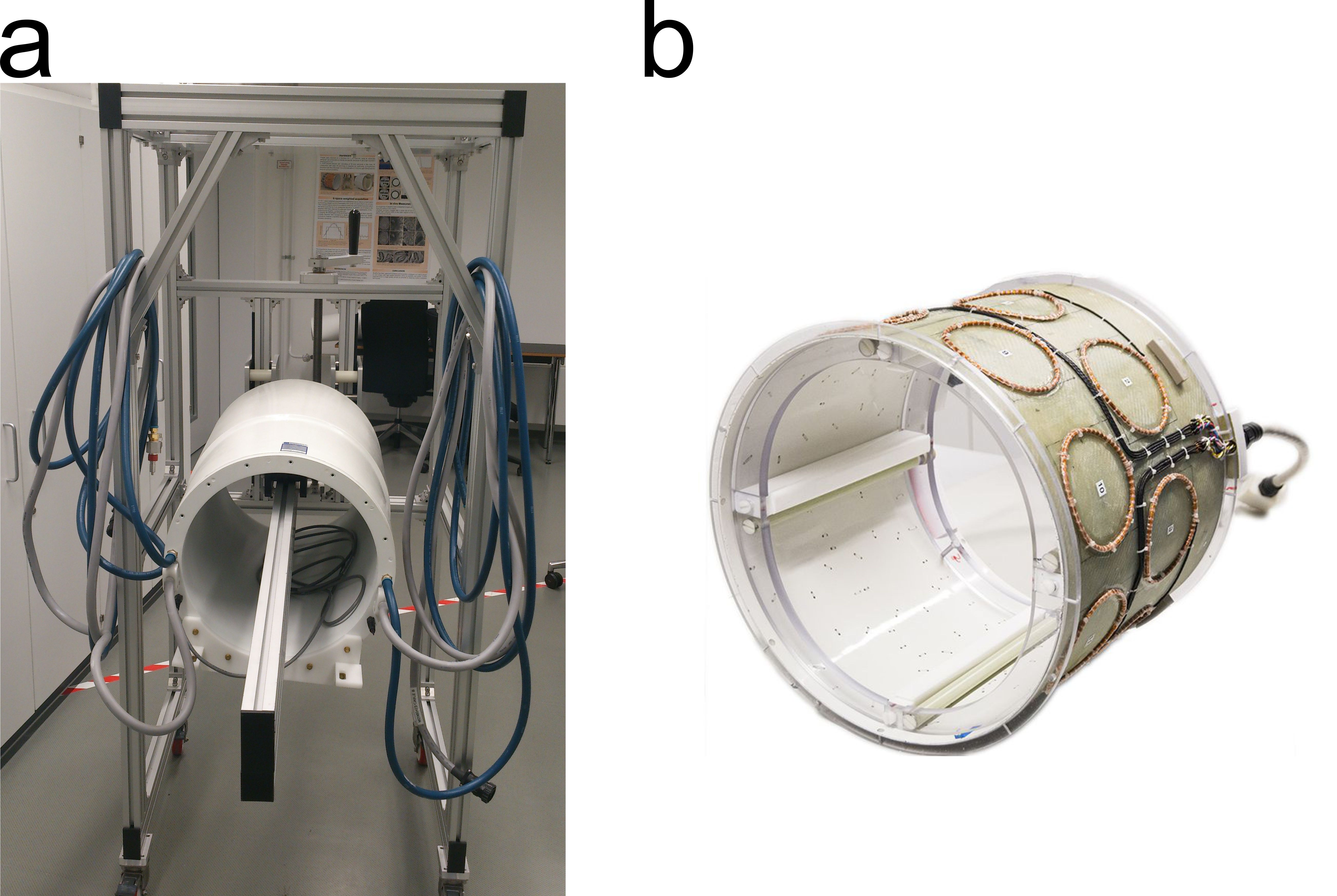
Figure 1. Photograph of the two setups used in the study. a) 28-channel insert shim setup which generates spherical harmonic terms, b) 16-channel multi-coil setup
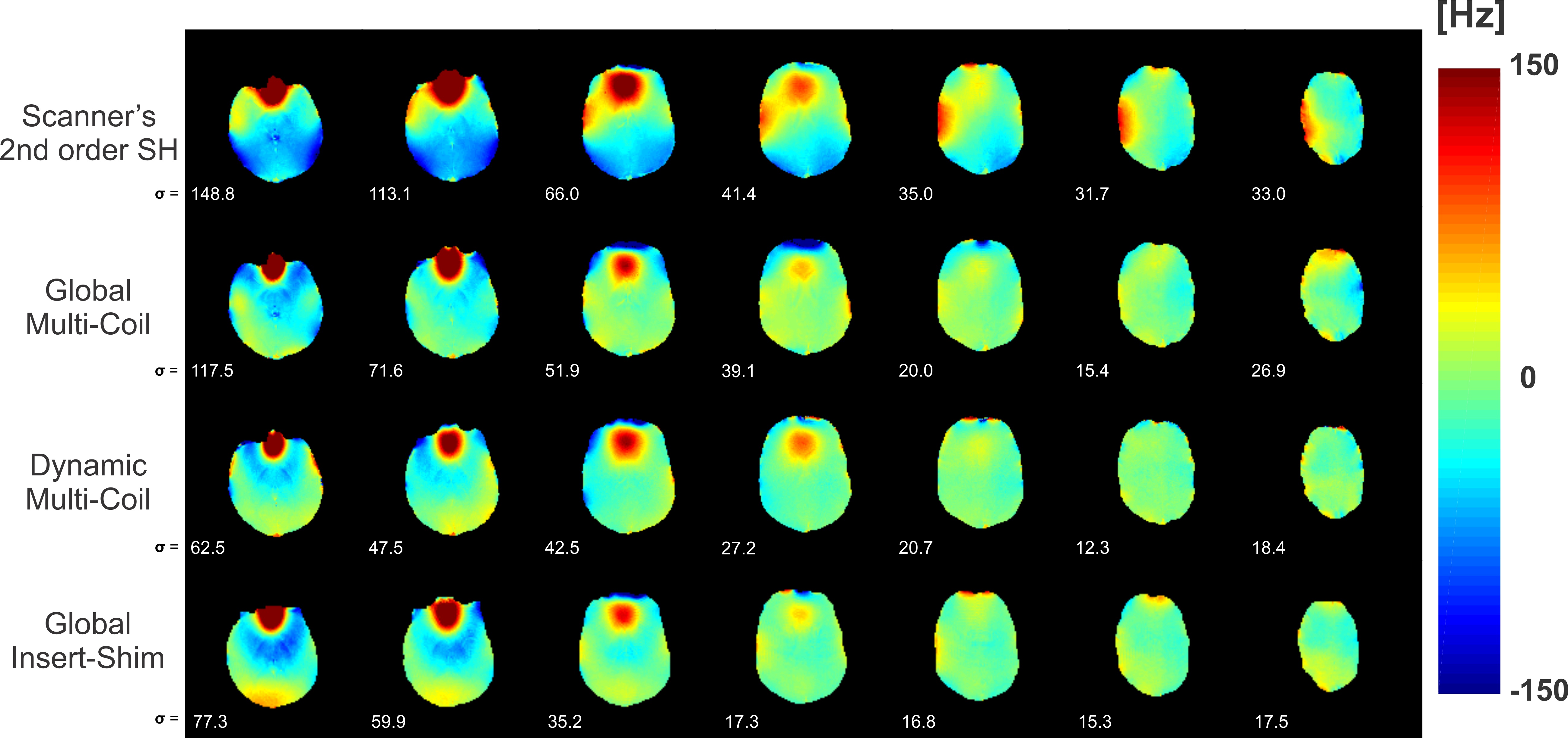
Figure 2. Comparison of the B0 shimming performance among scanner's 2nd order SH shim setup, 16-channel multi-coil and insert-shim which is measured with dual-echo GRE sequence. For lower slices of the brain which have stronger inhomogeneity, dynamic multi-coil shimming (row 3) performed better, while for middle and top slices, insert-shim (row 4) was more efficient. Subject was repositioned due to setup change, hence the slices in the last row are slightly shifted. For the whole ROI, a standard deviation of 68.2, 49.2, 37.3 and 37.5 Hz were obtained for shimming with scanner’s 2nd order SH, global multi-coil, dynamic multi-coil and global insert-shim respectively.