4409
Computer-aided design and simulation of a double-nuclear parallel transmit RF coil array for proton MRI and carbon-13 MRS of human extremities at 7T1Radiological Sciences Laboratory, Department of Radiology, School of Medicine, Stanford University, Stanford, CA, United States
Synopsis
13C MR spectroscopy can offer important metabolic information in the musculoskeletal system without the use of ionizing radiation. In this work, we design and simulate a novel double-nuclear pTx RF coil array (8-channel per frequency) with ICE decoupling coils for proton MRI and 13C MRS of human extremities. Coupling was compared between the RF coil array with and without ICE elements demonstrating the significant decoupling effect of the ICE coils. Calculated B1+ field maps for both the proton MTLs and the 13C loops with ICE elements show sufficient transverse magnetization for MRI and MRS applications. Through the use of switch-tuned ICE decoupling elements, we have demonstrated the feasibility of dual-frequency pTx arrays for 1H imaging and 13C spectroscopy of human extremities at 7T.
Introduction
Combined MR imaging and spectroscopy at ultra-high field strengths is known to have important applications in the musculoskeletal system for high resolution anatomical and high sensitivity metabolic imaging [1]; however, efficient data acquisition requires dual-tuned or switch-tuned array-based RF coils which are not commercially available and are challenging to design. 13C MR spectroscopy can offer important metabolic information in the musculoskeletal system without the use of ionizing radiation, but there have only been a few coils specifically designed for extremities, and none are volumetric. [2,3,4,5] In previous work, a double-nuclear surface coil design capable of proton MRI and 13C MRS at 7T was shown to have a high Q-factor and excellent decoupling between the two coils. [6] It was suggested that this design could be used as the basis for an array, though coupling between elements would need to be addressed. In this work, we propose and demonstrate (by simulation) a double-nuclear double parallel transmit (pTx) RF coil array (8 Tx/Rx elements at each frequency) with induced current elimination (ICE) decoupling coils [7] for proton MRI and 13C MRS of human extremities.Methods
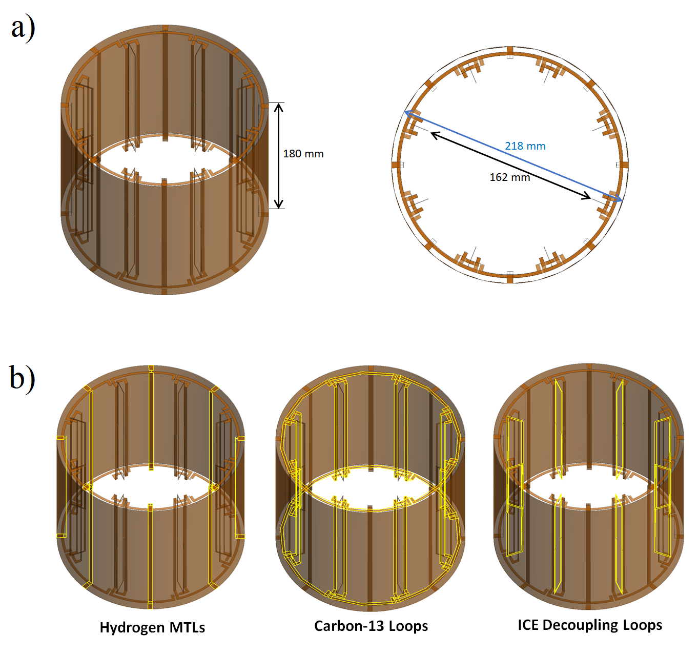
Our concept utilizes a microstrip transmission line (MTL) array for 1H, an overlapping loop array for 13C, and common ICE elements that operate in switch-tuned mode to serve the decoupling requirements at both resonant frequencies (298MHz and 75MHz). The coil array was modeled using the computational electromagnetics software package FEKO Suite 7.0.2 (Altair Engineering Inc; Troy, MI). We used CADFEKO to generate a cylindrical coil geometry with internal diameter of 162 mm, external diameter of 218 mm, and length of 180 mm (Figure 1a). Each of the 8 microstrips in the proton MTL array (strip width = 5.1 mm, strip length = 180 mm, displacement from shield = 11mm) was connected to a continuous copper cylinder (218 mm diameter), which served as the common shield. These MTL elements were positioned at 45˚ increments and utilized balanced tuning and matching capacitors. The rectangular loops of the 8-channel 13C lumped-element phased array (short diameter = 78 mm, long diameter = 170 mm, conductor width = 3.1 mm) were overlapped azimuthally to minimize mutual coupling and arranged at 45˚ increments, and used balanced tuning and matching capacitors. Decoupling of both arrays was achieved using rectangular ICE coils arranged radially between the overlapping 13C loops. Using a diode switch and balanced tuning capacitors, ICE tuning capacitance was switched between two states to achieve 1H MTL and 13C loop decoupling (Figure 1b). Lumped element optimization was performed using Advanced Design System 2016.01 (Keysight Technologies; Santa Rosa, CA). EM field modeling was computed using FEKO Solver. Results were visualized and exported using POSTFEKO.Results
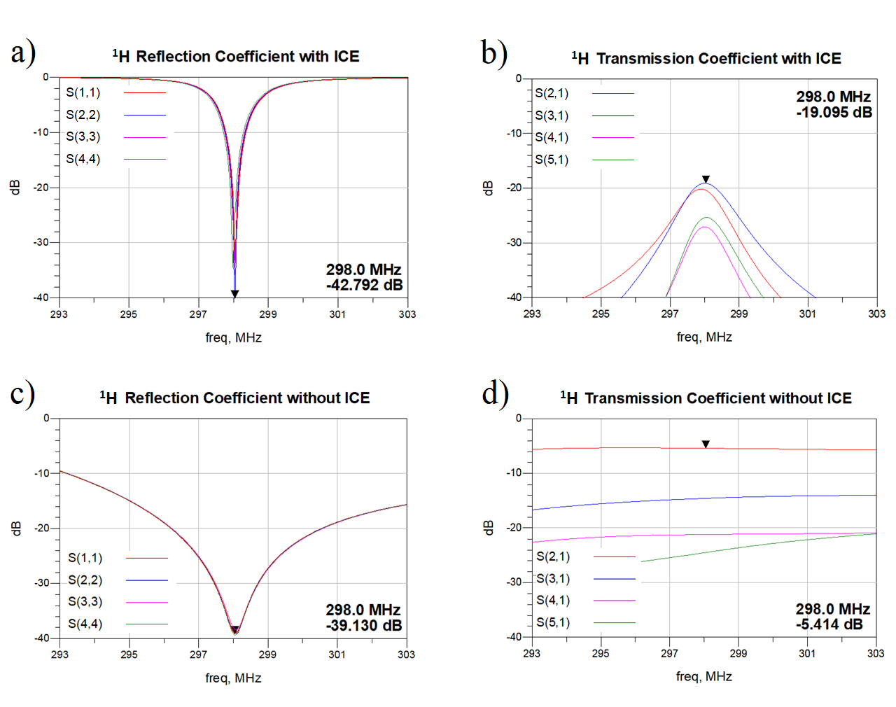
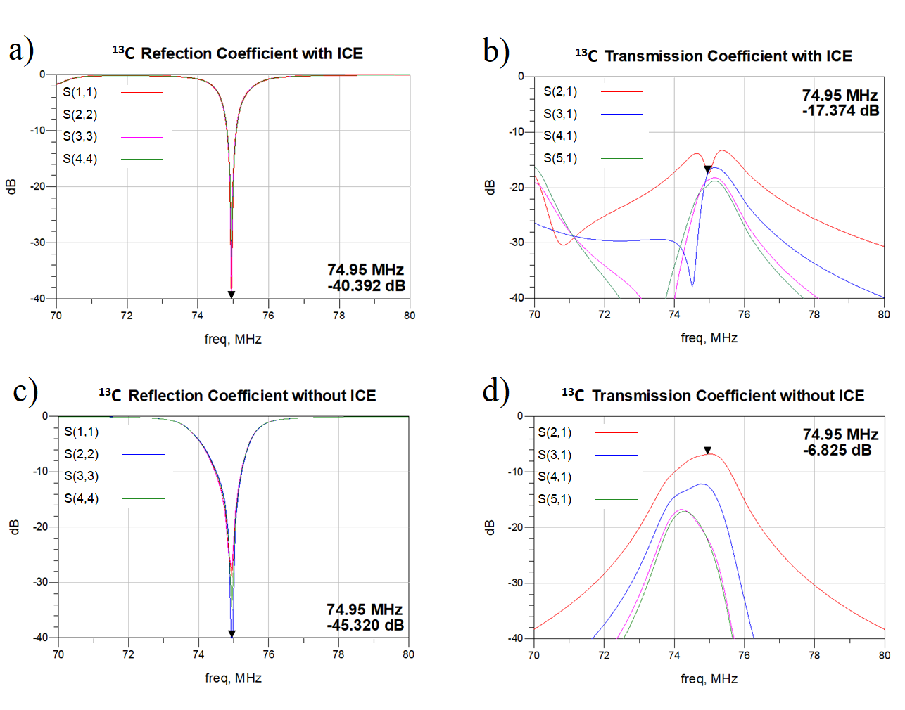
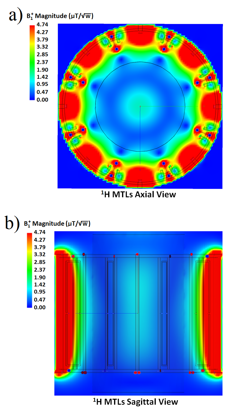
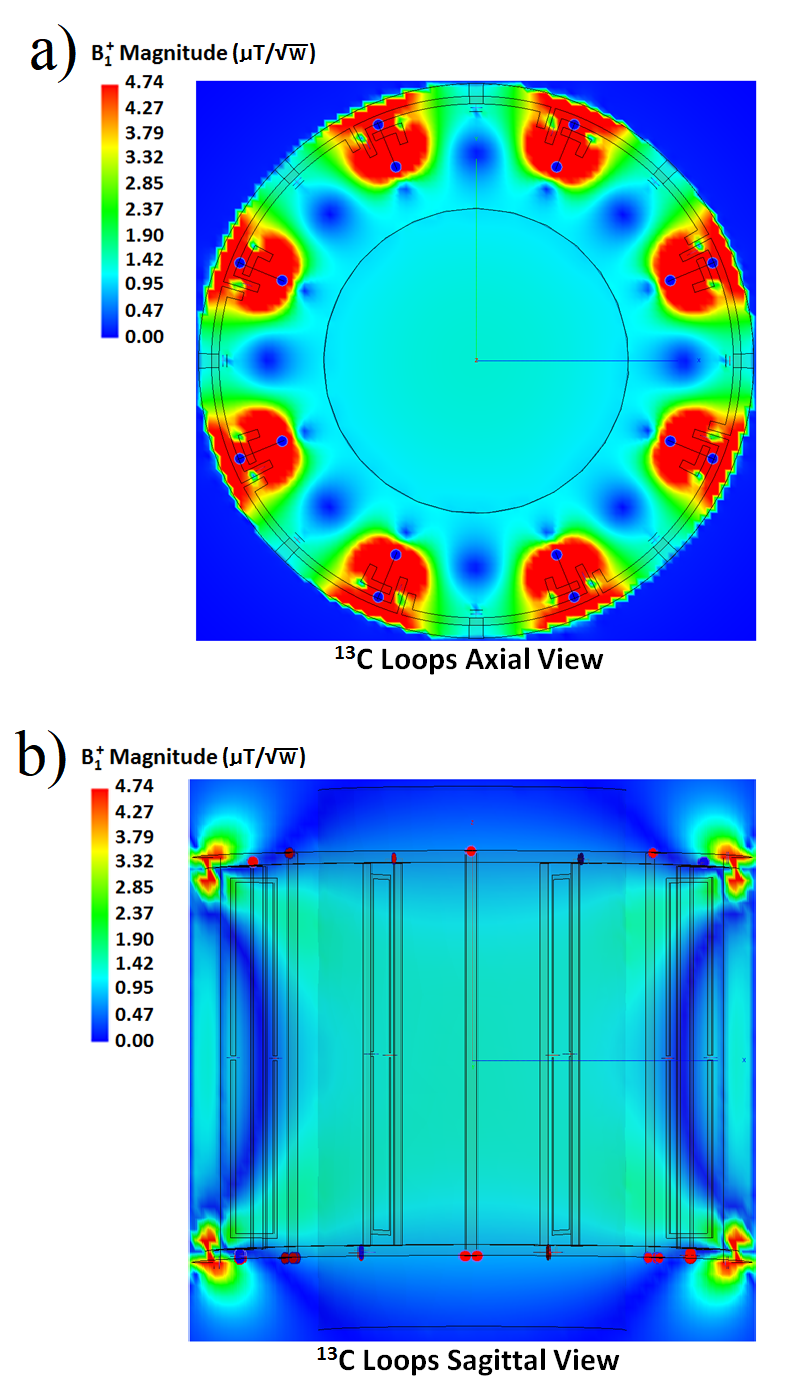
Bench test simulations are summarized in Figures 2 and 3. MTLs demonstrated a high Q-factor at -42dB (Figure 2a). By using ICE coils, good decoupling (< -19dB) between all pairs of elements of the MTL array was achieved (Figure 2b). In contrast, without the ICE coils, the Q-factor of the MTLs was poor at -39dB (Figure 2c), and S21 indicates significant coupling in the MTL array with the worst performance at -5dB. The 13C loops with ICE coils demonstrate a high Q response with S11 vales in the range of -40dB (Figure 3a). Figure 3b displays S21 responses for the 13C loop array incorporating ICE coils, demonstrating excellent decoupling (< -17dB) between all elements. Without the ICE loops, the 13C loops exhibit lower Q (Figure 3c) and significant coil coupling as high as -6dB (Figure 3d). Calculated B1+ field maps show high efficiency and good uniformity, with peak/mean B1+/√P values of 1.49/1.03 µT/√W at the 1H resonance and 1.39/1.23 µT/√W at the 13C resonance (Figures 4 & 5).Discussion
While the use of MTLs and superimposed loops was shown in prior work to have favorable decoupling properties due to inherently perpendicular fields [6], coupling between coil elements in an array configuration remained a problem to be solved. In the present work, we found that passive ICE loops, after design optimization and careful tuning, performed extremely well as “magnetic walls” that suppressed coupling to nearest as well as farther elements for both 1H and 13C arrays. Switch-tuning of the ICE loops allowed a common set of loops to decouple both 1H and 13C arrays. This highly successful decoupling behavior was not possible without the ICE elements.Conclusion
Through the use of switch-tuned common ICE decoupling elements, we have demonstrated the feasibility of dual-frequency pTx arrays for 1H / 13C imaging / spectroscopy of human extremities at 7T.Acknowledgements
The authors would like to acknowledge research support by GE Healthcare and by NIH P41 EB015891.References
1. Henning A. Proton and multinuclear magnetic resonance spectroscopy in the human brain at ultra-high field strength: A review. Neuroimage. 2017; in press.
2. Stephenson MC, Gunner F, Napolitano A, et al. Applications of multi-nuclear magnetic resonance spectroscopy at 7T. World J Radiol. 2011; 3(4):105-113.
3. Heinicke K, Dimitrov I, Romain N, et al. Reproducibility and Absolute Quantification of Muscle Glycogen in Patients with Glycogen Storage Disease by 13C NMR Spectroscopy at 7 Tesla. PLoS One. 2014;9(10):e108706.
4. Meyerspeer M, Roig E, Gruetter R, Magill A. An Improved Trap Design for Decoupling Multinuclear RF Coils. Magn Reson Med. 2014;72(2):584-90.
5. Roig E, Magill A, Donati G, et al. A Double-Quadrature Radiofrequency Coil Design for Proton-Decoupled Carbon-13 Magnetic Resonance Spectroscopy in Humans at 7T. Magn Reson Med. 2015;73(2):894-900.
6. Rutledge O, Kwak T, Cao P, Zhang X. Design and test of a double-nuclear RF coil for 1H MRI and 13C MRSI at 7T. J Magn Reson. 2016;267:15-21.
7. Li Y, Xie Z, Pang Y, et al. ICE decoupling technique for RF coil array designs. Med Phys. 2011;38(7):4086-4093.
Figures