4408
Development of an Asymmetrical Birdcage Design towards Homogeneous Volume Excitation for Hyperpolarized Xenon-129 Lung Imaging at 3T1Imaging Research Center, Cincinnati Children's Hospital Medical Center, Cincinnati, OH, United States, 2Center for Pulmonary Imaging Research, Cincinnati Children's Hospital Medical Center, Cincinnati, OH, United States, 3Philips Healthcare, Gainesville, FL, United States, 4Philips Healthcare, Best, Netherlands
Synopsis
Hyperpolarized 129Xe imaging is increasingly viewed as a viable tool for assessing lung structure and function. 129Xe MRI requires that large flip angle and frequency selective RF pulses be applied homogeneously across the entire thorax. The transmit performance of three common multi-nuclear whole-body birdcage designs was assessed under a loaded condition at different locations inside the RF shield with electromagnetic field simulations. The acquired data was used to develop a novel, optimized birdcage design for which the transmit performance was analyzed and compared to the three common designs.
Introduction
Hyperpolarized 129Xe imaging is increasingly viewed as a viable tool for assessing lung structure and function in patients1. Additionally, HP 129Xe is moderately soluble in tissues and possesses a large (>200 ppm) in vivo chemical shift range, making dissolved-phase 129Xe MR uniquely sensitive to regional gas-exchange dynamics2-4. Dissolved 129Xe MRI requires that large flip angle and frequency selective RF pulses be applied homogeneously across the entire thorax to avoid unintentionally exciting the gas-phase 129Xe magnetization, while confining dissolved HP 129Xe magnetization to the gas exchange tissues. Birdcage coils are traditionally constructed using a cylindrical birdcage design with equidistant rung-spacing centered in the magnet’s RF shield. Asymmetrical birdcage coils have been developed for non-proton nuclei imaging, allowing the multi-nuclear coil to be inserted into the magnet riding on top of the patient table5-7. However, the homogeneity of theses designs was evaluated with the coil unloaded and only within the central slice locations. Thus it remains unclear if these designs are suitable for truly quantitative dissolved-phase 129Xe MRI. To develop a suitable large and homogeneous 129Xe 3T birdcage coil, we analyzed the volume excitation performance of multiple coil designs with electromagnetic simulations. The influence of the shield size and position of the coils were analyzed in detail. Results of the analysis were used towards creating two optimized designs for homogeneous volume excitation necessary for 129Xe imaging.Methods
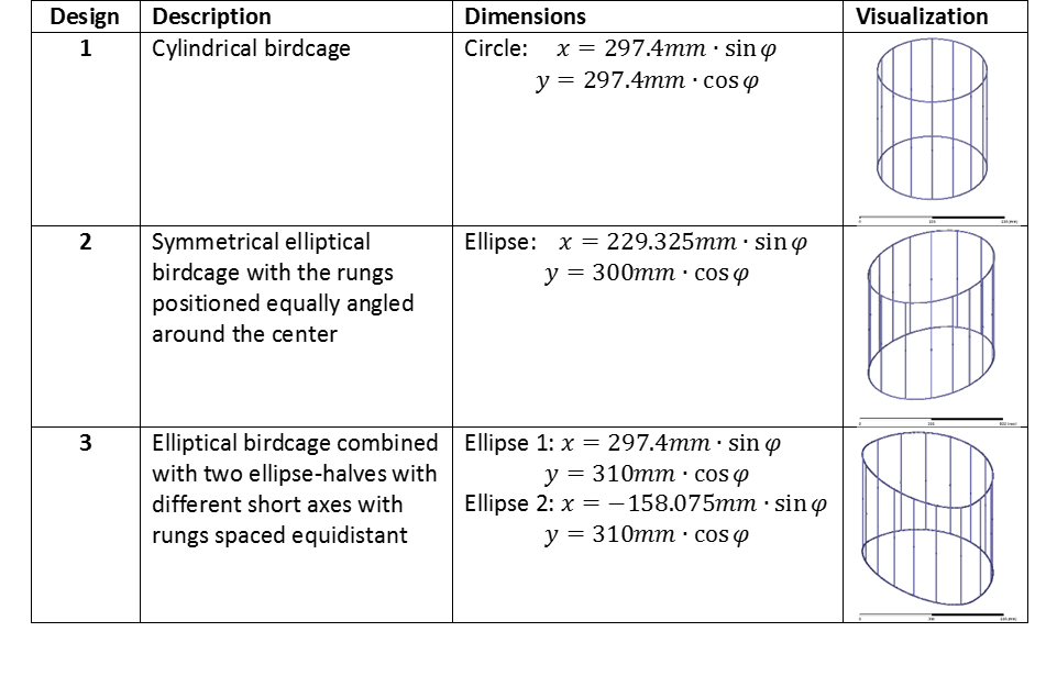
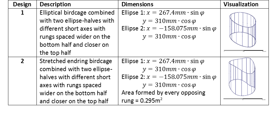
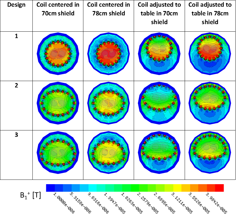
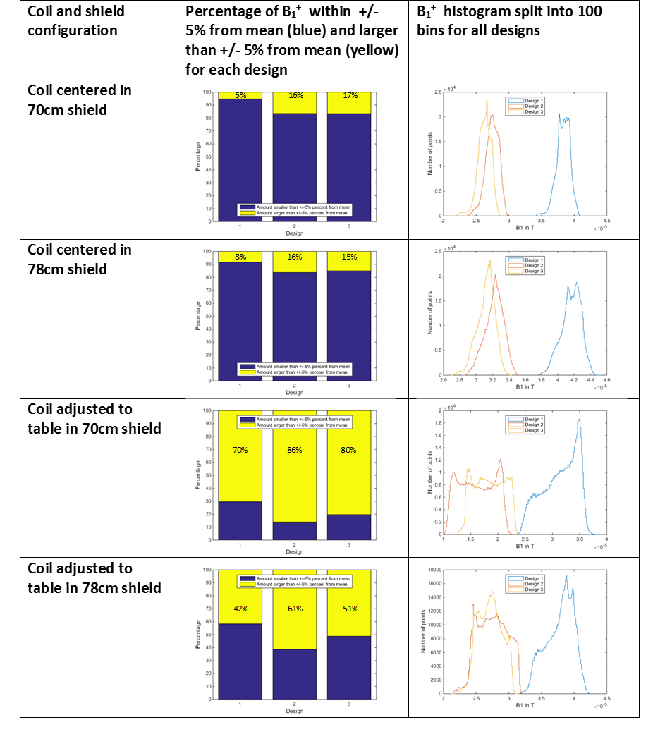
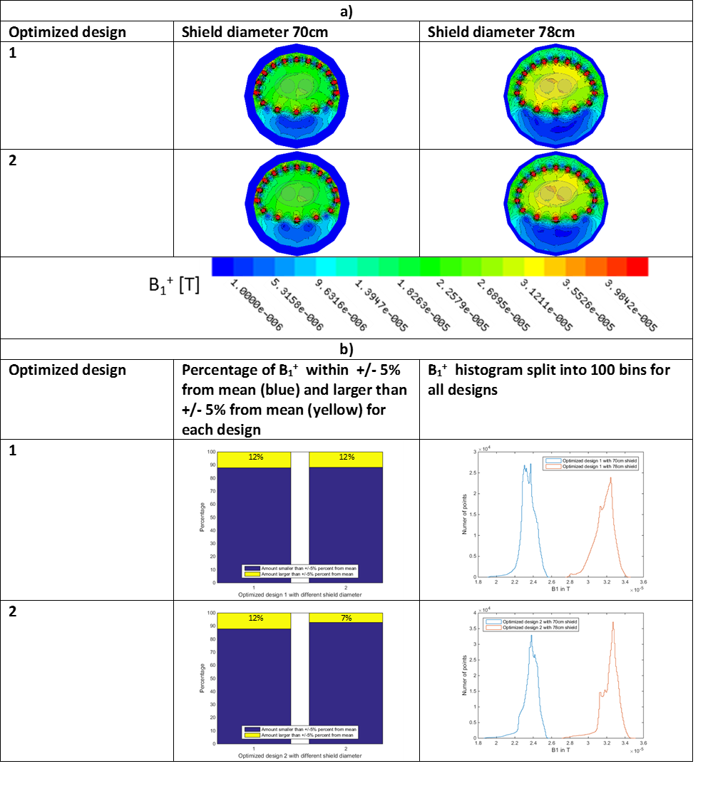
Electromagnetic field simulations were performed for three different 16-rung birdcage coil designs in HFSS (ANSYS), described in Table 1. Each coil design was simulated with two RF shield diameters, 70cm and 78cm, at two locations, centered and adjusted to the patient table inside the RF shield. Coils were loaded with a centered torso-shaped elliptical phantom with two cylinders inside the phantom representing adult-sized xenon-air filled lungs. Birdcages were excited with 35.3MHz, 6A current sources placed at the center of each rung with phase shift increments of 22.5°. Field maps of the center axial slice and B1+ histograms for a 17cm x 25cm x 25cm VOI centered inside the phantom were used for analytical comparison. A preferred deviation range inside the VOI of +/-5% from the mean B1+ was chosen as the target margin for comparison. To improve B1+ homogeneity inside the VOI two optimized designs, described in Table 2, were analyzed adjusted to the patient table for both RF shield diameters.Results
Transmit field maps and bar graphs with histograms for the birdcage designs are presented in Table 3 and 4 respectively. Axial center slices show a homogeneous B1+ for all centered designs with dielectric effects inside the phantom. Designs adjusted to the table position, however, showed highly inhomogeneous excitation profiles. All birdcage designs for the centered shield case show a homogeneity >83% for the target margin of +/-5% from the mean B1+ inside the VOI. Design 1 has the highest homogeneous profile at 95%, in addition to being the most efficient of the designs. The larger shield diameter improved VOI homogeneity for design 3. Coil mean B1+ efficiency improved by 3.3µT for design 1, 4.8µT for design 2, and 4.9µT for design 3. In the patient table configuration, design 1 retained the highest homogeneous B1+ profile with 30% inside the margin for the 70cm shield. Whereas the two other designs dropped to 14% and 20%. The 78cm shield increased the homogeneity to 58%, 39%, and 49%, respectively, and the mean B1+ increased by 6.4µT for design 1, 11.3µT for design 2, and 8.7µT for design 3. Iterative movements of rung positions and decreasing the short axis of the top-half ellipse improved the B1+ homogeneity to 88% for the smallest and largest shield. Stretching the endings and maintaining equal areas of opposing rungs improved the homogeneity for the larger shield size, increasing to 93% inside the VOI. The mean B1+ increased by 0.2µT-0.6µT.Conclusion and Discussion
Analysis of the three common birdcage designs showed B1+ homogeneity and efficiency is strongly dependent on the position of the birdcage within the RF shield and the distance between the rungs and the shield. Optimization of the rungs’ position and the coil dimensions homogenized the field distribution significantly under loaded condition. Furthermore, end-ring stretching improved the field homogeneity, approaching similar homogeneity for the desired target margin as on a traditional centered cylindrical birdcage, at the cost of efficiency and A-P dimensions. Data analysis of commonly used birdcage designs provided the insight in field pattern distribution under a loaded condition including dielectric effects. This resulted in optimizing rung spacing and end-ring shape for homogeneous B1+ excitation profiles across the entire thorax volume and ultimately identifying two potential designs for construction.Acknowledgements
No acknowledgement found.References
1. Walkup L L, Woods J C. Translational applications of hyperpolarized 3He and 129Xe. NMR in Biomed Volume 27, Issue 12 December 2014 Pages 1429–1438.
2. Stewart N J ,Horn F C, Norquay G, et al. Reproducibility of Quantitative Indices of Lung Function and Microstructure from 129Xe Chemical Shift Saturation Recovery (CSSR) MR Spectroscopy. Magn Reson Med 77:6 (2017) 2107-2113.
3. Kaushik S S, Robertson S H, Freeman MS et al., Single-Breath Clinical Imaging of Hyperpolarized 129Xe in the Airspaces, Barrier, and Red Blood Cells Using an Interleaved 3D Radial 1-Point Dixon Acquisition. Magn Reson Med 75:4 (2016) 1424-1443.
4. Ruppert K, Altes T, Mata J F, et al., Detecting Pulmonary Capillary Blood Pulsations Using Hyperpolarized Xenon-129 Chemical Shift Saturation Recovery (CSSR) MR Spectroscopy. Magn Reson Med 75:4 (2016) 1771-1780.
5. Wetterling F, Corteville D M, Kalayciyan R,et al. Whole body sodium MRI at 3T using an asymmetric birdcage resonator and short echo time sequence: first images of a male volunteer. Phys. Med. Biol. 57 (2012) 4555–4567.
6. Zanche N D, Chhina N, Teh K, et al. Asymmetric Quadrature Split Birdcage Coil for Hyperpolarized 3He Lung MRI at 1.5T. Magn Reson Med 60:431–438 (2008).
7. Dregely I, Ruset I C, 2 Wiggins G, et al. 32-Channel Phased-Array Receive with Asymmetric Birdcage Transmit Coil for Hyperpolarized Xenon-129 Lung Imaging. Magn Reson Med 70:576–583 (2013).
Figures