4398
Feasibility exploration of constructing volume RF coil with coupled dipole antennas at 9.4T1Institute of Biophysics, Chinese Academy of Sciences, Beijing, China, 2Lauterbur Imaging Research Center, Shenzhen Institutes of Advanced Technology, Chinese Academy of Sciences, Shenzhen, China, 3Shenzhen Key Laboratory for MRI, Shenzhen, China, 4Department of Radiology and Biomedical Imaging, University of California, San Francisco, CA, United States, 5UCSF/UC Berkeley Joint Graduate Group in Bioengineering, San Francisco, CA, United States
Synopsis
Dipole antenna, a simple resonator structure, has demonstrated a unique capability of achieving high resonant frequency and utility in MR imaging. Theoretically, this simple resonator structure could be advantageous for building non-array volume RF coil design for ultrahigh field MR imaging applications. In this work, we explore the feasibility of using dipole antenna as resonant element to design non-array volume coils for the ultrahigh field 9.4T MR imaging. The results show that the dipole antennas have sufficient coupling among the resonator elements to create multi-mode resonances and form uniform B1 field distribution within the volume coil at 400 MHz.
INTRODUCTION
Design of ultrahigh field RF volume coils could be technical challenging due to the required high operating frequency. Dipole antenna1,2,3, a simple resonator structure, has demonstrated a unique capability of achieving high resonant frequency and utility in MR imaging. Theoretically, this simple resonator structure could be advantageous for building non-array volume RF coil design for ultrahigh field MR imaging applications. In this work, we explore the feasibility of using dipole antenna as resonant element to design non-array volume coils for the ultrahigh field 9.4T MR imaging. The results show that the dipole antennas have sufficient coupling among the resonator elements to create multi-mode resonances and form uniform B1 field distribution within the volume coil at 400 MHz. This could open a new avenue to high frequency RF volume coil designs for MR imaging at high and ultrahigh fields.METHODS
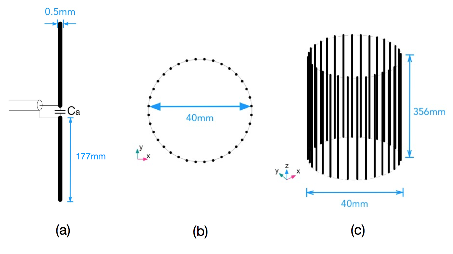
The dipole antennas will be used for building the RF volume coil is sketched in Figure 1 (a). The schematic of the volume coil which is expected to generate a uniform B1-field at 400MHz based on coupled dipole antennas is show in Figure.1 (b) and (c). Total 32 dipole antennas are equally placed in parallel to form a cylinder with a diameter of 40mm.The arm length is 177mm and the size of gap between two arms where the feed line is connected to is 2mm. The overall height of the formed volume coil is 356mm. The material of the arms of the dipole antennas is the annealed copper and its cross section is circle with radius of 0.25mm. As showed in Figure.1(a), on each dipole antenna, a capacitor of 10pF is connected in the middle of two conductive arms to enhance the coupling. To test the feasibility of this dipole volume coil setup, numerical simulations using the time domain solver CST Studio were performed to calculate the resonant frequency of the individual dipole antenna and also the dipole volume coil. The target resonant frequency of the volume coil was set to 400MHz for 9.4T imaging applications. B1 fields and electric fields of the volume coil were also numerically mapped. The distribution of linear field and quadrature or circularly polarized field was analyzed.
RESULTS
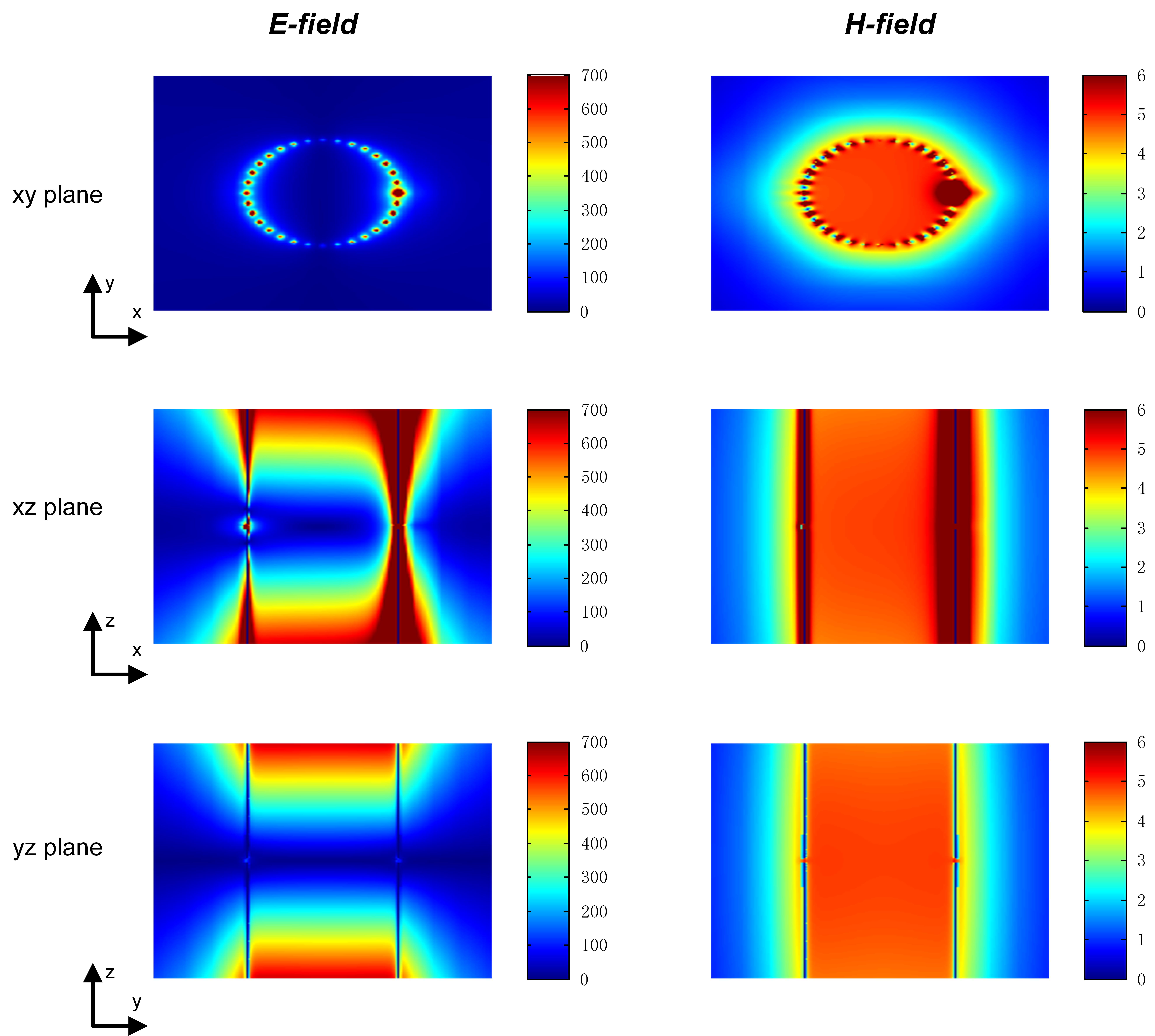
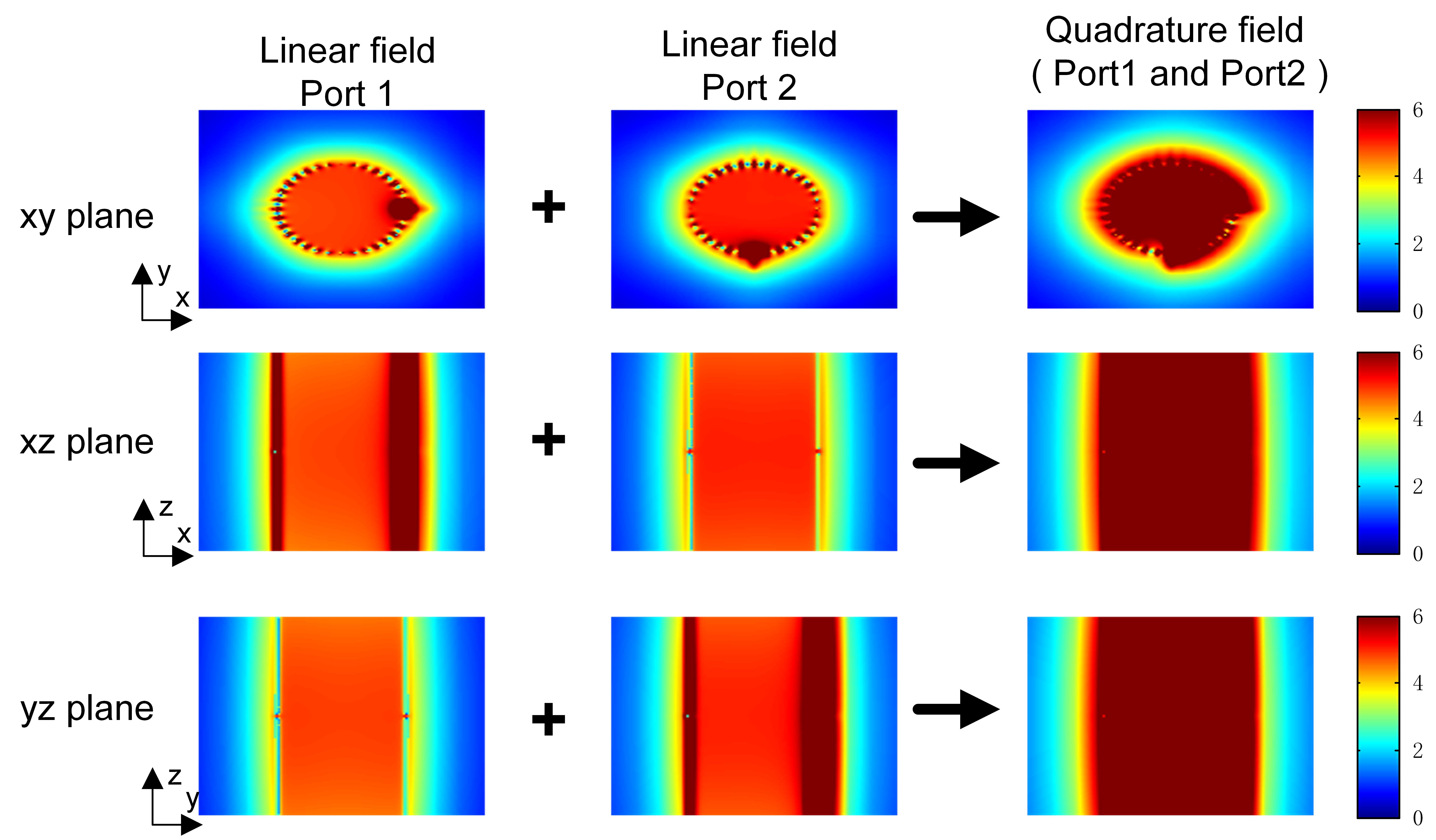
Electric and magnetic field distribution of the one port volume coil in the center horizontal xy plane and the center vertical xz/yz plane is showed in Figure.2, demonstrating a fairly homogeneous B1 field distribution. Although only one dipole in the volume coil is excited, the rest of dipoles are also excited due to the sufficient electromagnetic coupling among the dipoles. The quadrature magnetic field distribution of the volume coil generated by driving two orthogonal ports is showed in Figure.3. The relatively uniform distribution of the quadrature field exhibits that the dipole volume coil can also be used in quadrature.DISCUSSION/CONCLUSION
This study demonstrates that volume coil using coupled dipole antennas is feasible. With sufficient electromagnetic coupling among the resonant elements, homogeneous magnetic field can be generated. The distance between two adjacent dipoles is crucial to the coupling. The volume coil with large diameter may need more dipole antennas to enhance the required electromagnetic coupling. While the current design is based on standard dipole antennas of which the length of the dipoles cannot be arbitrarily changed to form a volume coil with desired coil length, more practical design can be achieved with appropriate treatments on individual dipole antennas.Acknowledgements
This work was supported in part by a 100-talent plan A-class award from Chinese Academy of Sciences, a NSFC grant under Grant No. 61571433, and a Pengcheng Scholar Award, and a grant from National Major Scientific Equipment Research and Developmental Project (ZDYZ2010-2).References
1. Raaijmakers A J E, Ipek O, Klomp D W J, et al. Design of a radiative surface coil array element at 7 T: the single‐side adapted dipole antenna[J]. Magnetic resonance in medicine, 2011, 66(5): 1488-1497.
2. Yan X, Zhang X, Wei L, et al. Design and test of magnetic wall decoupling for dipole transmit/receive array for MR imaging at the ultrahigh field of 7T[J]. Applied Magnetic Resonance, 2015, 46(1): 59-66.
3. Orzada S, Bahr A, Bolz T. A novel 7 T microstrip element using meanders to enhance decoupling[J]. Meander, 2008, 1(36): 10-7.
Figures