4395
An improved 32-channel loop-dipole transceiver array design for body imaging at 7.0 Tesla: Simulation study1Center for Magnetic Resonance Research, University of Minnesota, Minneapolis, MN, United States
Synopsis
An improved 32-channel transmit/receive (transceiver) body array (32LD) is designed by combining eight fractionated dipole elements with 24 loops, 3 each stacked lengthwise above each dipole. Performance of the 32LD is compared against a 16-channel loop-dipole (16LD) array through numerical modeling/simulations around the pelvis and torso of a male human model. The 32LD has ~10% SNR gain in deep centrally located tissues (prostate), >20% gain in medium-depth imaging targets (kidneys) and >30% near the surface. The 32LD has improved field-uniformity, higher transmit efficiency, comparable SAR to 16LD with phase-only RF shimming; and improved excitation fidelity with multi-band parallel transmit pulses.
Purpose
Compared to the use of dipole antenna elements alone[1], combining loops and dipoles have been demonstrated to improve imaging performance inside the body at 7.0T while maintaining good decoupling performance between channels[2,3]. Our earlier 32-channel loop-dipole array design had better signal-to-noise ratio (SNR) and parallel imaging performance but failed to improve parallel transmit (pTx) performance compared to a 16-channel loop-dipole array[2,3]. In this work, we redesigned the loop elements and updated the element placement layout of the 32-channel loop-dipole array[3] to improve parallel transmit and receive performance over existing loop-dipole designs[2,3].Methods
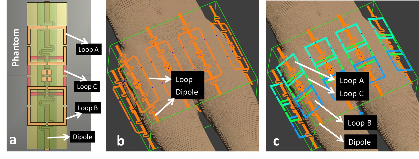
The new 32-channel combined loop-dipole transmit/receive (transceiver) array design (32LD) consists of 8 identical geometrically separated loop-dipole building blocks, with each block containing a fractionated dipole antenna[1] and three 8x8cm2 square loop coil elements (Figure 1.a). For comparison, the older design had three 6x10cm2 rectangular loops[3]. Loop-dipole blocks were distributed around the body with a center-to-center distance of ~11 cm (Figure 1.c). Three loop coils were centered on each dipole element along the z-axis overlapped by 12mm to minimize mutual coupling between nearest neighbors. Unlike previous loop-dipole designs[2-3], dipole elements were placed closest to the surface of the skin (~20mm lift-off) while loops were placed 1.6mm away from the dipoles. This change was motivated by practical implementation considerations, to move the loop coil cables away from dipoles.
The 32LD was modeled around the torso and pelvis of Duke model (Virtual-Family[4]). EM-field distributions of each coil element were computed using Sim4Life (ZurichMedTech, Zürich, Switzerland) and were imported to Matlab (Mathworks, Natick, MA) to investigate receive and pTx performance. The 32LD was compared against a 16-ch loop-dipole array (16LD)[2], which was modeled with same loop-dipole block geometrical placement configuration (Figure 1.b), simulated and analyzed using the numerical methods described herein.
SNR was quantified in a root sum-of-squares fashion inside prostate and kidneys. Phase-only RF shimming[5] yielding peak B1+ efficiency and efficiency-uniformity trade-off solutions were computed inside both targets; B1+ transmit efficiency, coefficient-of-variation of B1+ (CoV), peak 10g-averaged local SAR and SAR efficiency performance metrics were evaluated. Additionally, the performance of the arrays were compared by designing pTx multi-band (MB) pulses[6] with local and global SAR constraints for rapid imaging with large FOVs.
Results
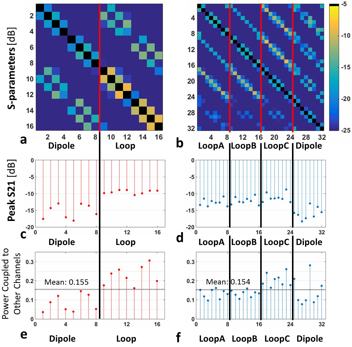
S-parameter matrices around the pelvis (Figure 2.a-d) show that highest coupling occurs between loop elements in neighboring block in 16LD. In contrast, highest coupling is between central (LoopC) and outside loops (LoopA and LoopB) in the same block. On average, around 15% of the accepted coil power couples to other array elements in both arrays (Figure 2.e-f). Despite doubling total channel count, 32LD achieves an inter-element decoupling performance on par with 16LD, solely using careful geometrical element placement.
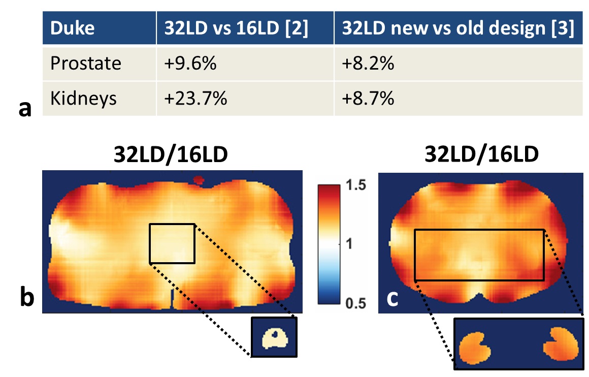
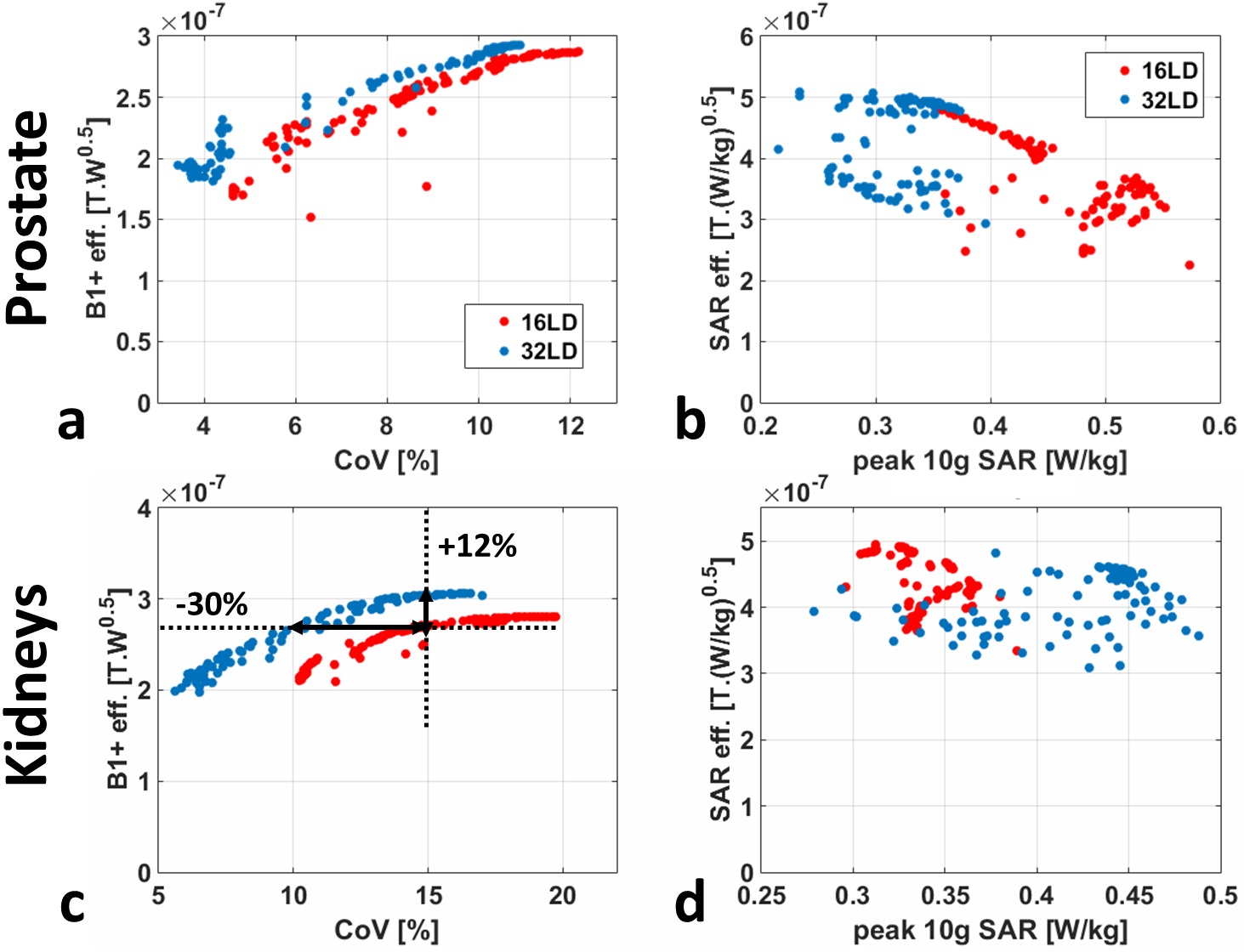
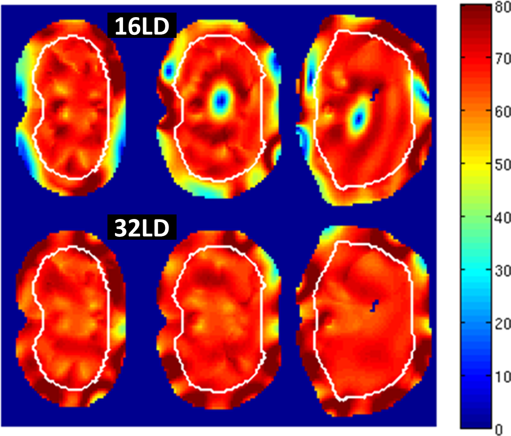
The 32LD achieves 9.6% and 23.7% higher SNR inside the prostate and kidneys, respectively (Figure 3.a). 32LD/16LD SNR ratios along axial slices in pelvis and torso (Figure 3.b-c) show that the SNR increase is higher around the periphery. The 32LD also achieves >8% SNR gain compared to the older 32-channel design (Figure 3.a). The 16LD and 32LD have similar B1+ efficiency and field uniformity inside the prostate (Figure 4.a), but 32LD can provide higher SAR efficiency for a variety of shim solutions owing to its lower peak SAR compared to 16LD (Figure 4.b). 32LD has 12% higher B1+ efficiency over 16LD at CoV=15% inside the kidneys and has 30% better field-uniformity at a chosen B1+ efficiency level (Figure 4.c). Despite having higher B1+ efficiency, 32LD has ~8% lower SAR efficiency compared to 16LD inside kidneys (Figure 4.d).
When designing pTx MB pulses to excite simultaneously three axial slices, the use of 32LD led to better excitation fidelity (better flip-angle uniformity with no black-holes) while complying with SAR limits (Figure 5).
Discussion/Conclusion
An improved 32-channel loop-dipole transceiver array was designed by tailoring loop coil dimensions and updating element placement layout to achieve acceptable decoupling performance without using any decoupling components/circuitries. The 32LD promises ~10% SNR gain in deep imaging targets (prostate), >20% gain in medium-depth targets (kidneys) and >30% gain near the surface of the body (Figure 3.b-c) compared to a state-of-the-art 16-channel array[2]. Even though not presented here, our earlier 32-channel body array design had improved parallel imaging performance and was capable of Z-axis accelerations due to Z-separation of loop coil elements[3]. We anticipate even higher parallel imaging gains with the new improved 32LD array design compared to previous work[3], and will be thoroughly investigated in future work.
In addition to improved SNR, the 32LD has improved field-uniformity and higher B1+ transmit efficiency compared to 16LD when using phase-only RF shimming. It also allows more uniform, black-hole-free flip angles to be created when designing pTx MB pulses for large FOV imaging.
Acknowledgements
Supported by: NCI R01 CA155268, NIBIB P41 EB015894.References
[1] Raaijmakers AJ et al. MRM 2016;75:1366-74.
[2] Ertürk MA et al. MRM 2017;77:884-94.
[3] Ertürk MA et al. ISMRM 2017;p1222.
[4] Christ, A et al. Phys Med Biol 2010;55(2):N23-38.
[5] Metzger, GJ et al. MRM 2008;59(2):396-409.
[6] Wu, X et al. MRM 2013;70(3):630-638.
Figures