4393
Comparison of Electric and B1+ Fields for Heterogeneous and Homogeneous Anthropomorphic Phantoms and Anatomical Models: Numerical Simulations and Experimental Findings1Bioengineering, University of Pittsburgh, Cranberry Twp, PA, United States, 2Bioengineering, University of Pittsburgh, Pittsburgh, PA, United States, 3University of Pittsburgh, Pittsburgh, PA, United States, 4Radiology, University of Pittsburgh, Pittsburgh, PA, United States
Synopsis
While numerical phantoms and detailed human tissue models have been available for some time in MR1-5, the development of experimental phantoms has yet to fully evolve. The growth of rapid prototyping and mimicry of tissues through tissue engineering is making it more possible for realistic electromagnetic phantoms that mimic human anatomy to be available to MR researchers and their collaborators. Wood et al. 6 fabricated a realistic head model (shown in Figure 1A) that can be used for a wide-variety of MR purposes and builds on the work of Gradel et al.7. However, the argument can still be made as to how beneficial it is to have a heterogeneous anthropomorphic phantom in place of a homogeneous anthropomorphic phantom. We hypothesize that the differences will be greatest in the electric field intensities and distribution as opposed to the B1+ field. In this work, we perform numerical studies and experimental 7T measurements that highlight the differences in the electric field and magnetic field of an anthropomorphic phantom (computer model as well as 3D printed) filled with homogeneous and heterogeneous media in comparison to a 10-tissue segmented head model. The phantom uses a resin plastic material that approximates the electromagnetic constitutive properties of a combination fat,
Synopsis
While numerical phantoms and detailed human tissue models have been available for some time in MR1-5, the development of experimental phantoms has yet to fully evolve. The growth of rapid prototyping and mimicry of tissues through tissue engineering is making it more possible for realistic electromagnetic phantoms that mimic human anatomy to be available to MR researchers and their collaborators. Wood et al. 6 fabricated a realistic head model (shown in Figure 1A) that can be used for a wide-variety of MR purposes and builds on the work of Gradel et al.7. However, the argument can still be made as to how beneficial it is to have a heterogeneous anthropomorphic phantom in place of a homogeneous anthropomorphic phantom. We hypothesize that the differences will be greatest in the electric field intensities and distribution as opposed to the B1+ field. In this work, we perform numerical studies and experimental 7T measurements that highlight the differences in the electric field and magnetic field of an anthropomorphic phantom (computer model as well as 3D printed) filled with homogeneous and heterogeneous media in comparison to a 10-tissue segmented head model. The phantom uses a resin plastic material that approximates the electromagnetic constitutive properties of a combination fat, bone and skin.Methods
Experimental B1+ Field Mapping.: Each phantom and the volunteer are centered within the TEM coil to acquire experimental 3D B1 maps using the saturated TurboFLASH (SatTFL) MR sequence with a 1ms rectangular RF pulse per 500V.
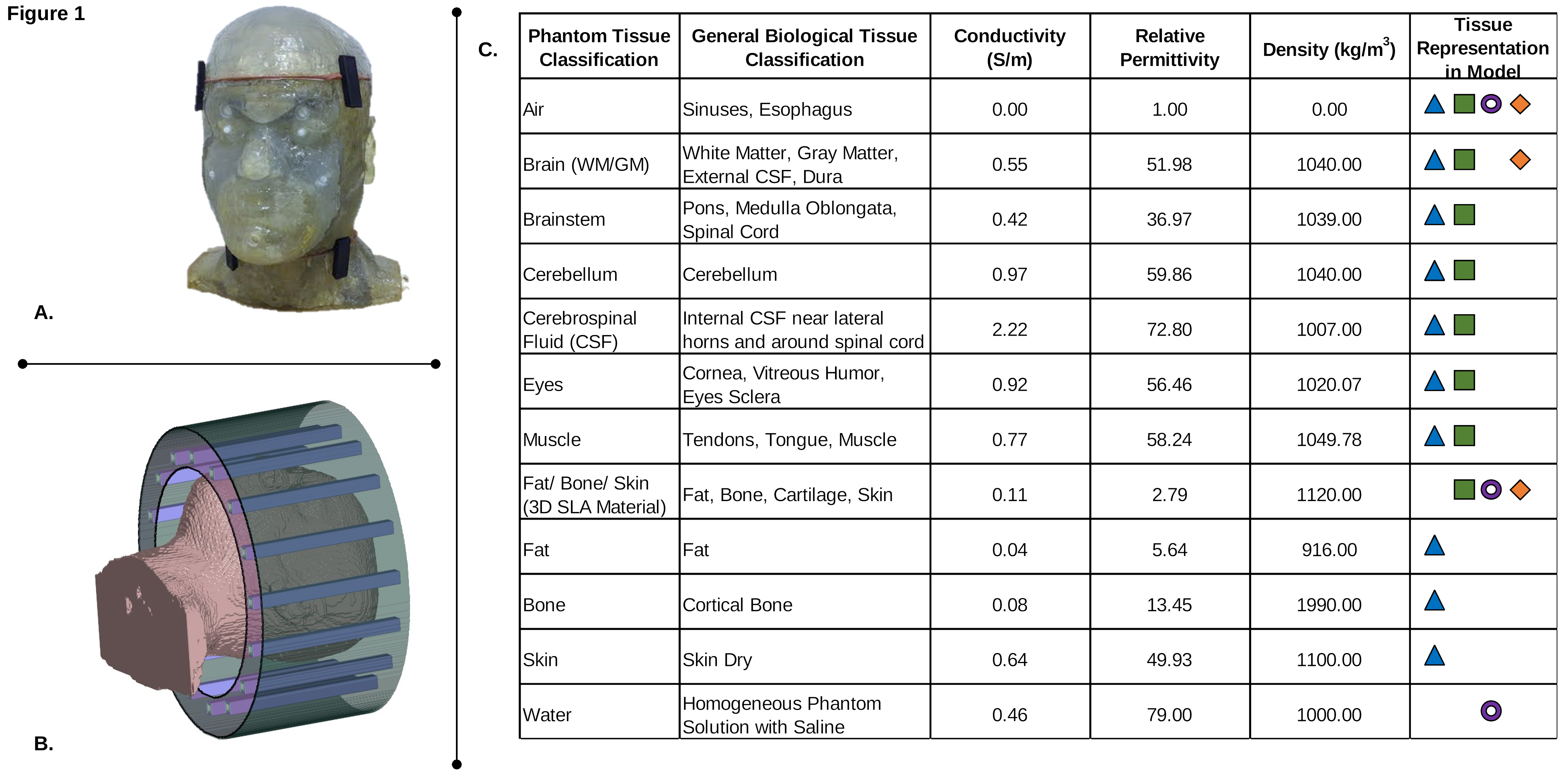
Numerical Modeling. Using the FDTD method8, an in-house numerical simulation software generates magnetic and electric (EM) fields at an operational frequency of 297.2 MHz. Four numerical models are used: 1) 10-tissue segmented head model (from which the phantom evolved), 2) anthropomorphic heterogeneous, 3-4) an anthropomorphic homogeneous water-doped and brain-doped head phantom head phantom (all constitutive properties are shown in Figure 1C). Each of the phantoms was loaded inside a model of a 16-strut TEM resonator9 as shown in Figure 1B). The coil was properly tuned and matched and the computational domain includes the head, neck and upper shoulders of each model.
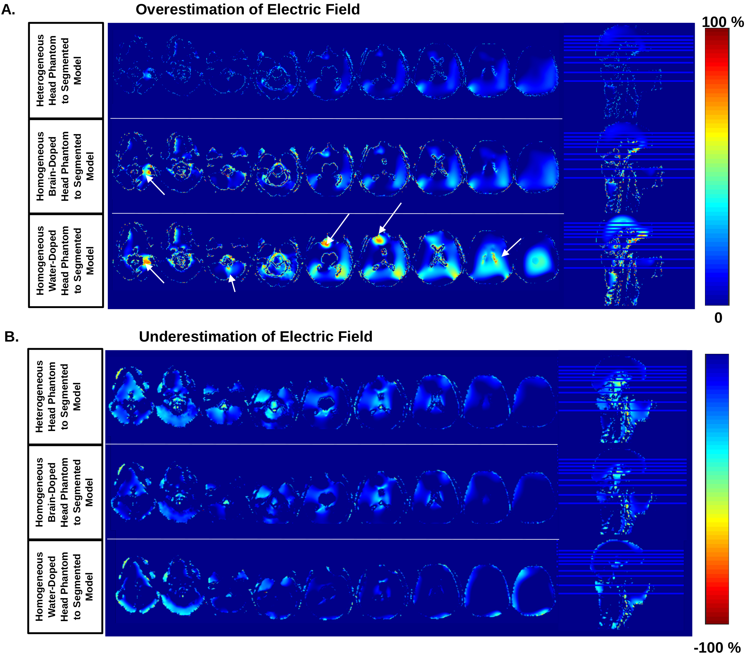
Comparisons of each phantom’s electric and B1+ fields are made to the respective values of the segmented model. For the B1+ field, the experimentally obtained and numerically calculated distributions were compared for all 4 phantoms. For the computer head models, the electric field is shown for the entire head and the upper shoulders. The electric field is analyzed by the percent change of the models compared to the 10-tissue segmented model (the baseline) and is shown in Figure 3. Each figure is scaled from 0 to -/+ 100% to show the negative/positive percent change. The plastic resin was removed to focus on the electric field only in the fillable tissue regions.
Results
Figure 2 shows the B1+ field distributions calculated using the FDTD method and measured experimentally at 7T using B1+ mapping technique for the 4 above-mentioned phantoms.
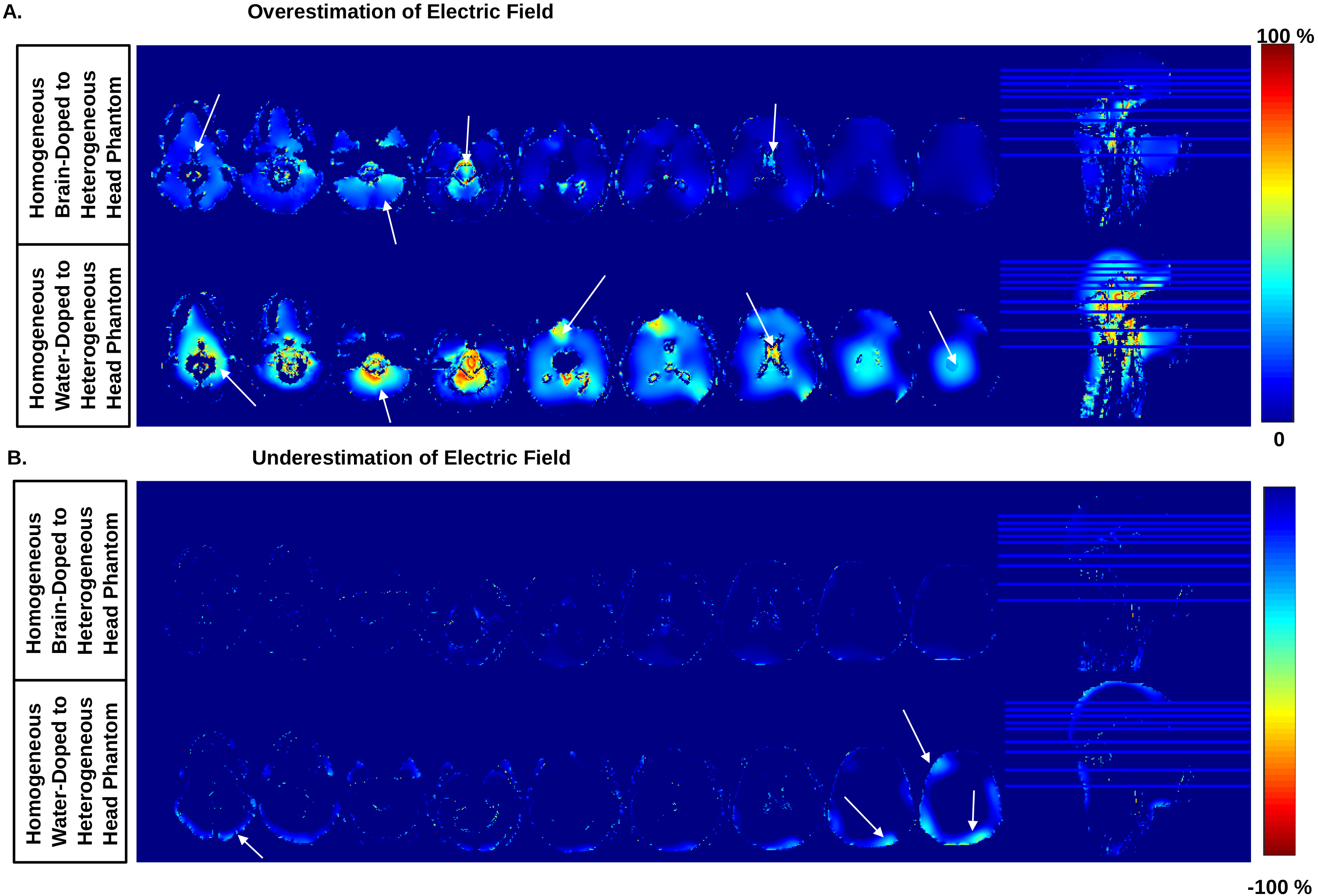
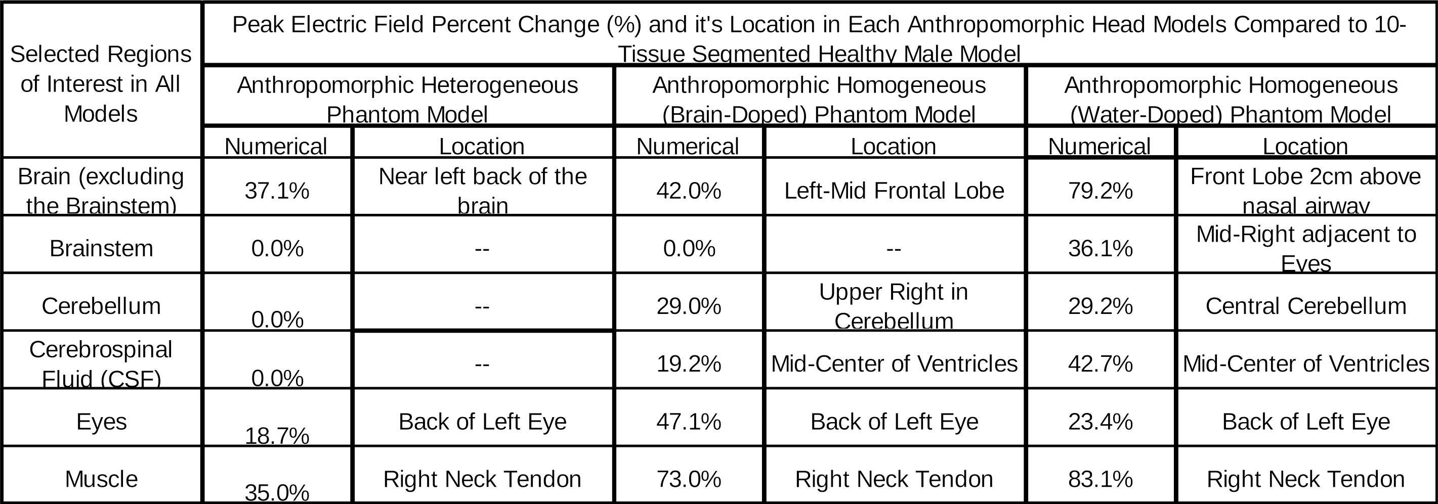
Figure 3 shows the results of the comparison of the electric field in each phantom media compared to the baseline. Figure 4 shows the percent change in the electric field in each homogeneous-doped media phantom compared to the anthropomorphic heterogeneous phantom to indicate the under/overestimation of the electric field. Arrows are used to highlight the peak locations of percent change. Table 1 highlights where the most significant peak change is based on the region in the brain.
Discussion
The B1+ field in the homogeneous brain-doped phantom is highly similar to the segmented model as well as the heterogeneous phantom. This observation holds true for both the experimental and numerical data.
While the electric field in the homogeneous brain-doped phantom is similar to the segmented model; differences are apparent in the brain region, the cerebellum and the muscle. The brain region electric field is higher than the heterogeneous phantom. The electric field in the heterogeneous phantom is most like the 10-tissue segmented phantom. While the heterogeneous phantom is most similar, Figure 3 highlights that there are subtle differences in the electric field. Most of those differences are in the brain region. The percent change in the electric field in the homogeneous water-doped phantom is highest. A comparison of the electric field percent change in the heterogeneous phantoms in comparison to the homogeneous-doped phantoms is shown in Figure 4. Table 1 illustrates the impact of the electric field distributions based on using the constitutive parameters highlighted in Figure 1C.
In conclusion, an anthropomorphic homogeneous brain-doped head phantom could be sufficient to predict the B1+ field distribution in-vivo; while the electric field and potentially SAR and well as temperature rises in the heterogeneous head model is most similar to the 10-tissue segmented model.
Acknowledgements
The work reported in this abstract was partially supported by NIH grants: 1R01MH111265-01 and F31EB019872.
References
1. Tang L, Hue YK, Ibrahim TS. Studies of RF Shimming Techniques with Minimization of RF Power Deposition and Their Associated Temperature Changes. Concepts Magn Reson Part B Magn Reson Eng. 2011;39B(1):11-25.
2. Curtis AT, Gilbert KM, Klassen LM, Gati JS, Menon RS. Slice-by-slice B1+ shimming at 7 T. Magn Reson Med. 2012;68(4):1109-1116.
3. Hetherington HP, Avdievich NI, Kuznetsov AM, Pan JW. RF shimming for spectroscopic localization in the human brain at 7 T. Magn Reson Med. 2010;63(1):9-19.
4. Zhao Y. Radio Frequency Antenna Designs and Methodologies for Human Brain Computer Interface and Ultrahigh Field Magnetic Resonance Imaging [Dissertation]: Bioengineering, University of Pittsburgh; 2015.
5. Zhao Y, Krishnamurthy N, Wood S, Zhao T, Raval SB, Ibrahim TS. 3D Eigenmodes Optimizations for 3D Imaging at 7T. Paper presented at: 23rd Annual Meeting of ISMRM2015; Toronto, Canada.
6. Wood S, Krishnamurthy N, Santini T, et al. Design and fabrication of a realistic anthropomorphic heterogeneous head phantom for MR purposes. PLoS One. 2017;12(8).
7. An anatomically realistic temperature phantom for radiofrequency heating measurements 2013. 8. Yee KS. Numerical solution of initial boundary value problems involving Maxwell's equations in isotropic media. IEEE Transactions on antennas and propagation. 1966;14(3):302-307.
9. Vaughan JT, Hetherington HP, Otu JO, Pan JW, Pohost GM. High frequency volume coils for clinical NMR imaging and spectroscopy. Magn Reson Med. 1994;32:206-218.
Figures