4384
3T Multiparametric MRI based detection of Prostate Cancer: features of detected and missed tumors base on PIRADS v2 in 429 patients- using whole mount histopathology reference1Abdominal Radiology, David Geffen School of Medicine at UCLA, Los Angeles, CA, United States, 2Radiology Sciences, David Geffen School of Medicine at UCLA, Los Angeles, CA, United States, 3Urology Department, David Geffen School of Medicine at UCLA, Los Angeles, CA, United States, 4Pathology Department, David Geffen School of Medicine at UCLA, Los Angeles, CA, United States
Synopsis
We evaluated the performance of the 3 Tesla multiparametric MRI (mp-MRI) for detection of prostate cancer (PCa) based on Prostate Imaging Reporting and Data System (PI-RADS) Version 2 in 429 patients with 874 lesions. The overall and index tumor detection rate of 3T mp-MRI was 49.3% and 77.9% respectively. The tumor detection rate based on
Introduction
Although some studies have already been published in order to the diagnosis performance of Prostate Imaging Reporting and Data System (PI-RADS) Version 2 in the detection of PCa, still there is some discrepancy between their results1. The purpose of this study was to determine the performance of 3T multiparametric MRI in prostate cancer, using PIRADS v2, and explain the characteristics of detected and missed tumors.
Methods
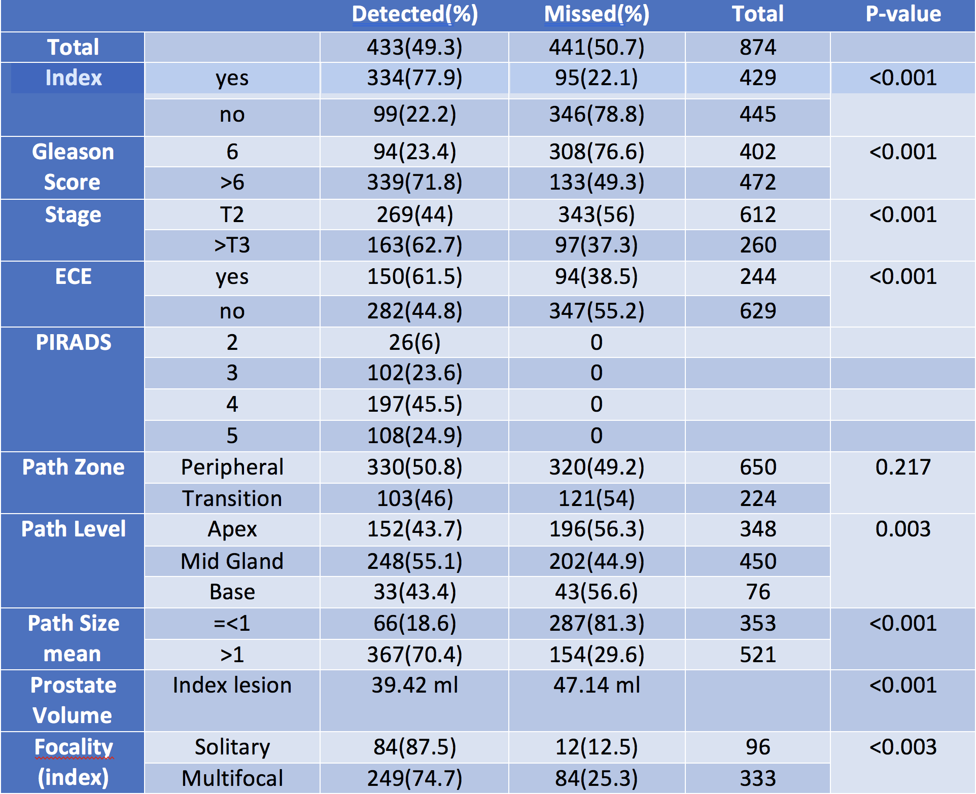
A retrospective study was performed of 429 consecutive men who underwent mp-MRI before radical prostatectomy at a single referral academic center between Dec 2009 and Jun 2016. Clinical, mp-MRI (i.e., T2-weighted imaging, diffusion-weighted imaging (DWI), and dynamic contrast-enhanced (DCE) imaging) and pathologic features were obtained. A structural report system, based on PI-RADS v2, was used for reporting qualitative and quantitative (DWI and DCE) features of each MRI detected lesion. MRI detected lesions were matched with previously finalized whole mount thin section histopathology (WMHP) in a joint session by a genitourinary (GU) radiologist and GU pathologist. MRI lesion detection rate was calculated. To assess statistically significance of normally distributed variables the chi-square analysis was performed. The t test and The Mann-Whitney U test were performed for normal and non-normal distribution categorical variables respectively, using SPSS v24.Results
Of 874 PCa lesions in 429 patients on WMHP, 3T MRI detected 443(49.3%) overall and 334 of 429 (77.9%) index lesions based on PIRADS v2. The 3T MRI detection rate was significantly higher in larger (70.4%, pathology size > 1cm) and higher grade (71.8%, Gleason score ≥7) PCa (p-value < 0.001). Of 441 missed lesions 308 (76.6%) had Gleason score ≤ 6 and 287 (81.3%) were ≤ 1 cm. Detection rate was highest in the mid gland 55.1% compared to the base and apex (p<0.001). The detection rate was not significantly different in the peripheral and transitional zones (50.8% vs 46% p =0.217). Sensitivity for index lesions detection was significantly higher in solitary (87.5%) vs multifocal tumors (74.7%) (p-value = 0.003). Prostate volume was significantly less in detected tumors (39.4cc), compared to missed tumors (47.1cc), p-value <0.001. PSA density was significantly higher in detected index lesions compared to the missed lesions.Discussion
The 2015 PI-RADSv2 classification attempted to standardize MRI nomenclature across institutions to provide uniformity for PCa detection and lesion characterization. In this study 70.1% of all detected lesions were PIRADSv2 4 or 5. There is a wide range of PCa detection rate for mp-MRI in much smaller prior studies. Both Tan and Le in a much smaller 3T cohort reported similar results2,3. Other smaller studies (with 34,49,150 subjects) have found a wide range of tumor detection from 48-92% of tumors 0.5 ml or greater on whole mount pathology4-6 .Tumor detection increases with tumor size and grade7,8. consistent with the present findings. Studies on the impact of multifocality on tumor detection by mp-MRI are limited. While Tan et al showed no impact of multifocal PCa for tumor detection2, we have demonstrated that multifocality decreased tumor detection rate to 74.7% from 87.5% for solitary tumors.Conclusion
In this large 3T Prostate MRI cohort with WMHP, tumor detection rate based on PIRADS v2 increased by PCa size, grade and stage was significantly higher for index lesions. Index and overall tumor detection rate was similar in the TZ and PZ and was higher with higher PSA density, in multifocal tumors and smaller prostate volumes.Acknowledgements
No acknowledgement found.References
1. Rosenkrantz AB, Oto A, Turkbey B, Westphalen AC. Prostate Imaging Reporting and Data System (PI-RADS), Version 2: A Critical Look. AJR Am J Roentgenol. 2016;206(6):1179-83.
2. Le JD, Tan N, Shkolyar E, et al. Multifocality and prostate cancer detection by multiparametric magnetic resonance imaging: correlation with whole-mount histopathology. Eur Urol. 2015;67(3):569-76.
3. Tan N, Margolis DJ, Lu DY, et al. Characteristics of Detected and Missed Prostate Cancer Foci on 3-T Multiparametric MRI Using an Endorectal Coil Correlated With Whole-Mount Thin-Section Histopathology. AJR Am J Roentgenol. 2015;205(1):W87-92.
4. Costa DN, Yuan Q, Xi Y, et al. Comparison of prostate cancer detection at 3-T MRI with and without an endorectal coil: A prospective, paired-patient study. Urol Oncol. 2016;34(6):255.e7-.e13.
5. Vargas HA, Hötker AM, Goldman DA, et al. Updated prostate imaging reporting and data system (PIRADS v2) recommendations for the detection of clinically significant prostate cancer using multiparametric MRI: critical evaluation using whole-mount pathology as standard of reference. Eur Radiol. 2016;26(6):1606-12.
6. Greer MD, Brown AM, Shih JH, et al. Accuracy and agreement of PIRADSv2 for prostate cancer mpMRI: A multireader study. J Magn Reson Imaging. 2017;45(2):579-85.
7. Bratan F, Niaf E, Melodelima C, et al. Influence of imaging and histological factors on prostate cancer detection and localisation on multiparametric MRI: a prospective study. Eur Radiol. 2013;23(7):2019-29.
8. Rosenkrantz AB, Mendrinos S, Babb JS, Taneja SS. Prostate cancer foci detected on multiparametric magnetic resonance imaging are histologically distinct from those not detected. J Urol. 2012;187(6):2032-8.
Figures