4335
Towards Fully Automated Breast MR Exams using Deep Learning1Global MR Applications & Workflow, GE Healthcare, Madison, WI, United States, 2MR Engineering, GE Healthcare, Waukesha, WI, United States, 3Radiology, University of Wisconsin-Madison, Madison, WI, United States, 4Medical Physics, University of Wisconsin-Madison, Madison, WI, United States, 5Carbone Cancer Center, University of Wisconsin-Madison, Madison, WI, United States, 6Global MR Applications & Workflow, GE Healthcare, Houston, TX, United States
Synopsis
Breast MR exams can be challenging for inexperienced MR technologists. For example, breast MRI typically requires the prescription of two carefully positioned and sized shim volumes, one for each breast, to improve the local B0 homogeneity and fat suppression. Normally, this procedure is performed manually, which requires an experienced MR technologist and can be challenging for new technologists. The goal of this project is to use deep learning to automate breast MR prescription, including placing the two shim volumes and imaging volumes automatically, to improve breast MR prescription consistency, quality, and to shorten the exam time.
INTRODUCTION
Breast MR exams can be challenging for inexperienced MR technologists. For example, breast MRI typically requires the prescription of two carefully positioned and sized shim volumes, one for each breast, to improve the local B0 homogeneity and fat suppression. Normally, this procedure is performed manually, which requires an experienced and vigilant MR technologist and can be challenging for new technologists. Machine learning methods, especially convolutional neural network-based deep learning (DL) technology, have been used in the medical imaging field to detect organs, regions and landmark localizations [2]. The goal of this project is to use DL to automate breast MR workflow by prescribing the two shim volumes automatically, to improve breast MR prescription consistency and exam quality and shorten total MR exam time.METHODS
A total of 164 clinical breast MRI exams were used for this IRB-approved, HIPAA compliant study. For each data set, the 3-plane localizer images were used as reference, and the shim volume information was extracted from a fat-suppressed axial T2-weighted scan. For all cases, the shim volumes were prescribed as purely orthogonal without any rotation. The size of the two shim volumes was the same along all directions. Therefore, for each data set, a total of 7 variables were used to represent shim volume information: (X1, Y1, Z1, X2, Y2, Z2, L), where (X1, Y1, Z1) and (X2, Y2, Z2) are the coordinates of the centers of the two shim volumes, and L is the length of the shim volume along a single direction.
A convolutional neural network was implemented in Keras with TensorFlow backend to identify the locations and size of the two shim volumes. The input to the network was the 3-plane localizer images, while the output was the shim volume information. After reviewing the user-prescribed shim volume information for all the cases, 130 data sets with good shim volume placement were used for “training”. The 3D volume data were then fed into a deep neural network with four layers of convolution neural networks and two layers of fully connected neural networks [2]. The standard mean squared error was used as the loss function. The remaining 34 data sets had suboptimal user-prescribed shim volume placement. The trained network was then applied to the 34 suboptimal data sets to see if DL could improve the shim volume placement.
RESULTS
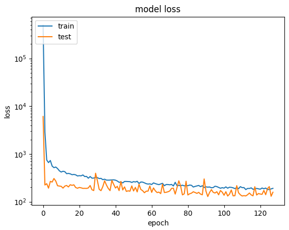
Figure 1 shows the loss curve from the training of the neural network.
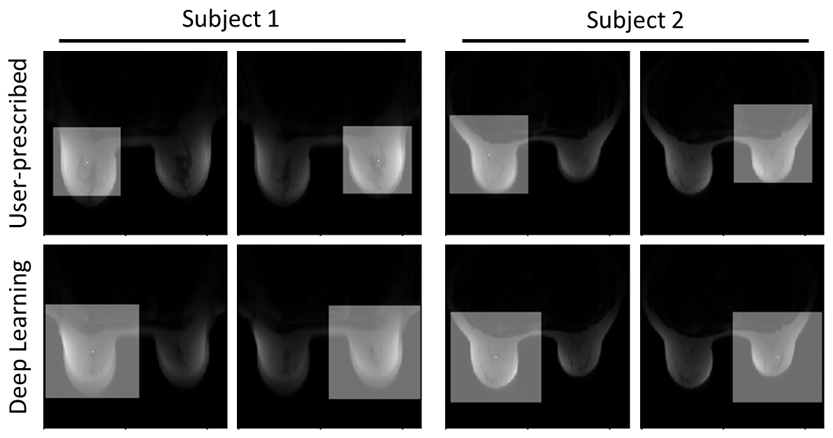
Figure 2 shows two examples of the shim volumes placed automatically by the DL algorithm, using only the axial localizer images. The DL algorithm is able to set the shim volume to the centers of the two breasts, and make them large enough to cover the entire breast. In contrast, the user-prescribed shim volumes are too small to cover the entire breast tissue.
The DL shim volume placements for the 34 data sets with suboptimal shim volume placement by clinical technologists were visually checked by an experienced breast MR researcher. The DL algorithm properly placed dual shim volumes for 29 out of the 34 data sets.
DISCUSSION
This preliminary work demonstrated that DL can assist and improve the placement of the dual shim volumes for breast MR exams. Five of 34 data sets with suboptimal placement by clinical technologists were also not placed well by DL. These were found to be from relatively large subjects. The failure is likely due to an insufficient number of similar data in the training set. Another possibility is that the all data sets were prescribed by different MR technologists, which lowers the consistency of the training set.
The results of the shim volume prediction suggest that it is feasible to auto-prescribe the imaging volume as well. For example, when prescribing a 3D bilateral axial scan, it is reasonable to assumed that users want to keep the size of the imaging field-of-view (FOV) fixed, so DL can be used to determine the center of the imaging FOV. The average of the two shim volume centers can be used as the FOV center. Note that frequency-encoding direction is always set to the A/P direction for reduction of cardiac motion artifacts. For the phase-encoding direction, oversampling is needed to avoid wrapping from the arms, and the oversampling factor can be automatically determined by comparing the prescription phase-FOV to the signal bound box along the phase direction.
CONCLUSION
In this work, we demonstrate the feasibility of using a DL approach to automate the prescription of dual shim volumes for breast MR exams. This has the potential to fully automate breast MR exams, increase consistency between technologists, assist inexperienced technologists, and ultimately improve exam quality.Acknowledgements
No acknowledgement found.References
- Kuhl CK, Schild HH, Morakkabati N. Dynamic bilateral contrast-enhanced MR imaging of the breast: Trade-off between spatial and temporal resolution. Radiology. 2005;236(3):789-800.
- Yang D, Zhang S, Yan Z, Tan C, Li K, Metaxas D. Automated anatomical landmark detection on distal femur surface using convolutional neural network. In: IEEE Int Symp Biomedical Imaging. 2015 pp. 17–21.
Figures