4333
Enhanced Ellipsoid Mapping of Diffusion Tensor Breast MRI for Improved Lesion ConspicuityMyra Shapiro-Feinberg1, Edna Furma-Haran2, Dov Grobgeld3, and Hadassa Degani4
1Meir Medical Center, Kfar Saba, Israel, 2Weizmann Institute of Sience, rehovot, Israel, 3Weizmann Institute of Science, Rehovot, Israel, 4Biological Regulation, Weizmann Institute of Science, Rehovot, Israel
Synopsis
Ellipsoid mapping of the breast with a specific colorization mode has been developed as a visualization means for evaluating the entire information embedded in breast Diffusion Tensor Imaging (DTI) and improve breast cancer detection. The 3D ellipsoid maps were displayed at voxel resolution with their shape and orientation determined by a respective eigenvalue-eigenvector pair of the associated diffusion tensor, followed by colorizing the ellipsoids according to the values of each diffusion tensor parameter. The results show that the enhanced ellipsoid mapping with λ1 colorization accentuating breast malignancy, may allow efficient differentiation of breast malignancy from normal breast tissue.
Purpose
To develop a means for visualizing multiple clinically important parameters derived from breast Diffusion Tensor Imaging (DTI) in a single map, enabling faster and more efficient detection and diagnosis of breast cancer.Introduction
Diffusion MRI has now been widely introduced into clinical breast imaging as an adjunct to Dynamic Contrast Enhanced (DCE) imaging, as many previous studies have demonstrated that lowered Apparent Diffusion Coefficients (ADC) are correlated with highly cellular regions such as breast cancer and can improve the overall specificity of the MRI exam1. We have previously shown that specific diffusion parameters of DTI in the breast, has exciting potential to serve as a method that may excludes the need for contrast injection2,3. DTI scan data is often presented as a diffusion ellipsoid map, depicting on a voxel-by-voxel basis the diffusion direction (eigenvectors) in three orthogonal axes of an ellipsoid shape that coincides with the diffusion frame of the tissue and the corresponding diffusion coefficients (eigenvalues λ1, λ2, λ3), as well as the diffusion anisotropy indices. As the information embedded in the ellipsoids metrics is clinically relevant, breast malignancies can be depicted by evaluating changes in the ellipsoids’ form and size in different regions of the breast. Here we show the potential of enhanced ellipsoid mapping with λ1 colorization accentuating breast malignancy and allowing differentiation of breast malignancy from normal breast tissue.Methods
This retrospective study was approved by the IRB of Meir Medical Center. Images were acquired on a 3 Tesla Trio scanner (Siemens). The MRI protocol included DTI sequence using twice refocused spin echo EPI and fat suppression, TE of 120ms, 60 slices with slice thickness = 2-2.5 mm, 1.9x1.9mm2 in-plane resolution, diffusion gradients at b values 0 and 700s/mm2 along 30 or 64 directions with respective scan times of 6 or 11 min. The DTI datasets were analyzed using a proprietary software2 that calculated the three diffusion coefficient at voxel resolution followed by a non-linear best fit regression algorithm to calculate the rank-2 symmetric diffusion tensor followed by diagonalization to yield three eigenvalue-eigenvector pairs. The 3D ellipsoid maps were displayed at voxel resolution with the length and orientation of each of the three principal axes of an ellipsoid determined by the respective eigenvalue-eigenvector pairs of the associated diffusion tensor. ROC curve analysis and calculation of contrast to noise ratio (CNR) were performed on datasets of twenty-four patients with pathology confirmed breast cancer. The ROC curves yielded upper threshold value for differentiating breast malignancy from normal tissue of each diffusion tensor parameter. The ellipsoids were colored according to the different diffusion parameters, using a color scale with the same upper threshold (0.017mm2/sec threshold of λ1) and unit scaling or using the upper threshold of each parameter for differentiating cancer from normal tissue as determined by the ROC curve analysis, keeping the same unit scaling.Results
The Ellipsoid maps revealed a reduced size and a more spherical shape of the ellipsoids in breast cancer lesions as compared to the surrounding fibroglandular tissue, indicating lower diffusion coefficients (λ1, λ2, λ3, mean diffusivity-MD), and reduced anisotropy, respectively, as displayed in Figure 1 for a typical invasive ductal carcinoma (IDC). In Fig.1A the color scale of the three diffusion coefficients is the same showing a decrease from λ1 to λ3 in normal ductal/glandular regions which reflects high maximal anisotropy (λ1- λ3), whereas in the cancer region, the three diffusion coefficients are similar and λ1- λ3 is low. Overall the CNR declined from λ1 to λ3 as demonstrated in Fig. 1A and CNRλ1 was higher than CNRMD as demonstrated in Fig. 1B. Ellipsoids’ maps with λ1 colorization also revealed the presence of small cancer lesions, with a size less than 5 mm, with detection efficiency similar to that of DCE as shown in Figure 2.Discussion
We developed the means for generating novel diffusion 3D ellipsoids’ maps from breast DTI scan data, providing a graphical representation of these maps. The fast and comprehensive visualization of the complete diffusion tensor information over the entire breast, at high spatial resolution, together with the emphasized coloring of λ1, the most instructive diffusion coefficient, provide a new means for efficiently characterizing breast tissue and identify breast malignancy. The clinical evaluation indicated that the features of malignancy in the ellipsoids maps with λ1 colorization were particularly exposed in dense breasts, where mammography is usually limited. Further quantitative evaluation of the diagnostic accuracy of the ellipsoids with λ1 colorization is underway.Acknowledgements
E. Furman-Haran holds the Calin and Elaine Rovinescu Research Fellow Chair for Brain Research. Prof. H. Degani holds the Fred and Andrea Fallek Chair for Breast Cancer ResearchReferences
1. Chen X, Li WL, Zhang YL, et al. Meta-analysis of quantitative diffusion-weighted MR imaging in the differential diagnosis of breast lesions. BMC Cancer, 2010 Dec 29; 10:693. doi: 10.1186/1471-2407-10-693. 2. Eyal E, Shapiro-Feinberg M, Furman-Haran E, et al. Parametric diffusion tensor imaging of the breast Invest. Radiol, 2012 May; 47(5):284-291. doi: 10.1097/RLI.0b013e3182438e5d. 3. Shapiro-Feinberg M, Weisenberg N, Zehavi T et al. Clinical results of DTI, Eur J Radiol. 2012 Sep; 81 Suppl 1:S151-2. doi: 10.1016/S0720-048X(12)70063-3. 4. Basser PJ, Mattiello J, LeBihan D. MR diffusion tensor spectroscopy and imaging Biophys J. 1994 Jan; 66(1):259-67.Figures
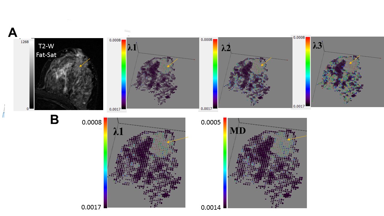
Enhanced ellipsoid maps
of invasive ductal carcinoma in a 40 YO patient. A. The same color scale for
the three diffusion coefficients; B. The upper thresholds of λ1 and
MD colorization are based on ROC curve analysis using equal unit scaling. All
diffusion coefficients are in mm2/sec units. Note in A reduction in
the value of the normal tissue diffusion coefficients from λ1 to λ3
and a reduction from CNRλ1 (2.37) to CNRλ2 (2.00) to CNRλ3 (1.5). Note in B
the lower CNRMD (2.05) than CNRλ1 (2.37) in the cancer region relative
to the normal tissue.
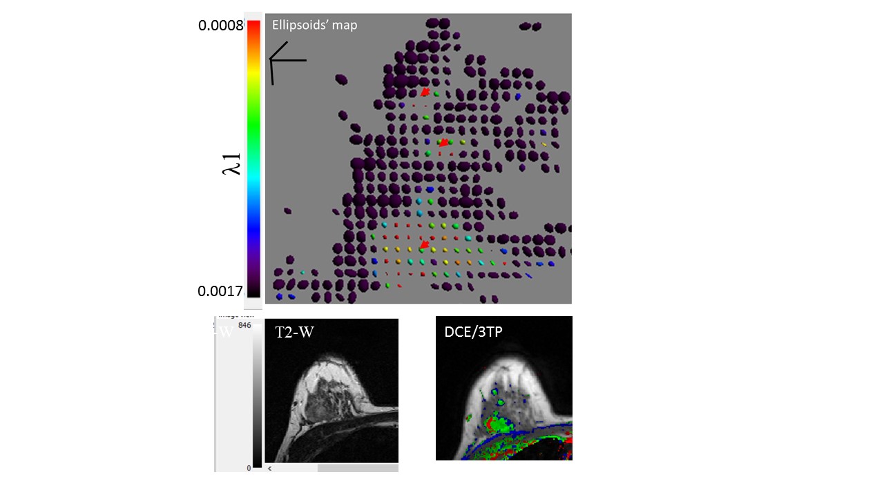
Ellipsoids’ map of a multi
focal invasive ductal carcinoma in a 38 YO patient. Note the congruence with
the DCE/3TP. λ1 is in mm2/s units.