4310
Initial Evaluation Utilizing Ultra-High Dielectric Constant (uHDC) Materials in Breast Imaging in 3TAvery Wang1, Sebastian Rupprecht1, Christopher T. Sica1, Angela Choe1, Susann Schetter1, Julie Mack1, and Qing X. Yang1
1Radiology, The Pennsylvania State University College of Medicine, Hershey, PA, United States
Synopsis
We explored the feasibility of using high dielectric constant materials to potentially further enhance signal-noise-ratio (SNR) in breast MRI. Through computer modelling at 3 Tesla (T), we showed that there can be great benefits of increasing sensitivity and specificity of breast MRI. As a side effect of using high dielectric constant materials, the RF safety can be improved by the largely increased transmit efficiency.
Purpose
To improve lesion morphology clarity in 3T breast MR. Breast MRI has high sensitivity in breast cancer detection, and the BI-RADS MRI lexicon was a step toward standardized description of lesions. However, it is estimated that about 70% of breast MRI findings are false-positives and lead to unnecessary biopsies. Numerous false-positive diagnoses occur due to an inadequate SNR, leading to circumstances where enhancements of the breast are considered breast cancer findings due to unclear shapes or small size. In order to avoid unnecessary biopsies, better detection of malignant versus benign morphology must be achieved.Methods
Previously, a passive shimming method using localized pads of high dielectric permittivity was used to improve image contrast and B1+ field homogeneity in 3T breast MRI (1). In this study, a similar shimming method consisting of local pads of high dielectric permittivity (ε0 = 600-1100) was studied in numerical simulations using xFDTD (Remcom Inc, PA, USA) at 125 MHz with the aim of improving SNR. Similar to a clinical system at 3T, a quadrature driven two-channel birdcage coil was used for transmit, while a clinical 16-channel breast coil was used to receive. The receive array was modeled after the Siemens 3T 16-channel Sentinelle breast coil, with 8 channels per breast, and it was utilized with a virtual model (Ella, Virtual Family, IT'IS Foundation, Switzerland) in prone position. Two breast models resembling average B cup breasts were attached to the female model, positioned so they were hanging freely in the coil. These phantoms were constructed with the electric properties of typical breast tissues. In addition to the baseline, a separate set of simulations were conducted employing the use of ultra-high dielectric constant materials (HDC). An 8-mm-thick layer of HDC material was added, conforming to the curvature of the coil structure and slightly displacing the outer 4 channels (Figure 1). Transmit efficiency and SNR maps were reconstructed from the acquired B and E fields acquired experimentally on a Siemens 3T Skyra clinical system to confirm the baseline RF numerical simulations. A brief clinical trial was conducted with the 3T 16-channel breast coil, utilizing four dielectric pads in a configuration similar to the simulated HDC material (Figure 2).Results and Discussion
We selected a permittivity of 1000 due to the homogenous transmit efficiency improvements within the breast. A strong improvement in transmit efficiency was observed, leading to a 6-9 fold increase in the inner breast, a 7-11 fold increase in the outer right breast, and a 12-16 fold increase in the outer left breast (Figure 3). SNR was also improved with a noticeable 1.5-2 fold increase in the middle of the breast (Figure 4). Though the largest improvement in SNR was observed with a permittivity of 1100, the drastic decrease in homogeneity and the dark voids within the calculated B1 field led us to favor a permittivity of 1000. The in-vivo trial we conducted produced an expected increase in transmit efficiency, leading to a 6-8 fold increase within the inner breast and a 3-4 fold increase within the rest of the breast (Figure 5). However, no noticeable increase in SNR was observed most likely due to a higher loss in the dielectric pad than anticipated.Conclusion
The utilization of a simple high permittivity layer on a standard clinical coil can potentially be used to improve sensitivity and specificity for breast MRI, to better separate malignant from benign morphology. While both further numerical and material optimizations are necessary, the initial results are encouraging. Additional benefits to be studied could be a strong B1 focus to the breast which could improve patient safety and potentially open breast scans for pregnant women as previously proposed in Reference 2.Acknowledgements
No acknowledgement found.References
Winkler SA, "Practical methods for improving B1+ homogeneity in 3 tesla breast imaging", JMRI 2005.
J.V. Hajnal, "Prediction of specific absorption rate in mother and fetus associated with MRI examinations during pregnancy", MRM 2006.
Figures
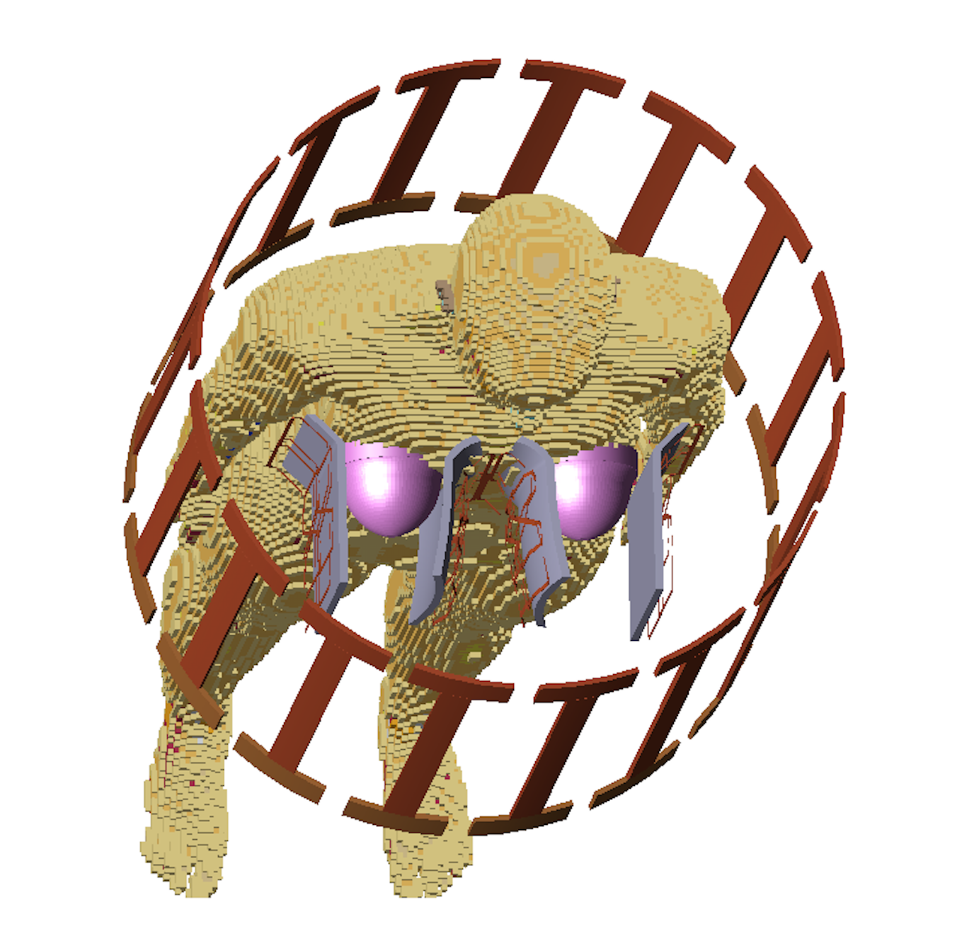
Figure 1. Experimental setup of Ella (Virtual Family, IT'IS Foundation, Switzerland) with a quadrature-driven two-channel-birdcage transmit coil and a 16-channel breast receive array positioned like the in-vivo experiment. (uHDC material-gray, breast tissue-pink)
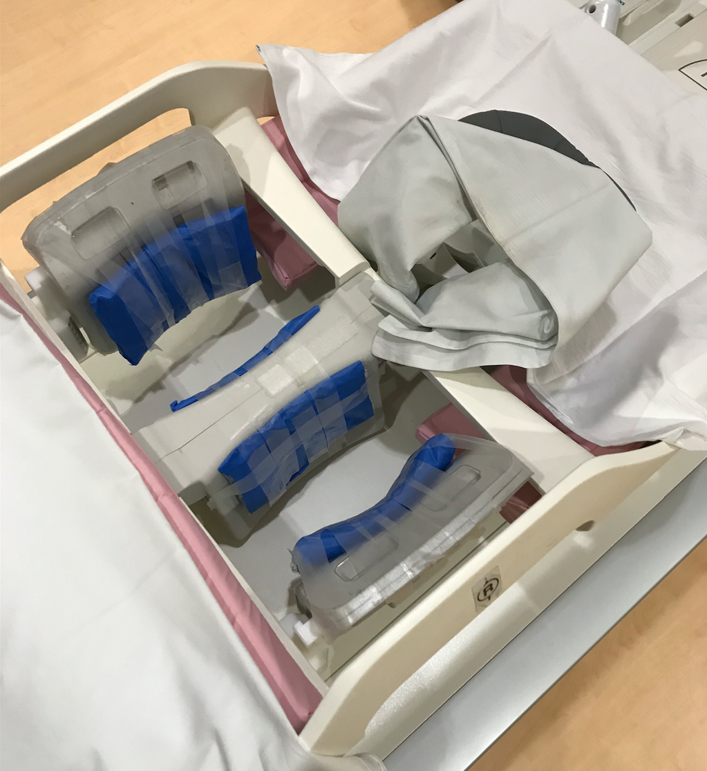
Figure 2. Siemens Sentinelle 3T 16-channel breast array lined with uHDC material (blue) as used in the in-vivo experiment.
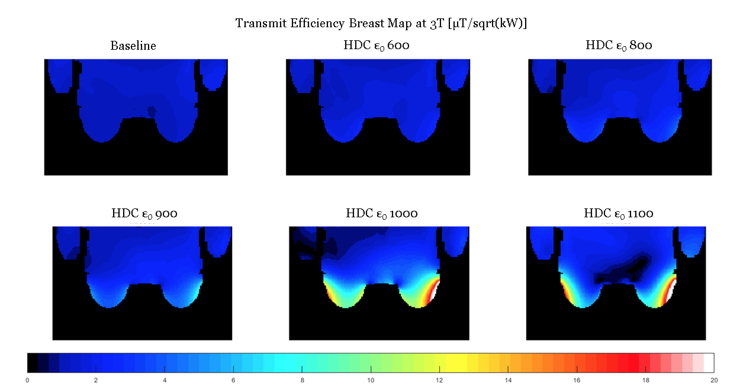
Figure 3. Simulated transmit efficiency maps showing homogenous and strong enhancement within the breast for a permittivity of 1000.
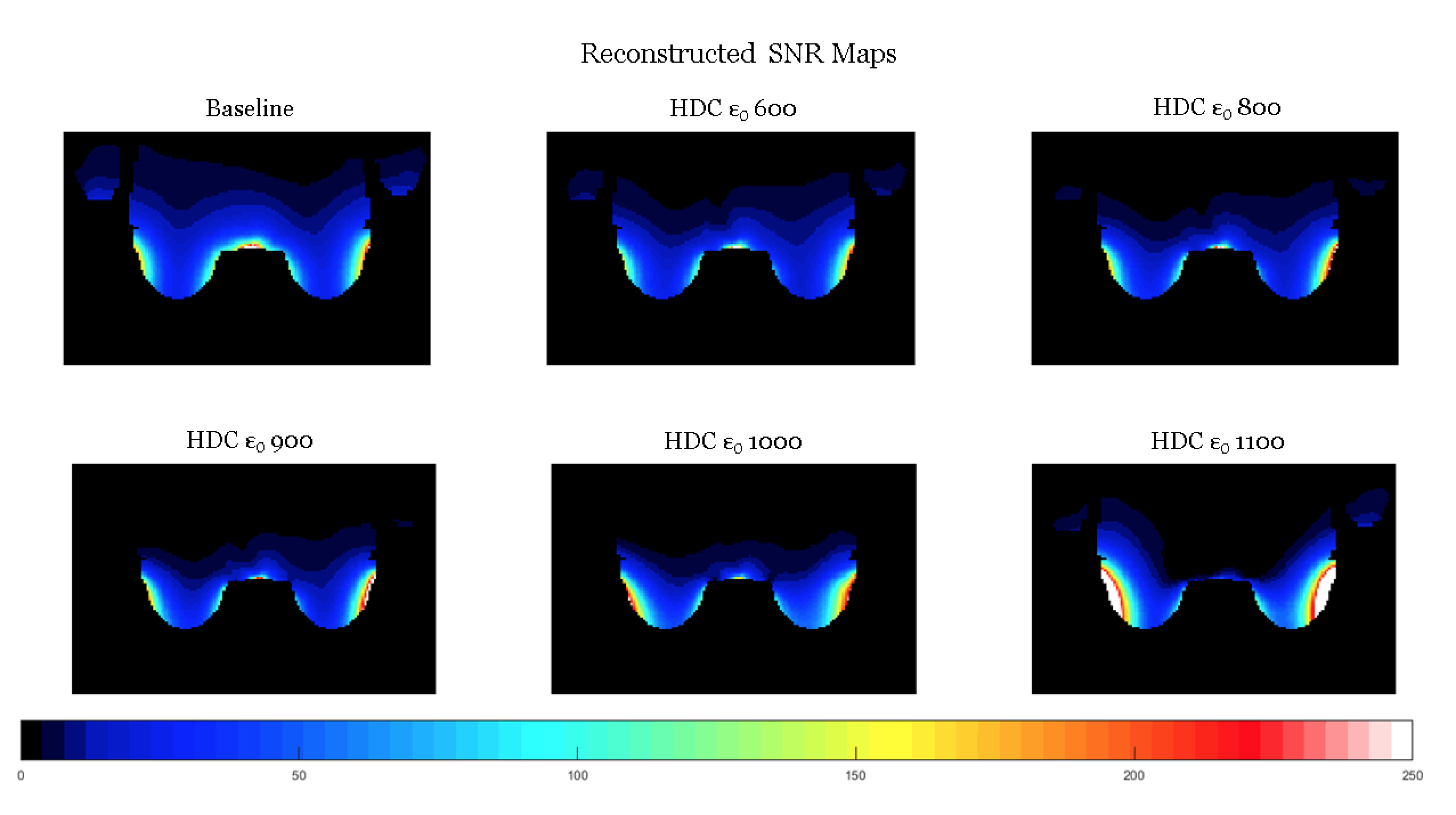
Figure 4. Simulated SNR maps showing the highest improvements with a permittivity of 1100.
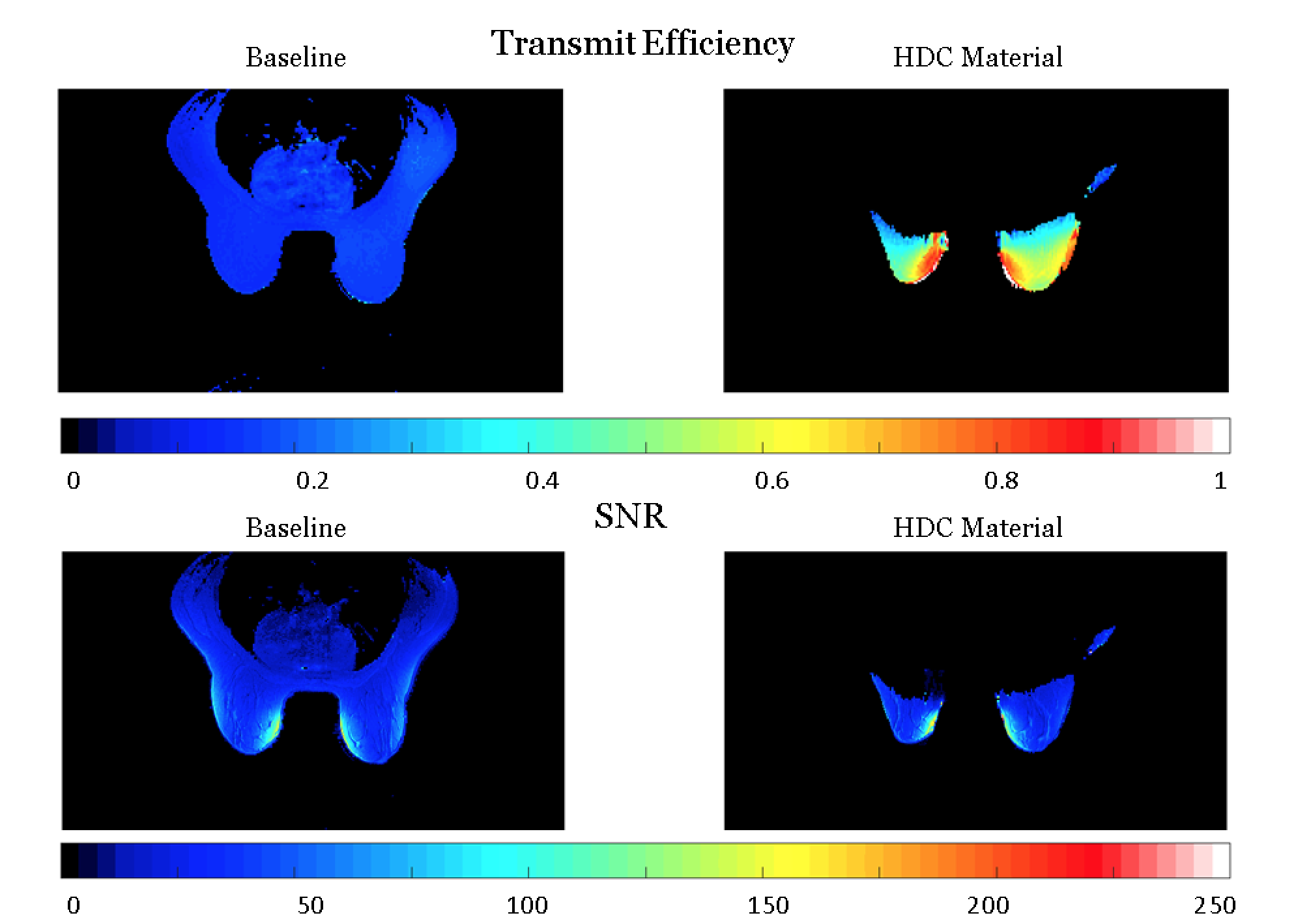
Figure 5. In-vivo results using a uHDC material pad with a permittivity of 1000.