4302
Evaluation of High-dielectric Pads for Macaque Brain Imaging at 7T: A Pilot StudyJie Zhao1,2, Wei Luo3, Meizhen Qian1,4, Yi Sun5, Ling Xia2,6, and Xiaotong Zhang1,2,6
1Interdisciplinary Institute of Neuroscience and Technology, Qiushi Academy for Advanced Studies, Zhejiang University, Hangzhou, China, 2College of Biomedical Engineering & Instrument Science, Zhejiang University, Hangzhou, China, 3Materials Research Institute, Pennsylvania State University, University Park, PA, United States, 4School of Medicine, Zhejiang University, Hangzhou, China, 5MR Collaboration NE Asia, Siemens Healthcare, Shanghai, China, 6Key Laboratory for Biomedical Engineering of Ministry of Education, Zhejiang University, Hangzhou, China
Synopsis
Non-human-primates are a valuable model for investigating the structure and function of the brain. The high-dielectric pads have been shown to increase B1 sensitivity and enhance image SNR and contrast locally in the human brain at high and ultra-high field. In this study, our pilot experiment results suggest that high-dielectric pads can effectively enhance macaque whole brain imaging at 7T, and in the meantime, do not increase the receive array element coupling, nor have any deterioration over the B0 homogeneity within the monkey brain.
Introduction
Non-human primates are a valuable model in investigating the structure and function of the brain. To achieve the requisite resolution to resolve fine anatomical detail and map localized brain activation requires high SNR. The high-dielectric pads have been shown to increase B1 sensitivity in areas of low signal intensity and locally enhance image signal-to-noise ratio (SNR) and contrast in the human brain at high and ultra-high field1,2. In this study, we demonstrate the benefits of dielectric pads for in vivo macaque brain imaging at 7T.Methods
All measurements were performed on a 7T research scanner (Siemens Healthcare, Erlangen, Germany) equipped with a 1Tx/28Rx QED knee coil (Mayfield Village, OH, USA). Images were acquired over a female macaque (2 years old, 3.5kg) that was maintained and anesthetized by 1.5~2% isoflurane – all procedures were in accordance with NIH standards and approved by our Institutional Animal Care Committee. The monkey was placed in the sphinx position with its head centered within the knee coil. Two pads (L=20/10cm, W=10cm, H=0.5cm) were used with the longer one placed on top of the head and covering the ears, while the cubic one covered the back of the head and the neck. The high-dielectric pads were made by HyQ Research Solutions LLC (State College, PA, US), in which powder slurry was made by mixing CaTiO3, BaTiO3, and distilled water, producing a conductivity of 0.1 S/m and relative permittivity of 120. Two datasets were acquired with and without the pads (same reference voltage and nominal flip angle were applied): 1) Flip angle maps acquired with a double-TR method3 and the voxel size of 1×1×1mm3, TR1/TR2/TE of 20/50/2.93ms, flip angle 60, 88 slices per slab, 2 averages, scan time 12’13”; SNR was measured4 with the voxel size of 0.5×0.5×3mm3. A noise correlation matrix was calculated from a noise scan in which the MRI signal is acquired while all gradients and RF pulses are turned off4. To evaluate susceptibility-induced artifacts, T2*-weighted 2D GRE images were acquired with a spatial resolution of 0.3×0.3×1mm3, TR/TE of 2480/25ms, GRAPPA = 2 in the F/H direction, FA = 50°, matrix size of 320×320×48, 3 averages, scan time 21’20”. Preceeding the measurement, whole-brain B0 shimming was performed up to the second order.Results
With pads added, B1+ efficiency and spatial SNR both show significant enhancement within the entire brain, especially at the center of the brain, as shown in Fig.1 and Fig.2. In the regions of, e.g., frontal lobe, sensory area, visual area, temporal lobe, cerebellum, and thalamus, the magnitude of the actual flip angle was elevated by approximately 5%, 30%, 30%, 15%, 15% and 16%, while the SNR improved in the order of 6%, 20%, 16%, 13%, 13% and 23%, respectively. Fig.3 shows the two noise correlation matrices without and with the dielectric pads in place, and the difference between the two correlation matrices, which suggests that those elements closest to the pads caused very trivial coupling from the additional magnetic field. Fig.4 shows the axial map of the brain using GRE T2*-weighted scan without pads and with pads. There was no apparent degradation in image quality in terms of B0-induced signal loss or distortions; on the contrary, improved B1+ efficiency and SNR resulted in moderately increased signal intensity and tissue contrast.Discussion and Conclusion
The study suggests that high-dielectric pads can be effective in enhancing macaque whole-brain imaging at 7T. Our preliminary results show that the use of such pads does not increase the receive array element coupling, nor have any deterioration over the B0 homogeneity within the monkey brain – such findings are consistent with human brain imaging with high-dielectric pads added. However, due to the smaller size of a macaque brain compared to human’s, we observed significant global elevations in transmit efficiency and spatial SNR within the entire macaque brain, especially at deep brain regions such as ventricles and thalamus. While it is anticipated that the high-dielectric pads may serve as a complementing tool in investigating tissue anatomy and neuronal activities over non-human primate models at ultra-high field, ongoing research needs to be conducted in areas such as pad optimization(geometry, size, etc.) and multiple metrics evaluation (structural and functional measurements).Acknowledgements
We would like to thank Mr. Guohua Xu, Bin Xu, and Dengfeng Zhou for technical support. This work was supported in part by National Natural Science Foundation of China (81701774, 61771423) and Fundamental Research Funds for the Central Universities (2016QN81018).References
1. Teeuwisse M, Brink M, Webb G, et al. Quantitative assessment of the effects of high-permittivity pads in 7 Tesla MRI of the brain. Magnetic Resonance in Medicine.2012; 67(5):1285-1293. 2. Luo W, Lanagan T, Sica T, et al. Permittivity and performance of dielectric pads with sintered ceramic beads in MRI: early experiments and simulations at 3 T. Magnetic Resonance in Medicine.2013;70(1):269-275. 3. Yarnykh V L. Actual flip-angle imaging in the pulsed steady state: a method for rapid three-dimensional mapping of the transmitted radiofrequency field.[J]. Magnetic Resonance in Medicine, 2007, 57(1):192. 4. Kellman P. Erratum: Image reconstruction in SNR units: A general method for SNR measurement (Magnetic Resonance in Medicine (2005) 54, (1439-1447))[J]. Magnetic Resonance in Medicine, 2007, 58(1):211-212.Figures
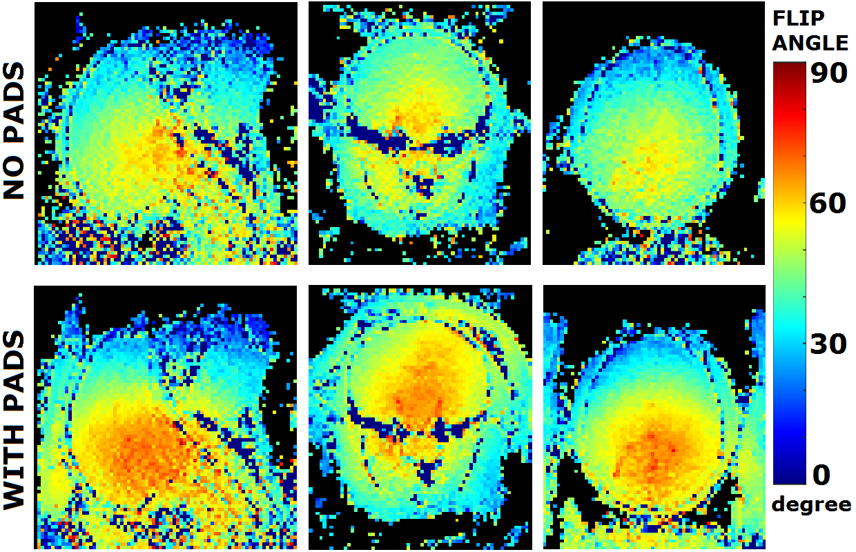
Fig.1. Comparison of actual flip
angle maps with and without high-dielectric pads in place.The
monkey was placed in the sphinx position with its head centered within the knee
coil. Two pads (L=20/10cm, W=10cm, H=0.5cm) were used with the longer one
placing on top of the head and covering the ears, while the cubic one covering
the back of the head and the neck.

Fig.2. Comparison of spatial SNR
maps with and without high-dielectric pads in place. Voxel size is 0.5×0.5×3 mm3.
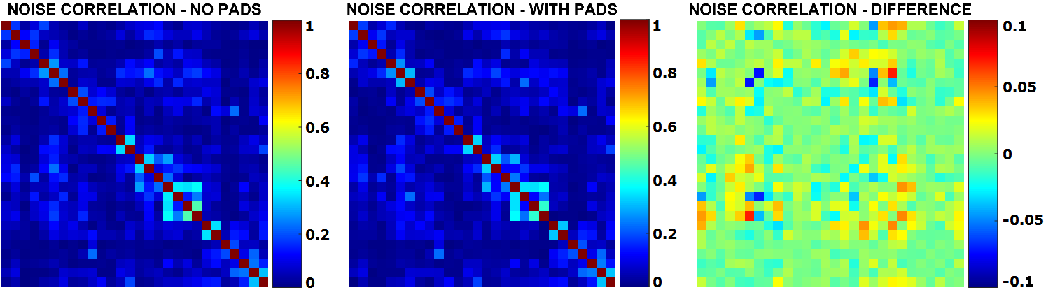
Fig.3. Noise correlation matrix
maps with and without high-dielectric pads in place and their difference map.
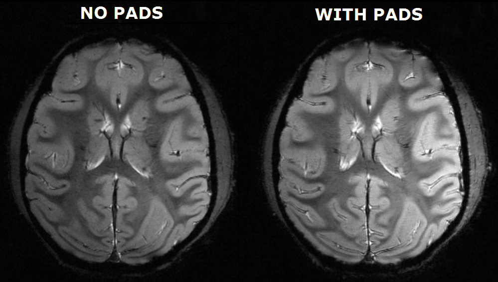
Fig.4. T2*-weighted GRE images to
investigate any effects on magnetic susceptibility-induced distortions of the
dielectric pads.