4295
Compact and adjustable metasurface-based resonator for in-vivo MRI1Nanophotonics and Metamaterials, ITMO University, Saint-Petersburg, Russian Federation
Synopsis
We have numerically and experimentally approved that a compact metasurface-based resonator with miniaturized capacitive load may efficiently operate as a wireless surface coil in 1.5 T MR scanner. Our results show that for a properly adjusted metasurface eigenmode resonance frequency it is possible to substantially increase the signal-to-noise ratio that was confirmed both with the phantom and in vivo experiments.
Introduction
Application of metamaterials in MRI is a novel and fast developing area. Previously, a signal-to-noise ratio (SNR) enhancement has been shown in 1.5 T field for a metasurface consisted of two arrays of parallel subwavelength brass wires terminated with the volumes of deionized water1, and for another design, where the similar wire array was fully immersed in deionized water2. Water, as a high-permittivity dielectric, created distributed capacitance and reduced the resonant length of the wires. The reduction of specific absorption rate (SAR) in simulations and two-fold enhancement of SNR in the area of human knee close to the resonator in MR-experiment were reported. In this work we propose a different type of capacitive load to substitute the inconvenient for in-vivo MRI experiments distilled water jars and minimize the metasurface size. Developed device can be used as a local wireless coil for in-vivo MRI.Methods and materials
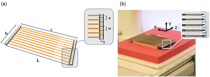
The developed prototype of metasurface based resonator (henceforth - «resonator») consisted of a flat array of 10 parallel telescopic, subwavelength brass wires. Each wire connected at both ends to capacitive loads, implemented as rectangular copper patches on a common grounded dielectric substrate based on Arlon AD1000 laminate (Arlon Microwave Materials). The length of the wires and dimensions of copper patches were optimized in 3D electromagnetic full-wave simulations (CST Microwave Studio 2017). The geometric parameters that tuned the resonator eigenmode with the highest penetration depth of $$$B_1$$$ field to the proton Larmor frequency at 1.5 T (f0=63.8 MHz) were used for fabrication of the prototype (Figure 1). B1+ and B1- fields were computed in a homogeneous rectangular lossy phantom. S11 measurements were performed inside a 1.5 T Siemens MR scanner. The resonator was placed in the isocentre of the scanner with wires perpendicular to the direction of the main magnetic field B0 (z-direction). This position has been chosen as the most convenient for the further in-vivo studies of wrist joint. A homogeneous phantom (GE Medical Systems, 46x62x162 mm) was located on top of the resonator. Gradient echo (GRE) pulse sequence (TR=1500 ms, TE=10.6 ms, FOV - 180x180 mm, matrix size - 256x256, slice thickness - 4 mm, FA=70°) was acquired for the following set-ups: (1) a body coil birdcage (BC) to transmit (Tx) and a clinical 4-channel flexible coil (Siemens Healthcare GmbH, 366x174 mm) to receive MR signal (Rx); (2) BC Tx and Rx with the resonator placed below the phantom. In-vivo MR experiments with the constructed resonator were performed at the Federal Almazov North-West Medical Research Centre (Saint-Petersburg, Russian Federation) and approved by the local medical ethics committee. T1-weighted turbo-spin echo coronal images of the wrist joint of a volunteer (TR=580 ms, TE=17 ms, FoV - 100x100 mm, matrix size - 256x320, slice thickness - 3 mm, FA=150°) were acquired in “superman” position for three set-ups: (1) BC Tx and 4-channel flexible coil Rx; BC TxRx with (2) and without (3) the resonator placed below the hand.Results and discussion
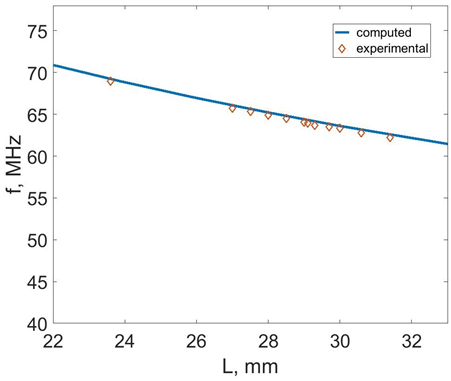
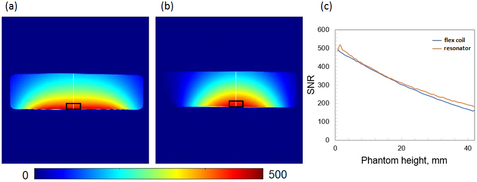
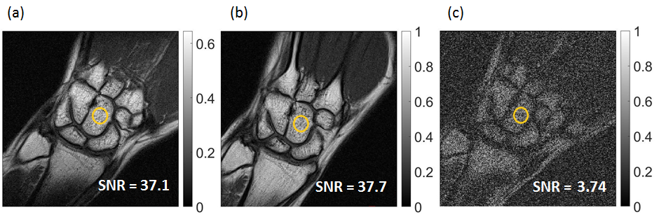
The simulated and measured dependence of the resonator first-order eigenmode frequencies on the wires length are plotted on Figure 2. The curves agree with each other and demonstrate an inverse dependency (predicted by a theory) of the frequency on the wires length. B1+ and B1- fields have a close to sinusoidal distribution in the central cutting plane perpendicular to the wires (Figure 3(a)) with maximum in the middle of it and minimum at the edges of the phantom. SNR maps based on GRE images of the phantom are shown on Figure 4 (a, b). For the region of interest (ROI) closest to the receive flex coil or resonator, the SNR was 443 or 438, consequently. The profiles of SNR in y direction are shown on Figure 4 (c). Imaging of the wrist in a presence of manufactured resonator allowed to obtain almost the same SNR in a ROI located in a capitate bone as the receive-only 4-channel flex coil and 10-fold enhancement of SNR when compare with BC-only receive image (Figure 5(a, b, c)). At the same time the voltage of excitation pulse dropped by 82% with the resonator.
Conclusion
The proposed metasurface-based resonator may be used as a pad (local wireless coil) and placed close to the area of interest, due to it’s compact and cordless implementation. It provides the same SNR efficiency as a 4-channel flex coil but with 32-fold less power that could be used to reduce acquisition time of clinical MR sequences containing power intensive RF pulses such as spin echo, PRESS, SPECIAL, as now these sequences require long TR because of RF safety constraints.Acknowledgements
This research was supported by Ministry of Education and Science of the Russian Federation (Zadanie No. 3.2465.2017/4.6).The authors are grateful to G. Solomakha for the help in the metasurface fabrication process and A. Mikhailovskaya, Dr. A. V. Sokolov, Dr. A. Yu.Efimtsev for assistance with measurements and to Dr. Redha Abdeddaim for the idea of using telescopic wires for mechanical tuning of wire metasurface structures.References
1. Shchelokova A, Slobozhanyuk A, Melchakova I, Wireless coil based on meta-technologies for MRI implementations. Proc. Intl. Soc. Mag. Reson. Med., 25 (2017 ), 0761;
2. Saha S, Shchelokova A, Sotiriou I, et al. Evaluation of a metasurface resonator for in vivo imaging at 1.5T. Proc. Intl. Soc. Mag. Reson. Med. 25 (2017 ), 2702.
Figures