4292
Establishing MR angiography of the mouse eye1Bio Engineering, Pennsylvania State University, University Park, PA, United States, 2Biology, Pennsylvania State University, University Park, PA, United States, 3Huck Institutes of the Life Sciences, Pennsylvania State University, University Park, PA, United States
Synopsis
High field Magnetic Resonance Angiography on mouse eye can be used to investigate the vasculature near the mouse eye. One of the difficulties with using on mice is the induced moving artifact. To overcome this obstacle, a custom mouse restrainer containing both a tooth bar and ear bars was designed and constructed for mouse eye imaging. The restrainer, in combination with a rigid surface coil, enabled three-dimensional mouse eye images to be captured with 30 um isotropic resolution. A three-dimensional mouse angiography was created by segmenting ocular blood vessels from these images.
Introduction
Imaging the mouse head, especially the mouse eye is often hampered due to motion artifacts. ‘Hiccup’ breathing that may occur in deep anesthesia impedes motion artifact free imaging even more. In previous work in [1], a T1 contrast agent was used to create an MR angiogram of the mouse eye. Some isolated retinal micro-vessels were successfully resolved, even in the presence of slight motion. However, the use of contrast agent involves intravenous injection that complicates the setup and the half-life of the contrast agent only allows imaging for a limited amount of time. This work focuses on the design and construction of a setup suitable for vertical wide bore system that restrains the movement of the head of the mouse allowing ocular angiography without the use of contrast agents.Methods
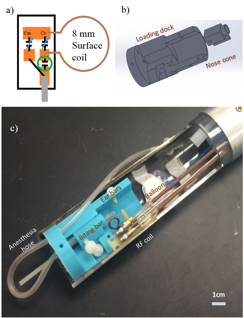
An 8 mm surface coil was fabricated as indicated by the schematic in figure 1 a. Printed circuit board was used to mechanically and electrically stabilize the coil and the coupling network. The restrainer for the mouse head shown in figure 1 b was designed using SolidWorks (Dassault Systemes SOLIDWORKS Corp. MA, USA). The main function of the restrainer was to restrain the movement of the mouse head due to possible ‘hiccup’ breathing and other motion. The restrainer was printed using PolyLactic Acid (PLA) with a Lulzbot TAZ5 3D printer (Aleph Objects Inc., CO, USA). The surface coil and the restrainer were assembled in a home built probe as shown in figure 1 c.
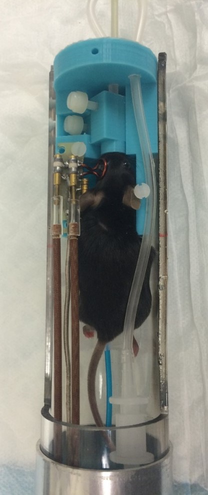
The mouse preparation for this particular set up was performed similarly as in previous work [1]. The only addition to this set up were the ear bars. The ear bars were inserted and anchored to the zygomatic arch hole to inhibit translation movement of the head. Proper set up of nose cone, biting bar, and ear bars allowed to restrain the mouse head from any movement (figure 2).The fabricated setup was tested at 14.1 T. Two-dimensional gradient echo imaging was performed and compared with the previous setup [1].
MR angiography of the mouse eye was performed three-dimensionally using a gradient echo sequence with 6mm x 7.2mm x 7.2mm Field Of View (FOV) and 200 x 240 x 240 pixel sizes (30 um isotropic pixel resolution). 40 ms repetition time (TR) and 5 ms echo time (TE) were used to create the contrast between blood vessels and surrounding tissues. The total experiment time for the scan was 38 mins 24 secs. The blood vessels were segmented and the shape of the eye was volume rendered using Avizo (Thermo Fisher, OR, USA).
Results and Discussion
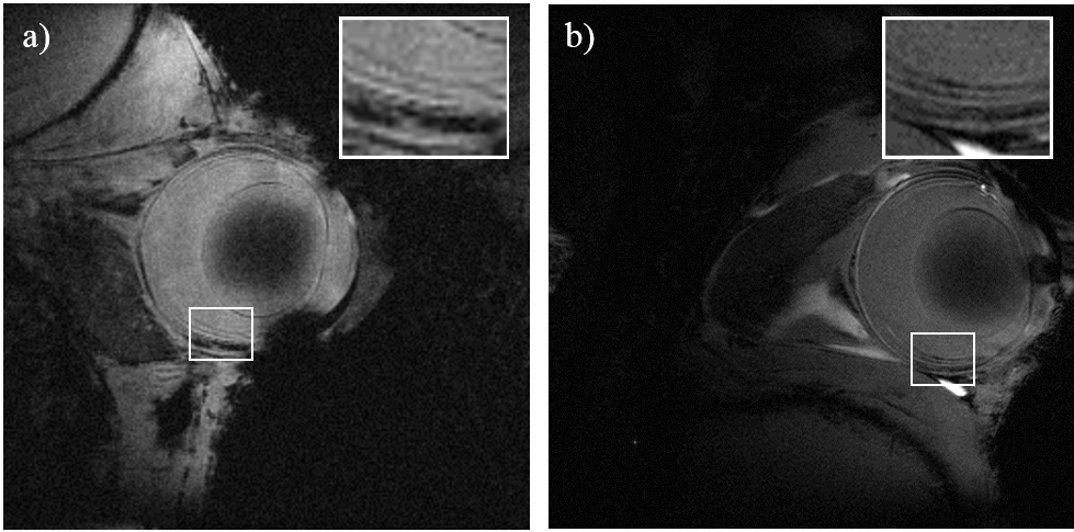
Figure 3 shows the two dimensional retinal images using the old set up (a) and the new set up with restrainer (b). The inlays in figure 3 show an enlarged part of the retina. The contrast of the layers seen with the restrainer is much clearer than the one acquired with the old set up.
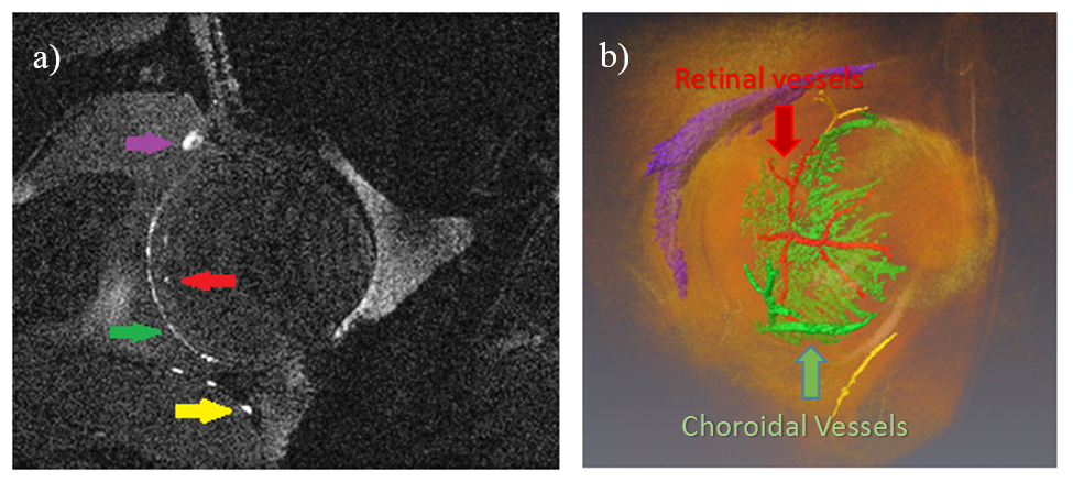
The middle slice of the three-dimensional MRI of the mouse eye is shown in figure 4 a. The micro-vessels that are represented by only a few pixels in this slide are well-resolved. The segmented vessels are shown in figure 4 b. The retinal blood vessels, color coded in red, are exhibited in respect to the position of the volume rendered eye.The choroidal blood flow and other blood vessels are also shown.
Both the previous [1] and the current work succeeded in determining the isolated retinal vessels. The outline of the choroidal vasculature was only shown in the current work. Choroidal vasculature edges were smeared in the previous work.
Conclusion
This work presented three dimensional high resolution MRI of the mouse eye using a 14.1 T micro imaging system. The angiography was created by segmenting the vasculature of the image. Future research includes combining the restrainer and contrast agent to image the ocular vasculatrue and finding the application of this method to mouse models with ocular diseases.Acknowledgements
No acknowledgement found.References
[1] G. Lee, M. Kim, and T. Neuberger, “High Field Magnetic Resonance Angiogram of the Mouse Eye,” in proceedings international society magnetic resonance medicine 23, 2015.Figures