4285
A 3He/1H Switched Frequency High-Pass Birdcage Coil for Hyperpolarized 3He Imaging in Neonates at 1.5 T1Imaging Research Center/Radiology, Cincinnati Children's Hospital Medical Center, Cincinnati, OH, United States, 2Department of Biomedical Engineering, University of Cincinnati, Cincinnati, OH, United States, 3CanonUSA, Cambridge, MA, United States, 4Pulmonary Imaging Research Center/Radiology, Cincinnati Children's Hospital Medical Center, Cincinnati, OH, United States, 5Department of Physics, Washington University in St. Louis, St. Louis, MO, United States, 6Departments of Radiology and BioEngineering, University of Missouri-Columbia, Columbia, MO, United States, 7Division of Pulmonary Medicine, Cincinnati Children's Hospital Medical Center, Cincinnati, OH, United States
Synopsis
We report details on the fabrication and performance of a transmit/receive 3He/1H switched frequency 16 rung high-pass birdcage coil for neonatal imaging at 1.5 Tesla. The coil enables collection of 3He images of neonatal lungs inflated with hyperpolarized 3He gas and ultra-short echo time (UTE) 1H images. Rotation of a shaft in the coil housing shifts the coil between 3He and 1H operating modes. 3He and UTE 1H images of explanted lungs inflated with hyperpolarized 3He gas collected using the coil demonstrate the excellent image quality achieved with the coil.
Introduction:
The production of hyperpolarized 3He and 129Xe has opened exciting possibilities for imaging lung structure and function using MRI1-4. Our institution has built a hyperpolarizer with the goal of using hyperpolarized 3He gas for MR imaging in neonatal lungs in a dedicated Neonatal Intensive Care Unit (NICU) 1.5T MR scanner developed at our institution5-6. With this goal in mind, it is essential that the MRI coil used for imaging operate at both 3He and 1H frequencies without a SNR penalty at either frequency and without the necessity to move the patient between 3He and 1H scans.
Designs for transmit/receive (T/R) 3He/1H dual tuned volume coils have been described in literature7-9. However, in these designs, coil performance at one or both frequencies is reduced by addition of inductive and capacitive elements to improve isolation between the two circuits. We have implemented a novel switched frequency (SF) transmit/receive RF coil design for a 16 rung high-pass birdcage coil that can be used to collect high quality images at both 3He and 1H frequencies (48.65 MHz and 63.86 MHz respectively at 1.5 Tesla). The SF coil described here is relatively simple with a common set of current carrying inductors used for both frequencies. The design does not include reactive elements between two tuned circuits. Advantages of this coil design include: 1) quadrature excitation and reception at both frequencies, 2) good SNR and B1 homogeneity at both frequencies, and 3) 5-10 second switching times between 3He and 1H operation. This report provides details of the coil fabrication. Images of hyperpolarized 3He and ultra-short echo time (UTE) 1H of ex-vivo explanted lungs demonstrate the excellent image quality obtained with the coil.
Materials and Methods:
The SF birdcage coil was constructed on a G10 (garolite) cylinder. The coil has an inner diameter of 7.0 in (178 mm) which will accommodate more than 90% of the neonates in our NICU. The coil rung length is 5.5 inches, which is adequate to cover the neonatal lung. A second G10 cylinder supports an integrated RF shield and serves as the outer shell of the coil.
Figure 1 shows the coil without its cover. A central component of the assembly is a white plastic carrier ring (red arrow) that holds 16 rectangular fiberglass strips (green arrow) with beryllium-copper contacts located at each end (blue arrows). As the carrier ring is rotated (black arrow), the contacts bridge gaps on the birdcage printed circuit board thereby adding additional capacitors in parallel with the birdcage coil’s main capacitors. This shifts the resonance frequency of the coil from 1H down to 3He. Manually rotating a control shaft (yellow arrow) 180 degrees rotates the carrier ring inside the coil. As the shaft rotates, two Kevlar strings (purple arrows) attached to fiberglass strips pull the contact ring assembly, causing it to rotate. Clockwise rotation of the shaft adds the capacitance needed to operate the coil at the 3He frequency. Counter clockwise rotation removes the added capacitance and returns the coil back to 1H frequency operation. After tuning the 1H and 3He modes, the measured S parameter values were - 1H mode: I -29dB S11, Q -28dB S11, -32dB S21. 3He mode: I -20dB S11, Q -14dB S11, -27dB S21.
Results and Outlook:
The coil’s 1H SNR performance was assessed using a daily quality assurance (QA) phantom. A spin echo protocol was used to collect the image data. For SNR comparison, the same QA phantom was imaged with a 180 mm ID 16 rung low-pass birdcage product coil delivered with the scanner using the identical imaging protocol. Image results are shown in Figure 2A (1H/3He coil) and 2B (product coil). The measured proton image SNR was 657.5 and 719.9 respectively for the two coils.
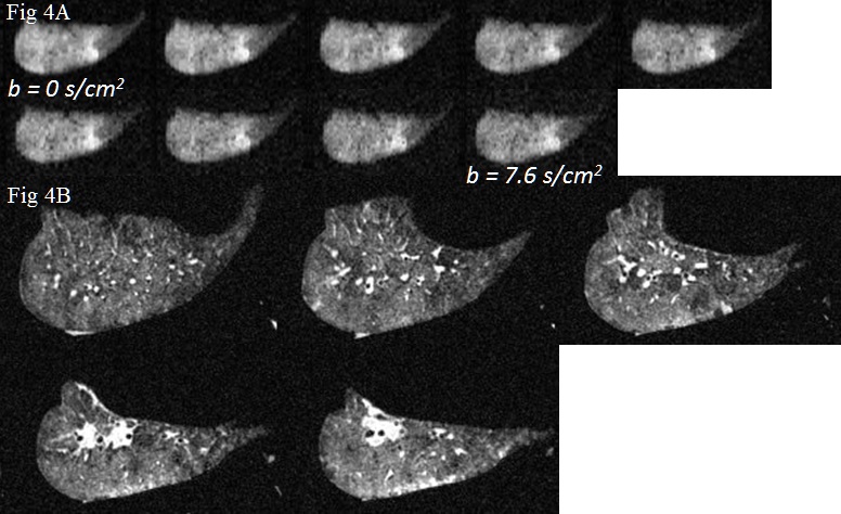
Ex-vivo imaging: Figure 3A shows 3He diffusion weighted images (DWIs) (one slice) of an explanted whole rabbit lung using nine b values incrementing in equal steps from 0 to 7.6 s/cm2. Figure 3B shows five 1H UTE images for the explanted whole rabbit lungs. Figure 4A shows 3He DWIs (one slice) of an explanted left lung of a 3 year old human using the same b values as used for the rabbit lung 3He images. Figure 3B shows five 1H UTE images for the explanted left lung of a 3 year old human.
We have shown the 1H/3He coil design described in this report provides excellent image quality and good SNR for both 3He and 1H imaging.
Acknowledgements
No acknowledgement found.References
1. Chang YV, Quirk JD, Ruset IC, Atkinson JJ, Hersman FW, Woods JC; MRM 2014 Vol 71(1); 339-344.
2. Thomen RP, Sheshadri A, Kozlowski J, Ellison HD, Szczesniak RD, Castro M, Woods JC; Radiology 2014 Aug 19:140080.
3. Pennati F, Quirk JD, Yablonskiy DA, Castro M, Aliverti A, Woods JC; Radiology 2014 Jan 15:132470
4. Walkup LL, Woods JC. 2014. “Translational applications of hyperpolarized 3He and 129Xe”. NMR Biomed 27:1429-1438.
5. Tkach JA, Merhar SL, Kline-Fath BM, Pratt RG, Loew WM, Daniels BR, Giaquinto RO, Rattan MS, Jones BV, Taylor MD, Tiefermann JM, Tully LM, Murphy EC, Wolf-Severs RN, LaRuffa AA, Dumoulin CL; Am J Roentgenol 2014 Jan;202(1):w95-W105
6. Tkach JA, Hillman NJ, Jobe AH, Loew WM, Pratt RG, Daniels BR, Kallapur SG, Kline-Fath BM, Merhar SL, Giaquinto RO, Winter PM, Li Y, Ikegami M, Whitsett JA, Dumoulin CL; Pediatr Radiol 2012 Nov 27:42(11):1347-56.
7. De Zanche N, Chhina N, The K, Randell C, Pruessmann KP, Wild JM. 2008. “Asymmetric quadrature split birdcage coil for hyperpolarized 3He lung MRI at 1.5 T”. Magn Reson Med 60:431–438.
8. Lim H, Thind K, Martinez-Santiesteban FM, Scholl TJ. 2014. “Construction and evaluation of a switch-tuned 13C-1H birdcage radiofrequency coil for imaging the metabolism of hyperpolarized 13C-enriched compounds”. J Magn Reson Imaging 40: 1082–1090.
9. Wang J-X, Boskamp EB, Santyr GE, Rutt BK. 2008. An efficient switched double-frequency birdcage coil for 3He and 1H imaging. In: 16th Proceedings of the International Society for Magnetic Resonance in Medicine. p 1117.
Figures
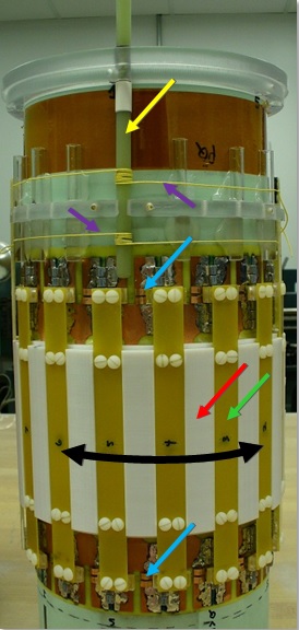
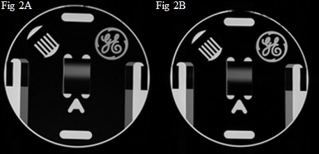
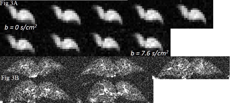
Fig. 3 (A) 3He DWIs of explanted whole rabbit lungs (one slice) for nine b values. A 3D gradient echo (3DGE) sequence was used; Voxel size: 1.7 x 1.7 x 10 mm; Matrix size: 64 (freq) x 40 (phase); FOV: 11 cm (freq) x 6.8 cm (phase); FA: 6°; TR/TE: 8.0/3.9 msec; Gas concentration: 1:1 3He/N2; NA: 1; SNR b0 image: ~50. (B) 1H radial 3D UTE images in explanted lungs: Voxel size: 0.7 x 0.7 x 0.7 mm; FOV: 18 x 18 cm; FA: 5°; TR/TE: 5.8/0.2 msec; NA: 4; SNR: ~ 3