4282
Development of a dedicated laryngeal surface coil for improved high resolution imaging in laryngeal cancer1Leiden University Medical Center, Leiden, Netherlands
Synopsis
A dedicated 4-channel larynx coil at 3T has been developed, allowing for high-resolution (1 mm isotropic) imaging of the larynx. The coil is flexible and can therefore be used on a radiotherapy mask to allow in-mask scanning of the larynx for radiotherapy planning purposes. Breathing and swallowing artefacts were eliminated using respiratory triggering and artefacts arising from pulsatile flow were suppressed using a saturation pulse. The improved image quality and resolution helps in assessing whether transoral surgical approaches on laryngeal tumors are possible.
Introduction
Advances in transoral surgical approaches in laryngeal cancer have put increasing demands on imaging for accurate evaluation of tumor involvement of the vocal cord, pre- and paraglottic space and cartilage in preoperative assessment and differentiation between scar tissue, edema and recurrent tumor postoperatively. Current imaging strategies primarily use (two) general purpose local surface coils to provide higher spatial resolution and a small decrease in acquisition time using parallel imaging techniques. While reasonable images have been obtained1,2,3, these coils are not specifically designed for laryngeal imaging (including the possibility of scanning the patient optimally in a radiotherapy mask) and have a relatively small field of view preventing simultaneous imaging of the entire larynx. Additionally, significant motion artefacts are present due to relatively long acquisition times since usually only two coils are used.
In this work we show results of high resolution imaging of the larynx using a specifically-designed four-element phased array receiver with an optimized imaging sequence using respiratory triggering and presaturation to reduce motion and flow image artifacts. The RF coil design allows a high degree of parallel imaging, thus shortening the total imaging time while maintaining high signal-to-noise, is flexible to allow imaging of different neck sizes, and can be contoured around a radiotherapy mask.Methods
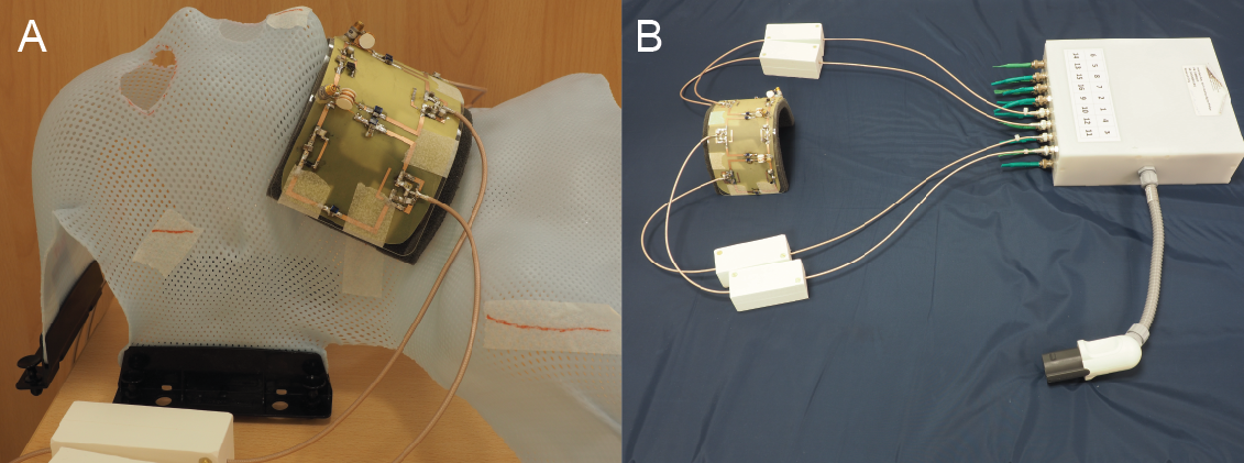
A dedicated four-channel receive-only surface coil for the larynx was developed on-site. The coil consists of four adjacent loops (60x60 mm2 each, in a 1x4 array) which are non-overlapped and induced current elimination (ICE)4 and pre-amp decoupled to allow efficient parallel imaging performance. The next nearest neighbour decoupling of the elements is lower than -30 dB. The geometry of the coil is chosen to fit closely but comfortably around the neck and is flexible enough to accommodate different neck sizes. Due to its flexibility, the coil also fits on a mask, which is used to secure the head and neck during radiotherapy. This allows for perfect matching of the diagnostic MR scan and the radiotherapy treatment planning. Scans are performed using a 3T Philips Ingenia dual transmit system and the four-channel phased array receive coil is interfaced to the scanner through a dedicated receive interface box (MR Coils, Zaltbommel, NL).
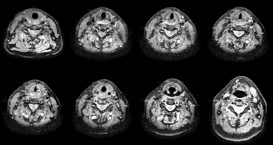
In order to reduce movement artefacts from breathing, swallowing and vascular pulsation, the scan is respiratory triggered and a 60 mm wide spatial saturation pulse is placed caudal to the larynx. The acquisition time for the multi-slice two-dimensional images was reduced by using parallel imaging with a SENSE reduction factor of 3 in the right-left direction. Image resolution was 1x1x1 mm3 isotropic.
Results
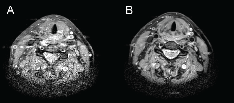
The dedicated larynx coil has been used on multiple volunteers. Any breathing or swallowing artefacts seen in non-triggered images (Figure 3A) were eliminated in the images acquired with respiratory triggering, as shown in Figure 3B. The saturation pulse eliminated most image artefacts arising from pulsatile flow. Patient and coil positioning was quick and easy due to the simple and flexible design of the coil. Volunteers indicated that the design was comfortable during the relatively short (20 minute) imaging protocol.Conclusion
High resolution (1 mm isotropic) MR images of the full larynx have been obtained without any breathing or swallowing artefacts. In order to achieve this, a dedicated 4-channel larynx surface coil was developed in-house. This coil allows efficient parallel imaging, which is used to speed up data acquisition. Furthermore, the coil is flexible and therefore easily accommodates different neck sizes. Scanning can also be performed with a subject fixated in a radiotherapy mask, which enables radiotherapy treatment planning using MRI. Additionally, the short total protocol time for patients and the fact that they can continue swallowing during the scans, reduces the burden of these scans for patients. We anticipate that this improved image quality will lead to better treatment planning and counselling in patients with laryngeal tumors.Acknowledgements
This work was funded by an ERC Advanced grant #670629 and an NWO Open Technology grant #13783.References
- Maroldi, R., Ravanelli, M., & Farina, D. (2014). Magnetic resonance for laryngeal cancer. Current opinion in otolaryngology & head and neck surgery, 22(2), 131-139.
- Preda, L., Conte, G., Bonello, L., Giannitto, C., Tagliabue, E., Raimondi, S., Ansarin, M., De Benedetto, L., Cattaneo, A., Maffini, F., Bellomi, M. (2017). Diagnostic accuracy of surface coil MRI in assessing cartilaginous invasion in laryngeal tumours: Do we need contrast-agent administration?. European Radiology, 1-9.
- Casselman J.W. (2007) High-Resolution Imaging of the Skull Base and Larynx. In: Schoenberg S.O., Dietrich O., Reiser M.F. (eds) Parallel Imaging in Clinical MR Applications. Medical Radiology (Diagnostic Imaging). Springer, Berlin, Heidelberg
- Yan, X., Gore, J. C., & Grissom, W. A. (2017). New resonator geometries for ICE decoupling of loop arrays. Journal of Magnetic Resonance, 277, 59-67.
Figures