4276
SNR Improvement of In Vivo 31P MRSI in Human Brain using Head-Shape Ultrahigh Dielectric Constant Material at 7T1Center for Magnetic Resonance Research, Department of Radiology, University of Minnesota Medical School, Minneapolis, MN, United States, 2Center for Nuclear Magnetic Resonance Research, Department of Radiology, The Penn State College of Medicine, Hershey, PA, United States, 3Department of Engineering and Science and Mechanics, The Penn State College of Engineering, University Park, PA, United States
Synopsis
Increased RF power (thus, higher SAR) and inadequate detection sensitivity (or SNR) even at high/ultrahigh field are the major challenges for X-nuclei MRS imaging (MRSI) for human applications. In this work, we demonstrate that using the ultrahigh dielectric constant material (uHDM) conformed to the human head incorporated into the RF head volume coil, improved detection sensitivity and reduced demand of RF transmit power were achieved across an entire object for testing phantom and human brain 31P MRSI at 7T. Therefore, incorporating optimized geometry of uHDM with RF coil can significantly boost SNR and reduce SAR in X-nuclei MRS applications, ultimately, improve spatiotemporal resolution.
Introduction
The advanced technology of in vivo MRS and MRI has provided a wealth of knowledge about brain function, connectivity, neurochemistry and neuroenergetics in the human brain. However, due to the lower cerebral metabolite concentrations, the need for higher sensitivity, spatiotemporal and spectral resolutions is growing for in vivo 31P MRS imaging applications even at ultrahigh field to address questions of interest. In addition, the increased RF transmit power at ultrahigh field raises a major safety concern in the human brain research. Recent development of high dielectric constant materials (HDM) incorporated with RF coils has shown significant improvements in the RF transmit field (B1+) and reception field (B1-) for MRI applications [1-4]. Due to the relatively low Larmor frequency of X-nucleus, the translation of the HDM technique into in vivo X-nucleus application requires an ultrahigh dielectric constant material (uHDM) [5] with optimal geometry conformed to a target object. Thus, our aim in this work was to test a proof-of-concept design of a human head-shaped uHDM for improving detection sensitivity of in vivo 31P MRS imaging at 7T.Methods
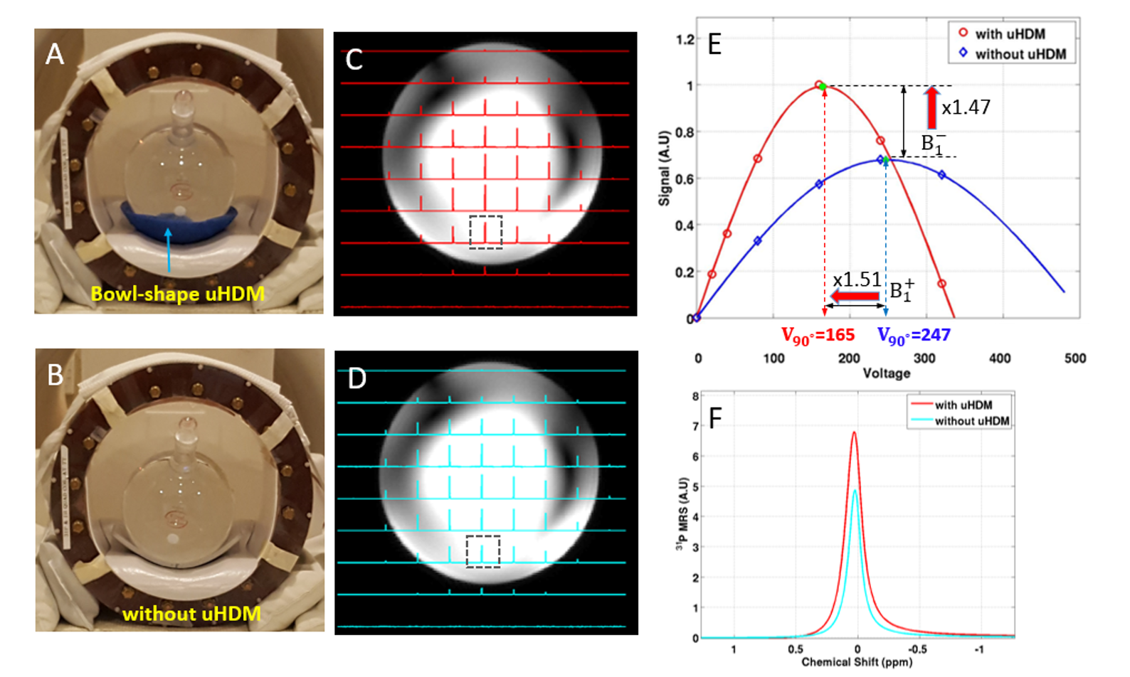
A bowl-shaped uHDM former (BS-uHDM) was designed and fabricated to conform the human head shape. It was made of low lossy (0.05 loss tangent) PZT (Pb(Zr, Ti)O3) ceramics (effective permittivity: εeff = 800, thickness = 0.8 cm, and diameter = 8 cm) (Fig. 1A). 31P MRS studies were carried out on a Siemens 7.0T/90 cm human scanner (Magnetom, Germany) using a 31P-1H dual-channel RF head TEM volume coil. All measurements were repeated with or without use of the uHDM former on a spherical phantom (2 liter, 50 mM inorganic phosphate (Pi)) and healthy human subject. Multiple 3D 31P CSI data with varied RF pulse powers (voltages) were acquired for estimating B1+ and B1- maps based on the Pi resonance in the phantom and phosphocreatine (PCr) resonance in the human brain.Results
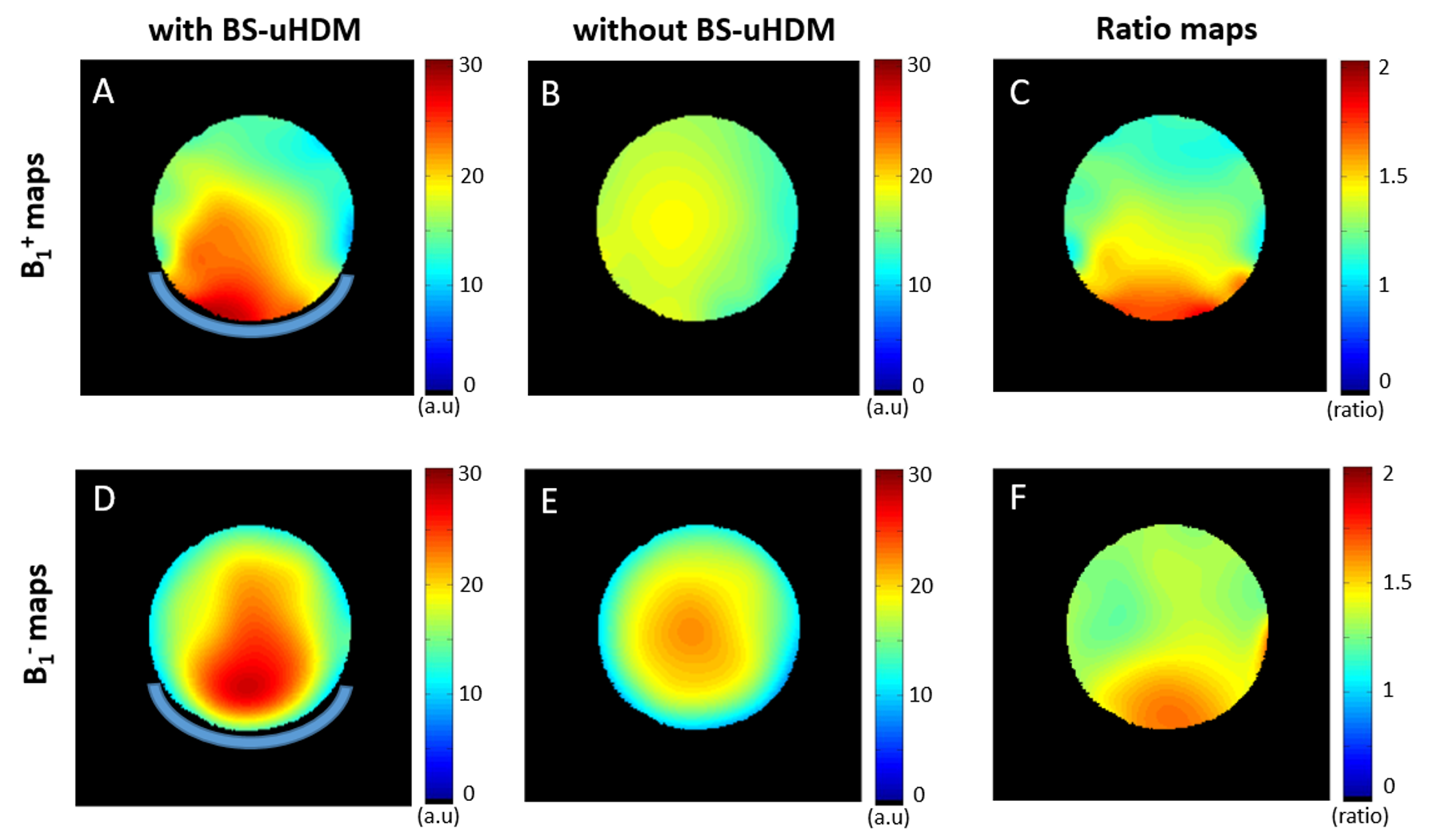
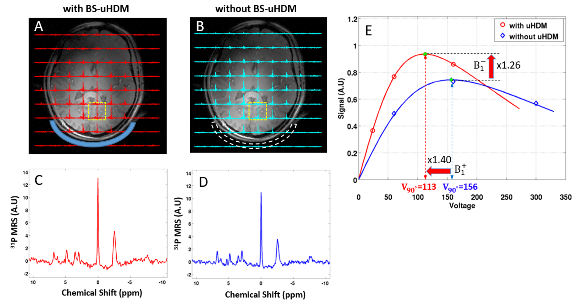
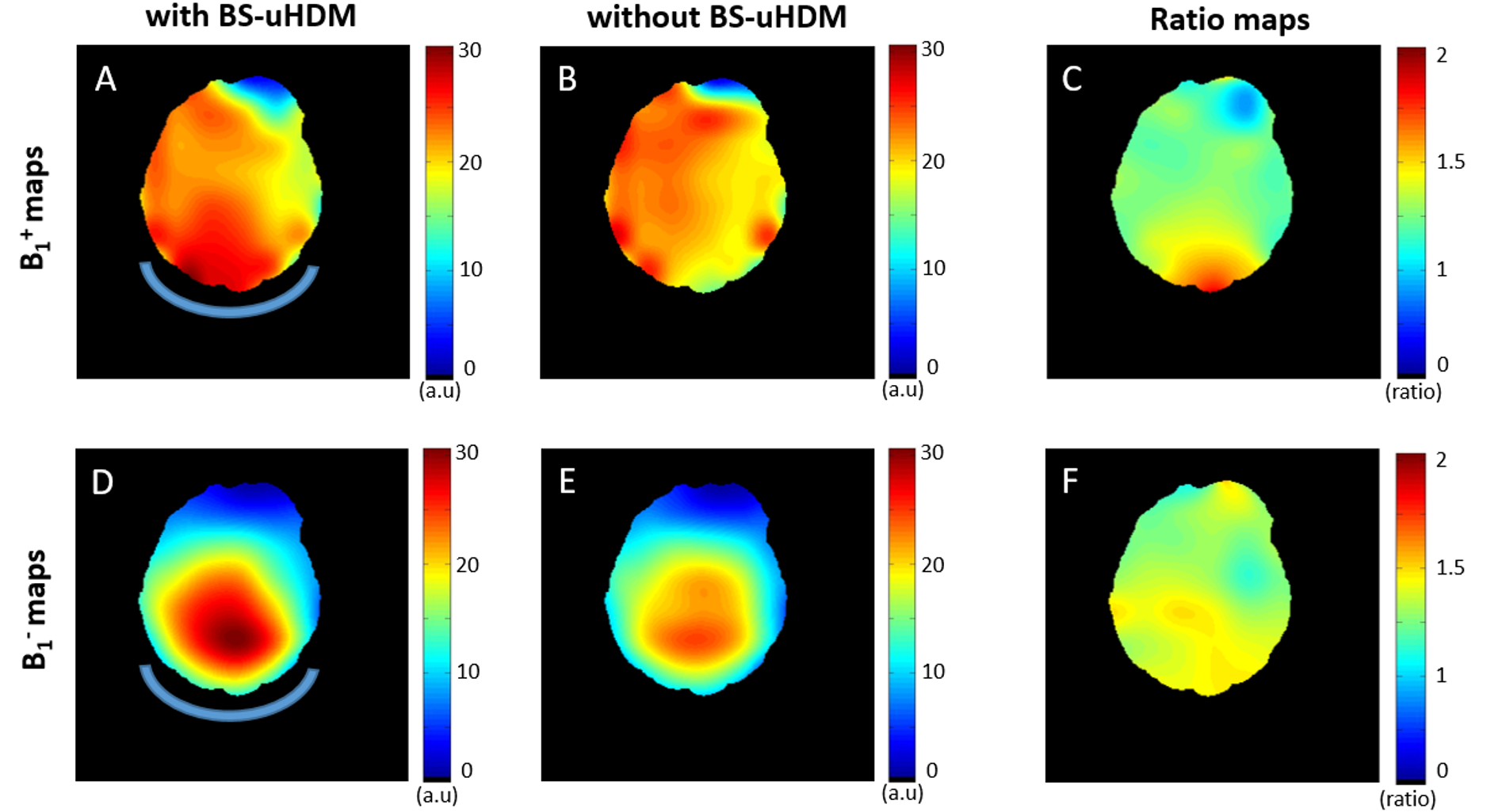
Figures 1 and 3 display the 31P CSI spectra of the phantom and in vivo human brain acquired with a reference voltage, respectively. The 31P CSI data clearly demonstrate the apparent B1 improvements in the presence of the BS-uHDM across the entire object, showing a higher signal intensity with a lower reference voltage. Figures 2 and 4 show the quantitative comparisons of the 31P B1 results in the representative central CSI slice for the phantom and in vivo human brain, respectively. In general, the use of the BS-uHDM former in the 31P imaging led to overall B1+ and B1- enhancement across the entire object. The B1 ratio map on the phantom shows the significant improvement using the BS-uHDM former reaching up to 60% for both B1+ and B1- (Fig. 2C and 2F). In Fig. 4, the BS-uHDM former for in vivo 31P human brain also improved the B1 efficiency, reaching up to 53% for B1+ and 37% for B1-, which is consistent with phantom results by considering the loss of detection sensitivity due to the presence of a gap between human skull and brain tissue.Discussion
The results provide compelling evidence of the uHDM benefits for effectively improving both the B1+ and B1- efficiency, consequentially leading to a large increase in 31P imaging sensitivity in the human brain at 7T. The degree of the B1 improvements and their spatial distribution are sensitive to the uHDM design including the ceramic permittivity constant, loss tangent, and geometry for optimizing the performance and maximizing the efficacy [6]. The merit of a high-quality uHDM with large coverage further boosts the SNR for X-nuclear MRS imaging applications. These findings open new and exciting opportunities for more research that integrates the MRI/MRS technology with RF engineering and material science.Conclusion
Our study demonstrates that the novel design of uHDM technology can significantly improve both RF transmitter efficiency and detection sensitivity (i.e., SNR) for human brain application of in vivo 31P MRS at ultrahigh field. The uHDM technology provides an important and cost-effective engineering solution for advancing MRI and in vivo MRS techniques at high/ultrahigh field, which will provide enormous benefits for in vivo human applications in particular for brain research. The same concept of the uHDM technology can be extended to other X-nuclear MRS or imaging applications beyond 31P and 1H spins or other organs beyond the brain, as demonstrated in this work, under healthy and diseased conditionAcknowledgements
NIH grants: R01 NS057560, NS070839, MH111447; R24 MH106049, MH106049 S1, S10RR026783, P41 EB015894 and P30 NS057091; the AHC Faculty Research Development (FRD) grant from the University of Minnesota.References
[1] Yang et al., JMRI 24, 197-202 (2006).
[2] Haines et al., JMR 203,
323-327 (2010).
[3] Yang et al., MMR 65,
358-362 (2011).
[4] Webb, Concepts in MR. 148-184 (2011).
[5] Lee et al., MRI 42,
158-163 (2017).
[6] Lee et al., Proc.
ISMRM; 24: 746 (2017)
Figures