4275
Experimental and numerical evaluations of simultaneously excitable Eigenmodes in a 20-channel transmit RF array for 7 Tesla human MRI1University of Pittsburgh, Pittsburgh, PA, United States, 2Siemens Medical Solutions, Tarrytown, NY, United States
Synopsis
The Eigenmodes of a 20-channel Tic-Tac-Toe RF array are presented and analyzed. The Eigenmodes were calculated for each excitation level in Z (static magnetic field) direction, resulting in 4 modes per level and 20 in total. FDTD simulations and in-vivo experiments were performed. Quadrature mode is the most efficient, and generally excites the center of the brain; Anti-quadrature mode excites the head
Introduction
The excitation of two modes has shown improvements in RF inhomogeneities 1 most especially for 7T imaging. In this work, the Eigenmodes of a 20-channel Tic-Tac-Toe (TTT) RF array for head imaging at 7T are investigated and accurately validated with in-vivo measurements.Methods
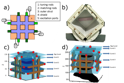
The TTT design is composed of eight transmission lines connected to each other in the Tic-Tac-Toe fashion (Figure 1a). The TTT head coil is built from 5 sets of TTT sides, totaling 20 transmission channels 2. The excitation points are positioned at different locations in Z (static magnetic field) direction, composing 5 levels of excitation: Top Level, Level 1, …, Level 4.
Simulations were performed using an in-house developed FDTD package with an accurate transmission line model 3. Each channel was simulated individually, and the transmission line model was used to tune and match the RF coil (by adjusting the tuning and matching inner rods, described in Figure 1a). Using the simulated data, the eigenmode of each excitation level was calculated. There are 4 excitation modes for each Level of the RF coil, named as: Mode 1 (quadrature, with regular phase increments of 90°), Mode 2 (opposite-phase, with regular phase increments of 180°), Mode 3 (anti-quadrature, with regular phase increments of 270°), and Mode 4 (zero-phase, without any phase increment). Using Eigenmode theory, the phase and amplitude errors are within 8% of the nominal values.
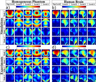
The experimental data were acquired in a 7T MRI scanner using the 8-channel pTx mode. The sequence used was the Turbo Flash, with the following parameters: TE/TR = 2.34/160ms, resolution 3.2mm isotropic, and flip angle 12 degrees. The sequence outputs the relative B1+ maps (normalized by the square root of the sum of the square of all transmitting channels). Since the system only contains 8-Tx channels, two levels were transmitting at the same time (while the other ones were only receiving signal), and Level 1 was always transmitting. This process was repeated 4 times.
Results
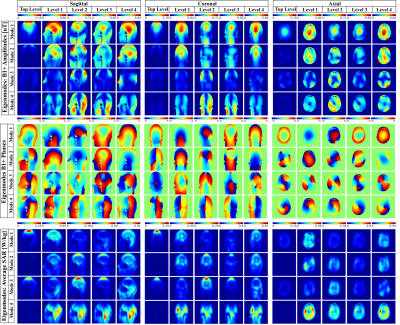
Figure 2 shows the B1+ field distribution (amplitudes and phases) and SAR distribution for the 20 Eigenmodes of the RF array. Both B1+ and SAR maps are normalized for 1W input power per channel (total of 4W per level). The scale used in the plots was from 0 to maximum in each level.
Figure 3 shows the compassion between the simulated and experimentally acquired B1+ maps in a homogeneous phantom and in-vivo.
Discussion and conclusions
The RF array produces 20 different Eigenmodes and 5 of these modes can be exited simultaneously. Based on the results, we can state that: 1) Mode 1 (quadrature) Levels 1 and 4 excites the center of the brain; 2) Mode 4 (zero-phase) Levels 2 and 3 excites brain periphery and cerebellum; 3) Mode 1 Top Level is the most efficient, but also produces higher SAR; 4) Mode 3 (anti-quadrature) usually excites the periphery of the head, having potential applications for fat saturation; and 5) Mode 2 (opposite-phase) Levels 1 and 4 also excites the center of the brain, however with lower efficiency when comparing with the Mode 1.Acknowledgements
This work was partially funded by the National Institutes of Health R01MH111265 and by the CAPES Foundation, Ministry of Education of Brazil, 13385/13-5.References
1. Orzada S, Maderwald S, Poser BA, et al. Time-interleaved acquisition of modes: an analysis of SAR and image contrast implications. Magn Reson Med. 2012;67(4):1033-1041.
2. Ibrahim T, Zhao Y, Krishnamurthy N, et al. 20-To-8 Channel Tx Array with 32-Channel Adjustable Receive-Only Insert for 7T Head Imaging. Paper presented at: International Society of Magnetic Resonance in Medicine2013; Salt Lake City, Utah.
3. Santini TK, Junghwan; Wood, Sossena; Krishnamurthy, Naraynan; Raval, Shailesh; Ibrahim, Tamer. A new RF coil for foot and ankle imaging at 7T MRI. Paper presented at: In Proc. of the 25th International Society of Magnetic Resonance in Medicine Annual Meeting2017; Honolulu, Hawaii, USA.
Figures


