4260
Fast parameter mapping at 7T with SSFP MR Fingerprinting using 3D radial trajectories1IMAGO7 Foundation, Pisa, Italy, 2National Institute for Nuclear Physics, Pisa, Italy, 3Technische Universitat Munchen, Munich, Germany, 4University of Pisa, Pisa, Italy, 5IRCCS Fondazione Stella Maris, Pisa, Italy, 6GE Healthcare, Munich, Germany
Synopsis
When using ultra-high field MRI scanners (UHF, B0>= 7T), quantitative imaging is challenging due to B0 and B1+ non-uniformities. Magnetic resonance fingerprinting (MRF) represents a great opportunity for quantitative imaging at UHF as it can estimate these effects at the same time of the parameters of interest. Here, we demonstrate in vivo at 7T a novel 3D MRF approach based on a three-dimensional radial k-space acquisition, estimating M0, T1, T2 and B1+ simultaneously in 5 minutes.
Purpose:
Recent technological advances have improved on the effectiveness of ultra-high field MRI scanners (UHF, B0>=7T). Although new contrasts have been exploited at high field, common approaches are inherently non-quantitative, mostly due to the difficulties in dealing with B0 and B1 non-uniformities. Magnetic resonance fingerprinting (MRF) represents a great opportunity for quantitative imaging at UHF as it does not require fields to be homogeneous. By including these nuisance parameters into the signal encoding model, these can be estimated at the same time of the parameters of interest. Recent approaches have studied 2D acquisitions at 7T with simultaneous B1+ estimation1,2. Here, we propose a novel 3D MRF approach based on a three-dimensional radial k-space acquisition, estimating M0, T1, T2 and B1+ simultaneously, demonstrating it in vivo at 7T.Methods:
Acquisition Parameters
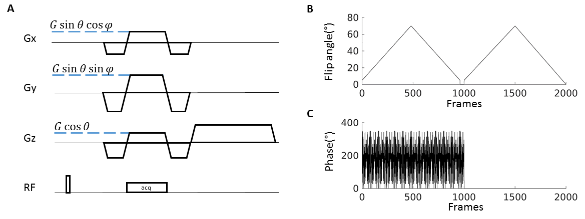
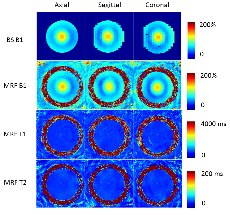
We obtained images of a uniform phantom (T1 600ms, T2 of 45ms) and a healthy volunteer using a Discovery MR950 7T MRI system (GE Healthcare, Milwaukee, WI, USA) equipped with a 2-channel transmit / 32-channel receive head coil (Nova Medical, Wilmington, MA, USA). The acquisition sequence was based on SSFP MRF3 (FOV 25.6x25.6x25.6cm2, 128x128x128 matrix), using hard pulses of 500 us length for excitation. We used a fixed TE/TR of 1.5/10ms, acquiring 16 repetitions of 2000 frames preceded by a 10ms-long hyperbolic secant adiabatic inversion pulse, using the flip angle and phase lists in Figure 1. To increase the capability of the sequence to discriminate T2 and B1+ effects, we introduced radiofrequency spoiling in the first part of the acquisition following Cloos et al.1. For comparison of the B1+ values, in the phantom we acquired a standard 2D Bloch Siegert B1+ map (TR=100ms, TE=13ms, Flip Angle=30°, 4ms Fermi pulse, 2kHz off-resonance).
3D Trajectory
Our trajectory included 3294 3D radial spokes. Each spoke was acquired symmetrically with respect to k-space center and achieved zero-moment nulling in x, y and z, after which a spoiler in z was added creating 2π dephasing across a 2-mm voxel (see Figure 1). To increase spatial and temporal incoherence, we randomly permutated the order of the radial spokes using a uniform probability distribution. Prior to reconstruction and matching, k-space data were combined using SVD compression and adaptive coil combination was performed in the image domain.
Dictionary
Simulations were performed using the extended phase graphs formalism4. The dictionary included T1 values ranging from 10ms to 2s with 50ms steps and from 2s to 6s with 250ms steps, T2 values ranging from 20ms to 100ms with 2ms steps, from 100 and 200ms with 5ms steps and between 200 and 600ms with 20ms steps. B1+ values were scaling factors to the nominal flip angle, ranging from 20% to 200% in 5% steps. Randomized SVD with rank k=8 was used5. MRF maps were obtained by inner-product pattern matching.
Results and discussion
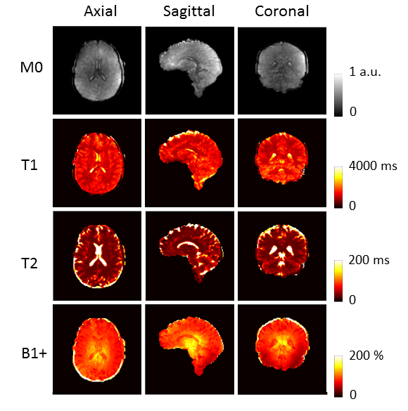
Figure 2 shows the comparison of the MRF B1+ map from our 5-minutes acquisition with a standard 2D Bloch-Siegert map acquired in 7 minutes, showing good agreement. Figure 3 shows representative parametric maps in one subject. It can be noted that T1 and T2 maps are free from B1+ artifacts. We could obtain an estimate of T1, T2, M0 and B1+ in 5 minutes at 7T. Importantly, our method did not require external B0 or B1+ calibrations, and T1 and T2 values were compatible with previous literature. Compared to other literature methods for 3D MRF based on stack of spirals6,7, the current approach offers more undersampling opportunities as all the spokes traverse the central region of k-space in each TR. Further optimised acquisitions can achieve higher spatial resolution and include other biophysical parameters of interest.Conclusion:
Our novel acquisition achieves a fast assessment of quantitative parameters at 7T in 3D, simultaneously estimating and eliminating the effect of B1+ non-uniformity.Acknowledgements
No acknowledgement found.References
1. Cloos MA, Knoll F, Zhao T, Block KT, Bruno M, Wiggins GC, et al. Multiparametric imaging with heterogeneous radiofrequency fields. Nat Commun 2016;7:12445. doi:10.1038/ncomms12445.
2. Buonincontri G, Schulte R, Cosottini M, Sawiak SJ, Tosetti M. Spiral MR fingerprinting at 7T with simultaneous B1 estimation. Magn Reson Imaging 2017. pii: S0730-725X(17)30074-7. doi: 10.1016/j.mri.2017.04.003.
3. Jiang Y, Ma D, Seiberlich N, Gulani V, Griswold MA. MR fingerprinting using fast imaging with steady state precession (FISP) with spiral readout. Magn Reson Med 2015;74:1621–1631
4. Weigel M. Extended phase graphs: dephasing, RF pulses, and echoes - pure and simple. J Magn Reson Imaging2015;41:266–295.
5. Yang, M., Ma, D., Jiang, Y., Hamilton, J., Seiberlich, N., Griswold, M. A., & McGivney, D. (2017). Low rank approximation methods for MR fingerprinting with large scale dictionaries. 2017 Aug 13. doi: 10.1002/mrm.26867
6. Ma, D., Jiang, Y., Chen, Y., McGivney, D., Mehta, B., Gulani, V. and Griswold, M. (2017), Fast 3D magnetic resonance fingerprinting for a whole-brain coverage. 2017 Magnetic Resonance in Medicine. doi:10.1002/mrm.26886
7. Congyu Liao, Berkin Bilgic, Mary Kate Manhard, Bo Zhao, Xiaozhi Cao, Jianhui Zhong, Lawrence L. Wald, Kawin Setsompop. (2017) 3D MR fingerprinting with accelerated stack-of-spirals and hybrid sliding-window and GRAPPA reconstruction. NeuroImage 162 (2017) 13–22
Figures