4218
Single echo radial random alternating TE acquisition for B0 field inhomogeneity robust fat suppression1Yonsei University, Seoul, Republic of Korea
Synopsis
A single echo method which provides B0 field inhomogeneity robust fat suppression was proposed using a radial acquisition with random alternating echo time. To validate this study, simulation and in vivo experiments were performed. This technique would be useful in fat suppressed applications (e.g., MSK, liver, ...) which are required fast scan and robustness of B0 field inhomogeneity.
Introduction
To overcome B0 field inhomogeneity is a classical challenge in fat suppressed MRI1. Therefore, many researches were proposed, however, most of the studies were based on multi-echo acquisition to figure out the B0 field distribution2-3. Herein, a single echo method which provides B0 field inhomogeneity robust fat suppression was proposed using a radial acquisition with random alternating echo time (TE).Method
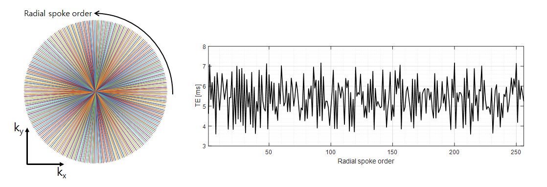
Previously, single echo fat suppression method using radial alternating TE (rAT) sequence was proposed4. Although, the previous method successfully suppressed fat component in on resonance situation, however, fat suppression can be failed when B0 field inhomogeneity exists. To overcome this problem, we proposed a radial acquisition method with random alternating TE (rRAT) as plotted in Figure 1.
The proposed TE pattern was generated by
$$TE(n^{th} spoke)=\begin{cases}TE_{desired} + τ(n^{th} spoke)\space [ms] & , \space for\space n^{th}\space spoke =1, 3, 5, …\\TE_{desired} + 1.2 + τ(n^{th} spoke) [ms] & , \space for\space n^{th}\space spoke =2, 4, 6, …\end{cases}$$
$$ -1.2 < τ(n^{th} spoke) < 1.2,\space\space τ(n^{th} spoke) \space\space follows\space a\space uniform\space random\space distribution$$
First, alternating TE (1.2 ms), same manner of the previous study4, was applied. Then, random TE (τ(nth spoke)) was also added to every spoke, as shown in Figure 1, to mitigate B0 field inhomogeneity effect.
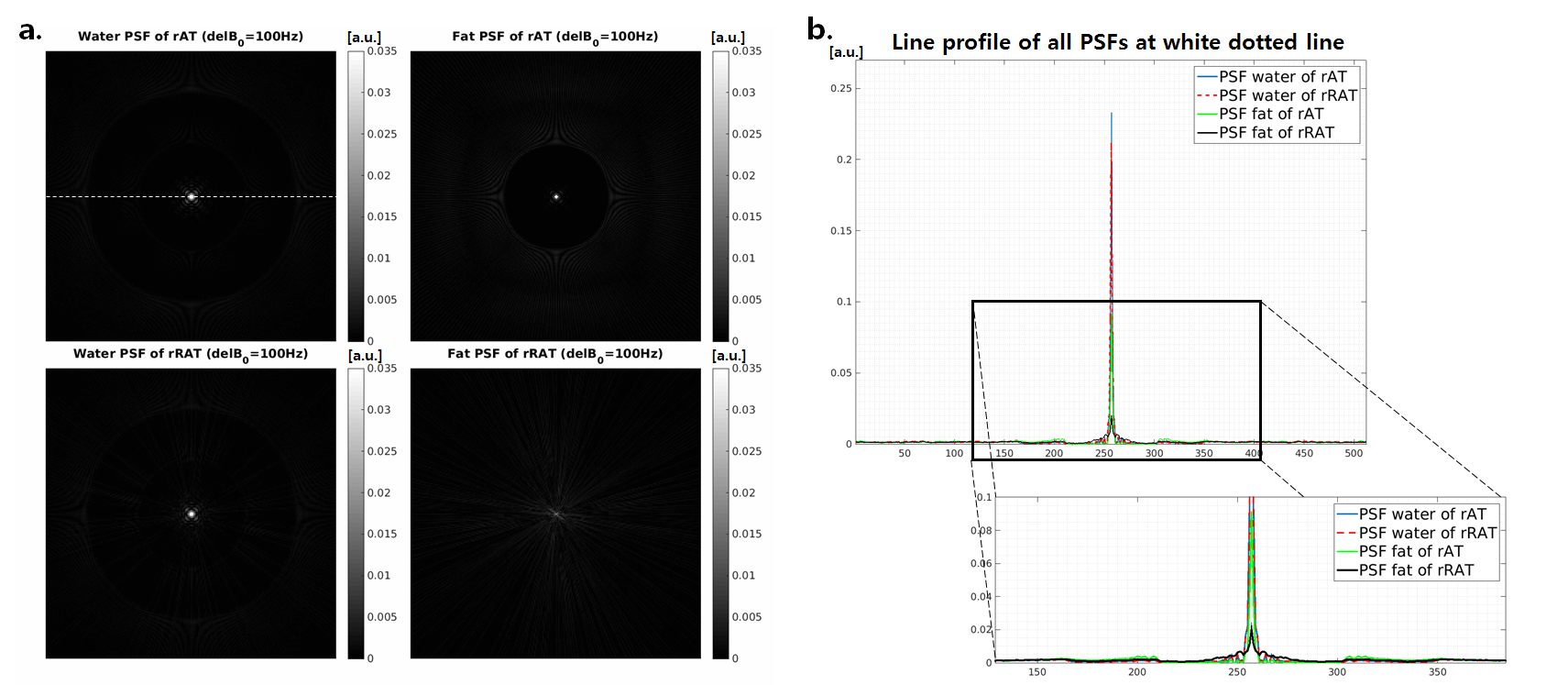
To validate this study, simulation and in vivo experiments were performed. For the simulation, a square which is composed with two parts, outer square (only water) and inner square (linearly varying water/fat mixed), was generated as shown in Figure 3. To figure out the effect of B0 field inhomogeneity in both rAT and rRAT method, delB0 map (100 Hz) was generated as shown in Figure 3e. For simulation, total 256 golden angle5 radial spokes were used and TEdesired was set to be 7.4ms. Point spread functions (PSF) were also generated within 100 Hz field inhomogeneity condition.
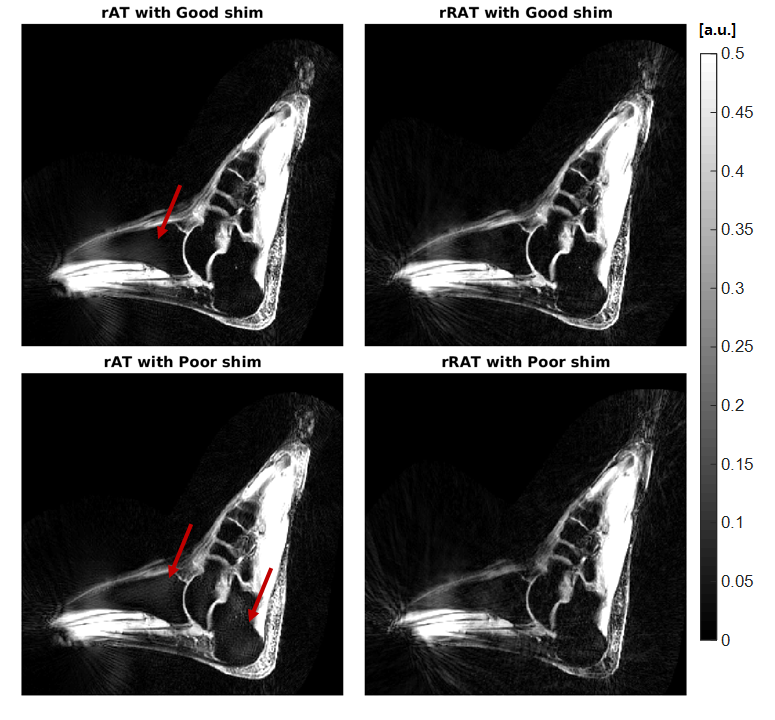
For the in vivo experiments, foot data from healthy volunteer (IRB approved) was collected using 16ch foot coil. Scan parameters : FA=25˚, TR=40ms, Resolution=1x1 mm2, Slice thickness=5mm, TEdesired=7.4ms, BW=390Hz/Px. Golden angle radial pulse sequence based on FLASH acquisition was generated (total 512 radial spokes were acquired). To validate the proposed method, in vivo data were acquired with both good shim and poor shim condition. To reduce the streaking artifacts, ESPIRiT, NUFFT and parallel imaging CS were applied to reconstruct acquired in vivo data using BART toolbox6.
Results
Figure 2 shows the water and fat PSFs of each method. Peak value of the rAT was 5 times higher than the peak value of the rRAT within 100 Hz field inhomogeneity condition, as shown in Figure 2b.
Simulation results were shown in Figure 3. Because of severe B0 field inhomogeneity (100Hz), rAT method which is based on 2-point Dixon method failed to suppress fat component. The proposed method which was applied random alternating TE (rRAT) shows much better fat suppression than the result of rAT. However, streaking artifact was increased.
These results were also plotted in the line profiles with different B0 field inhomogeneity values in Figure 3f. Fat suppression errors using rAT were increased as B0 field inhomogeneity become severe, however, the proposed method (rRAT) successfully suppressed fat components within various B0 field inhomogeneity values.
In vivo results were shown in Figure 4. Both methods show good fat suppression in good shim condition, except red arrowed region in the results of rAT. However, rAT method failed to suppress fat in some region (red arrow) within poor shim condition. The proposed method (rRAT) successfully suppressed the fat component for every region within the poor shim condition. Furthermore, severe steaking artifact due to the random TE was not shown in the in vivo result of the rRAT method, because parallel imaging CS (PICS) using BART toolbox were applied.
Discussion
PICS in BART tool box was applied to reduce the streaking artifact, however, further optimization or more advanced reconstruction technique would be our future work.
The min, max value of random TE and random distribution were decided in curious way. Therefore, further analysis or optimization would be also required.
In this study, the proposed pulse sequence was based on FLASH, however, we have planned to apply the method based on TSE or bSSFP pulse sequences. Furthermore, applying the proposed to other applications, such as spine and MSK with implant, would be our future work.
Conclusion
We have presented a single echo fat suppression method which have robustness in B0 field inhomogeneity using random alternating TE radial acquisition. This technique would be useful in fat suppressed applications (e.g., MSK, liver, ...) which are required fast scan and robustness of B0 field inhomogeneity.Acknowledgements
This research was supported by Global PH.D Fellowship Program through the National Research Foundation of Korea(NRF) funded by the Ministry of Education (NRF-2015H1A2A1030698) and Basic Science Research Program through the National Research Foundation of Korea(NRF) funded by the Ministry of Science, ICT and future Planning (NRF-2016R1A2B3016273).References
1. Dixon WT. Simple proton spectroscopic imaging. Radiology 1984; 153: 189-194.
2. Glover GH, Schneider E. Three-point Dixon technique for true water/fat decomposition with B0 inhomogeneity correction. Magnetic Resonance in Medicine. 1991;Apr;18(2):371-83.
3. Hernando D, Kellman P, Haldar J, Liang ZP. Robust water/fat separation in the presence of large field inhomogeneities using a graph cut algorithm. Magnetic Resonance in Medicine, 2010;63:79-90.
4. Chris A. F, Brian D, et al. Radial Alternating TE Sequence for Faster Fat Suppression. Magnetic Resonance in Medicine, 2003;50:1095–1099
5. Winkelmann S, Schaeffter T, Koehler T, Eggers H, Doessel O. An optimal radial profile order based on the Golden Ratio for time-resolved MRI. Medical Imaging, IEEE Transactions on 2007;26:68-76.
6. BART Toolbox for Computational Magnetic Resonance Imaging, DOI: 10.5281/zenodo.592960
Figures