4213
Accurate Slice-Selective MRI near Metallic Implants with FOCUSED-xSPEN1Chemical & Biological Physics, Weizmann Institute, Rehovot, Israel, 2Chemical & Biological Physics, Weizmann Institue, Rehovot, Israel
Synopsis
Metals frequently cause dramatic spatial inhomogeneities in the static field B0. This causes severe localization errors, including slice z-displacements and slice-thickness variations. Clinically used techniques often avoid such errors by adding a phase encoding loop in z, prolonging examination times. This study examines a novel Fully refOCUSED cross-term SPatio-temporal ENcoding (FOCUSED-xSPEN) approach, as a slice selection technique for 2D MRI in the presence of metal implants. The method relies on xSPEN’s proven immunity to in-plane distortions caused by B0 heterogeneities, to design a new excitation scheme that delivers faithful 2D slices near implants with reduced scan times.
Introduction
Non-ferromagnetic metal devices can cause dramatic spatial inhomogeneities in the static field B0, leading to severe localization errors during the slice-selection process. Clinically proven techniques such as Multi-Acquisition Variable Resonance Image Combination (MAVRIC) [1] or Slice Encoding for Metal Artifact Correction (SEMAC) [2] avoid such errors by adding either a phase encoding loop in z or a loop on the transmitter/receiver offsets. We explore here the consequence of relying on cross-term spatio-temporal encoding xSPEN [3], a recently proposed single-shot MRI technique with unprecedented robustness to B0 inhomogeneities, as basis for developing an inhomogeneity-insensitive slice-selective excitations. The resulting Fully refOCUSED cross-term SPatiotemporal ENcoding (FOCUSED-xSPEN) approach relies on the field inhomogeneities themselves to encode the desired slice (Figure 1a); when combined with other principles described below, this allows one to target a precise z-slice and enabling faster 2D imaging near the implant.Methods
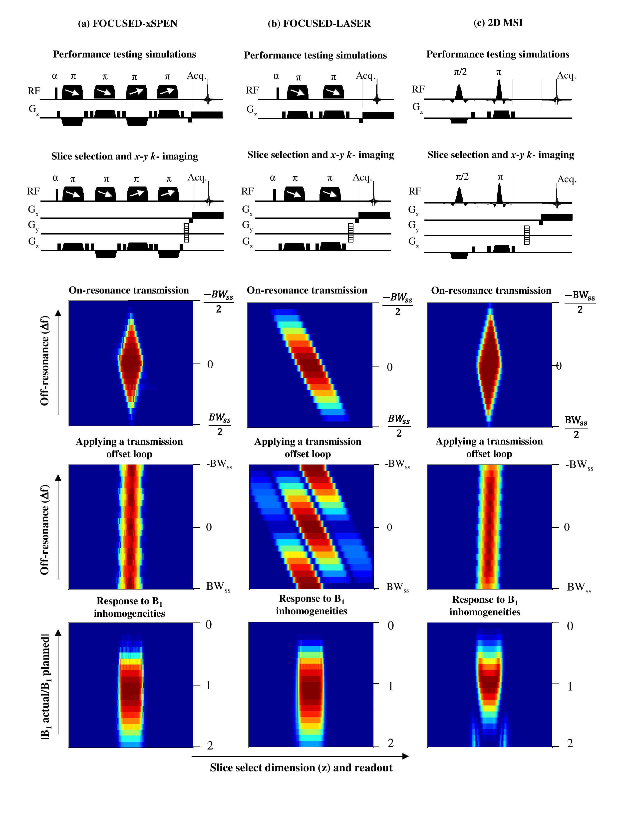
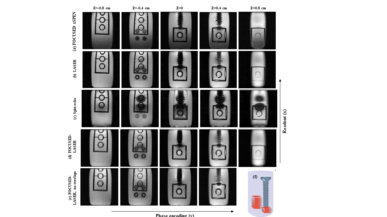
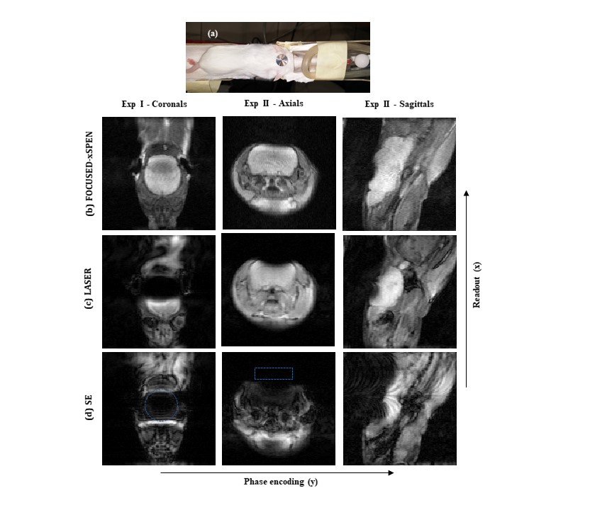
FOCUSED-xSPEN involves a non-selective excitation followed by adiabatic inversion pulses, linearly sweeping a frequency range associated with the slice selection bandwidth (BWss) while in the presence of alternating gradients. The first pair of adiabatic pulses, which are relatively insensitive to RF heterogeneities, impart an evolution phase that is linear in both the resonance offset Δf, and on position along the z-dimension. To remove this bilinear term for arbitrary resonance offsets an additional pair of swept pulses is added, identical to the first pair except for their reversed sweeping direction (Fig. 1a). The overall region that is selected by this cascade of pulses is a diamond in offset and space, coming from an intersection of the diagonal slabs that in Δf-z are subtended by the pulses. While the position of the desired slice is therefore accurately defined, the resulting approach is still narrowband in the sense of addressing up to BWss offsets. If BWss is chosen smaller than the a priori unknown inhomogeneity, this can be overcome by looping acquisitions over transmission and reception offsets in steps Δftrans=BWss/(2n). This produces several frequency binned images corresponding to shifted “Δf-z diamonds” along the off-resonance dimension. Summing all these images yields a “slice” centered in z and covering the off-resonance dimension. As shown in Figure 1, uniform response and a well-defined z-thickness then follows regardless of off-resonance. To evaluate FOCUSED-xSPEN in the presence of metal devices, we compared its performance to simpler alternatives including conventional Spin-Echo (SE), Localization by Adiabatic SElective Refocusing (LASER) [4], the recently proposed 2D Multi-Spectral Imaging (2D MSI) approach [5], and to a new technique we developed following the above-mentioned principles called FOCUSED-LASER that loops a LASER block over transmission offsets. Figure 1 shows simulated results of how all these sequences are expected to perform in single- and multiple-offset acquisitions. To verify these calculations experiments were carried out on a 7 T pre-clinical Agilent MRI using a phantom containing a titanium screw and Lego® pieces, and by placing a titanium disk on the head of a live mouse. LASER and FOCUSED-LASER were compared to FOCUSED-xSPEN under conditions of equal SAR by doubling their slice selection bandwidths; experimental times were equalized for multi-shot SE, 2D MSI and 2D FOCUSED-xSPEN comparisons.Results
Figure 1 demonstrates FOCUSED-xSPEN’s ability to deliver a uniform, well-localized slice across off-resonances, devoid of amplitude or displacement distortions. In-vitro experiments confirmed this, showing reduced shading artifacts for FOCUSED-xSPEN in the proximity of a metalling screw and along its edges (Figure 2 and Figure 3). So did in-vivo tests, where shading artifacts induced by a titanium disk were also greatly reduced for FOCUSED-xSPEN (Figure 4) compared to other techniques.Conclusions
A novel pulse sequence was introduced to select slices near metals. In a single-scan mode it affords a diamond-like excitation profile covering arbitrary off-resonance offsets, yet with non-uniform slice width. This can be solved by looping over transmission and reception offsets. Ways can be conceived whereby such repetitions are avoided by further tailoring the adiabatic refocusing pulses. The robustness of the method for achieving a good selection despite large B0 heterogeneities was confirmed. By using adiabatic pulses for the slice selection, the method also achieves good robustness against B1+ inhomogeneities lacking in other methods. Clear disadvantages of our new method rest in the more numerous swept pulses and longer selection times than all other alternatives; the consequences of this on clinical scenarios are under investigations.Acknowledgements
Funding from the Israel Science Foundation (#2508/17), EU ERC-2016-PoC grant # 751106, Minerva Foundation - Germany (#712277), Kimmel Institute for Magnetic Resonance & Perlman Family Foundation (Weizmann), are gratefully acknowledged. We are grateful to Zhiyong Zhang and Amir Seginer for useful discussions.References
[1] K.M. Koch, J.E. Lorbiecki, R.S. Hinks, K.F. King, A multispectral three-dimensional acquisition technique for imaging near metal implants, Magn. Reson. Med. 61 (2009) 381–390.
[2] W. Lu, K.B. Pauly, G.E. Gold, J.M. Pauly, B.A. Hargreaves, SEMAC: Slice encoding for metal artifact correction in MRI, Magn. Reson. Med. 62 (2009) 66–76.
[3] Z. Zhang, A. Seginer, L. Frydman, Single-scan MRI with exceptional resilience to field heterogeneities, Magn. Reson. Med. 77 (2017) 623–634.
[4] M. Garwood, L. DelaBarre, The Return of the Frequency Sweep: Designing Adiabatic Pulses for Contemporary NMR, J. Magn. Reson. 153 (2001) 155–177.
[5] B.A. Hargreaves, V. Taviani, D. V. Litwiller, D. Yoon, 2D multi-spectral imaging for fast MRI near metal, Magn. Reson. Med. 0 (2017) 1–6.
Figures