4196
STUDY OF SKIN ENHANCEMENT BY AGE IN WOMEN1Department of Diagnostic Imaging and Nuclear Medicine, Kyoto University, Graduate School of Medicine, Kyoto, Japan, 2Human Brain Research Center, Kyoto University, Graduate School of Medicine, Kyoto, Japan
Synopsis
In women, the head skin signal enhancing effect tends to decrease with age.
INTRODUCTION
We reported in the ISMRM of last year that the gadolinium based contrast agent (GBCA) shows a signal enhancing effect on the skin of the head (Figure 1). Even in dynamic contrast MRI analysis, the head skin showed a sharp signal rise.Hair loss is a major concern in women before and after menopause. Among alopecia of female type, those of male hormone (androgen) dependent type develop before and after menopause. In addition, it is reported that skin blood flow rate decreases around menopause. In this study, we hypothesized that the effect of female head skin contrast decreases with age, especially during the menopause.METHODS
In order to verify this hypothesis, the contrast head MRI was retrospectively studied. MRI equipment used in this study was as follows. 3-T or 1.5-T MRI MAGNETOM TRIO, SKYRA, PRISMA and AVANTO (Siemens Healthcare GmbH, Germany). Dedicated head receive coil was used in each MR examination. The subject patient using MRI, imaging of the head region was performed, including brain tissue and the surrounding skin tissue. Three-dimensional T1-weighted image (MPRAGE or VIBE) were taken before and after GBCA administration. Sagittal image was obtained in the same FOV size and position. Spatial resolution was 0.9mm isotropic. GBCA in this retrospective study the following five types were used. Gd-DTPA, Gd-DTPA-BMA, Gd-HP-DO3A, a Gd-BT-DO3A and Gd-DOTA. GBCA of the standard dose in Japan was used. Per body weight 1kg, it was 0.1mmol or 0.05mmol.Ten female patient data were retrospectively analyzed. Age ranges from 15 to 84 years. ROI analysis method was used for signal rise evaluation by head skin contrast effect (Figure 2). ROI was set for the white matter before and after contrast enhancement, and the average value of the signal values of these ROIs was obtained. Signal values of ROI set on the head skin were measured before and after contrast enhancement and the difference was obtained. A value obtained by dividing the difference between the signal values by the average white matter signal was obtained. In order to investigate the correlation between this value and age, Pearson's correlation coefficient was calculated.RESULTS
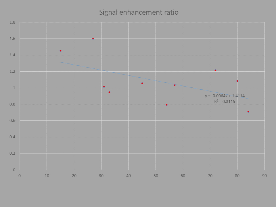
Signal enhancement ratio (value obtained by dividing difference by white matter signal value) showed the same degree as white matter signal in all cases (Average 1.09). With age, the value of this signal enhancement ratio was low. Pearson's correlation coefficient was -0.56. However, there was no sharp decline in the vicinity considered to be around menopause (Figure 3).DISCUSSION
After GBCA administration, a strong contrast effect on the skin was observed. In this study, the signal enhancement ratio of female head skin tended to decrease with age. There was no sharp decrease in contrast effect at the time of menopause.It has been reported that skin blood flow decreases with age. Distribution of GBCA to the skin tissue is thought to be due to nonspecific transfer from the vascular lumen. The result of this time is considered to be consistent with the conventional report.In menopause seen in women, sudden changes in female hormone secretion occur, it is known that various symptoms are caused in the whole body. Reduction of skin blood flow rate is one of them, but in this study, there was no sharp decrease. The fact that the number of cases examined is small may be the factor.In addition to the reduction in skin blood flow due to menopause, the action of androgen is also known for female pattern depilation. In this study, male and female hormone values are not measured. Also in men, it is known that hormone secretion changes similar to menopause, although not as much as women.CONCLUSION
In women, the head skin signal enhancing effect tends to decrease with age.Acknowledgements
No acknowledgement found.References
1. SKIN ENHANCEMENT WITH GADOLINIUM BASED CONTRAST AGENTS, Proceedings of annual meeting of ISMRM 2017, Honolulu, Hawai, E-Poster 5565
2. Camacho-Martinez FM. Hair loss in women. Seminars in cutaneous medicine and surgery. 2009;28(1):19-32.
3. Scheinfeld N. A review of hormonal therapy for female pattern (androgenic) alopecia. Dermatology online journal. 2008;14(3):1.
4. Raine-Fenning NJ, Brincat MP, Muscat-Baron Y. Skin aging and menopause : implications for treatment. American journal of clinical dermatology. 2003;4(6):371-8.
5. Phillips TJ, Demircay Z, Sahu M. Hormonal effects on skin aging. Clinics in geriatric medicine. 2001;17(4):661-72, vi.
6. Harvell J, Hussona-Saeed I, Maibach HI. Changes in transepidermal water loss and cutaneous blood flow during the menstrual cycle. Contact dermatitis. 1992;27(5):294-301.
Figures