4191
New occurrences of macroscopic myocardial fibrosis in thalassemia at long term by multiple follow-up1Fondazione G. Monasterio CNR-Regione Toscana, Pisa, Italy, 2"ARNAS" Civico, Di Cristina Benfratelli, Palermo, Italy, 3Presidio Ospedaliero “Giovanni Paolo II”, Lamezia Terme, Italy, 4Ospedale del Delta, Lagosanto (FE), Italy, 5Ospedale "Sandro Pertini", Roma, Italy, 6Ospedale “San Giovanni Di Dio”, Agrigento, Italy, 7Ospedale “S Maria alla Gruccia”, Montevarchi (AR), Italy, 8Ospedale “Giovanni Paolo II”, Olbia, Italy
Synopsis
We investigated the evolution of myocardial fibrosis in terms of new occurrences over a period of 6 years in thalassemia patients who underwent to multiple LGE CMR scans and we detected an high number of new occurrences of myocardial fibrosis. Our data suggest the importance of repeating the LGE CMR over time using 'low risk' macrocyclic agents.
Introduction
To date in thalassemia patients it is recommended to repeat cardiac magnetic resonance (CMR) scans for iron quantification every 1 or 2 years based on the myocardial iron overload (MIO)1. Also in these patients, late gadolinium enhancement (LGE) has been demonstrated to be a strong predictor for cardiac events2. However, many studies have shown an association between intravenous gadolinium based contrast agents (GBCA) exposure and neuronal tissue deposition3-7. So, it appears prudent at this time to revisit institutional protocols for GBCA administration, in particular in the follow up (FU) studies.
We investigated the evolution of myocardial fibrosis in terms of new occurrences over a period of 6 years in thalassemia patients who underwent to multiple FU.
Methods
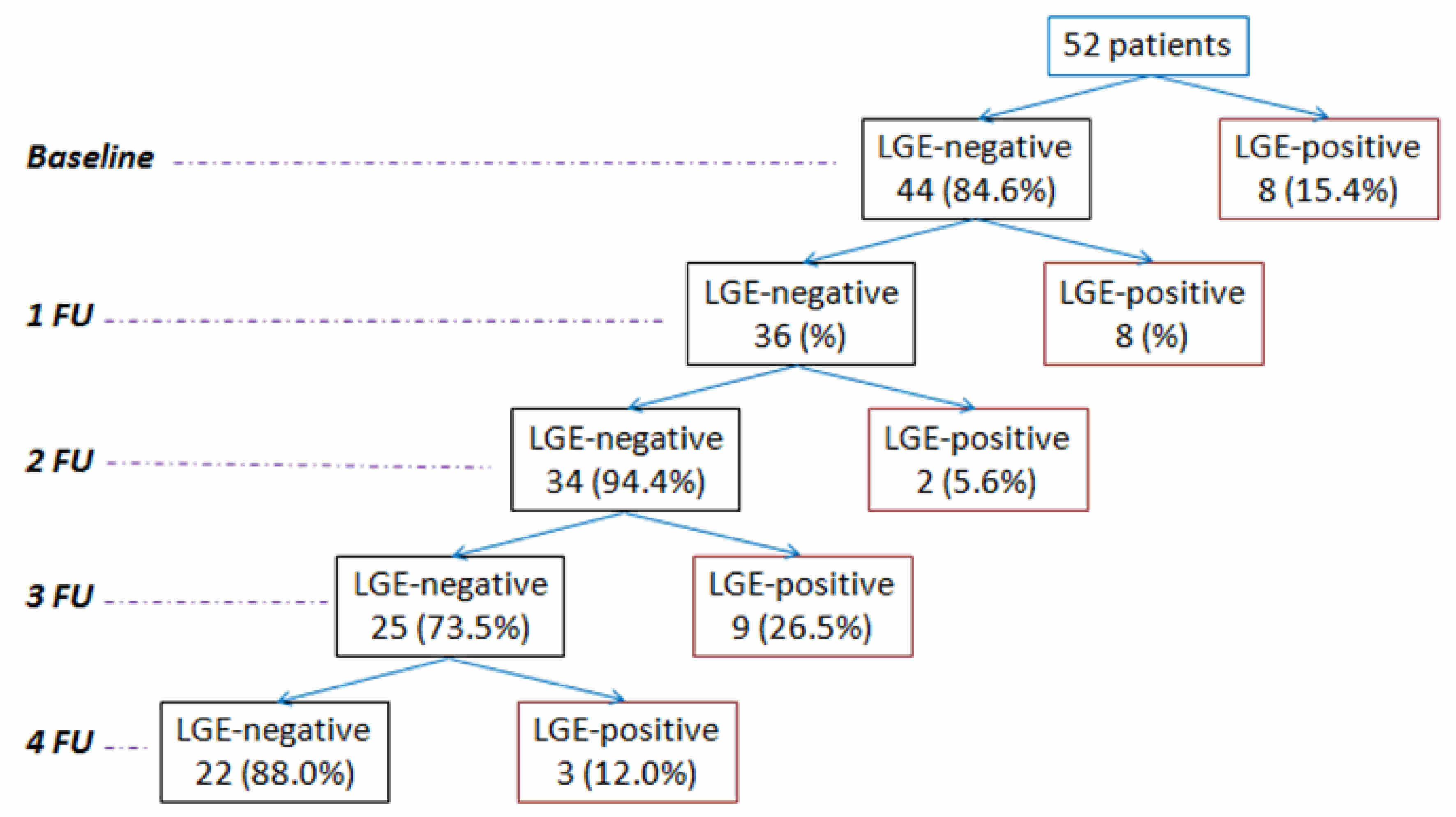
We considered 52 patients with thalassemia major (28.78±8.59 years; 28 females) consecutively enrolled in the Myocardial Iron Overload in Thalassemia (MIOT) Network8 who underwent 5 LGE CMRs (baseline + 4 follow-up) using Gadobutrol (0.2 mmoli/kg). The time interval between two subsequent scans was 18±3 months.Results
At the baseline CMR, 44 patients (84.6%) were LGE‐negative.
Eight new occurrences of myocardial fibrosis were detected at the first follow-up (FU). At the second FU, 2 out of the 36 previously LGE-negative patients had myocardial fibrosis. At the third FU, 9 new occurrences of myocardial fibrosis were detected. At the forth FU, 3 patients showed myocardial fibrosis for the first time. The figure shows a simplifying flow-chart.
The 22 patients who developed myocardial fibrosis during the follow-up showed comparable frequency of diabetes and HCV infection and comparable baseline cardiac iron than patients who remained always LGE-negative.
Conclusion
A serial monitoring of thalassemia patients revealed an high number of new occurrences of myocardial fibrosis, suggesting the importance of repeating the LGE CMR over time using 'low risk' macrocyclic agents.Acknowledgements
We would like to thank all the colleagues involved in the MIOT project (https://miot.ftgm.it/). We thank Claudia Santarlasci for her skillful secretarial work. We finally thank all patients for their cooperation.References
1. Pennell DJ, Udelson JE, Arai AE, et al. Cardiovascular function and treatment in beta-thalassemia major: a consensus statement from the American Heart Association. Circulation 2013;128(3):281-308.
2. Pepe A, Meloni A, Rossi G, et al. Prediction of cardiac complications for thalassemia major in the widespread cardiac magnetic resonance era: a prospective multicentre study by a multi-parametric approach. Eur Heart J Cardiovasc Imaging 2017.
3. Kanda T, Ishii K, Kawaguchi H, Kitajima K, Takenaka D. High signal intensity in the dentate nucleus and globus pallidus on unenhanced T1-weighted MR images: relationship with increasing cumulative dose of a gadolinium-based contrast material. Radiology 2014;270(3):834-841.
4. Errante Y, Cirimele V, Mallio CA, Di Lazzaro V, Zobel BB, Quattrocchi CC. Progressive increase of T1 signal intensity of the dentate nucleus on unenhanced magnetic resonance images is associated with cumulative doses of intravenously administered gadodiamide in patients with normal renal function, suggesting dechelation. Invest Radiol 2014;49(10):685-690.
5. Quattrocchi CC, Mallio CA, Errante Y, et al. Gadodiamide and Dentate Nucleus T1 Hyperintensity in Patients With Meningioma Evaluated by Multiple Follow-Up Contrast-Enhanced Magnetic Resonance Examinations With No Systemic Interval Therapy. Invest Radiol 2015;50(7):470-472.
6. Radbruch A, Weberling LD, Kieslich PJ, et al. Gadolinium retention in the dentate nucleus and globus pallidus is dependent on the class of contrast agent. Radiology 2015;275(3):783-791.
7. Zhang Y, Cao Y, Shih GL, Hecht EM, Prince MR. Extent of Signal Hyperintensity on Unenhanced T1-weighted Brain MR Images after More than 35 Administrations of Linear Gadolinium-based Contrast Agents. Radiology 2016;282(2):516-525.
8. Meloni A, Ramazzotti A, Positano V, et al. Evaluation of a web-based network for reproducible T2* MRI assessment of iron overload in thalassemia. Int J Med Inform 2009;78(8):503-512.
Figures