4188
Assessment of high dielectric material in different sizes for fetal MRI at 3T1Lauterbur Research Center for Biomedical Imaging, Shenzhen Institutes of Advanced Technology, Chinese Academy of Sciences, Shenzhen, China, 2Shenzhen Key Laboratory for MRI, Shenzhen, China, 3School of Basic Science, Guangzhou Medical University, Guangzhou, China, 4Department of Radiology and Biomedical Imaging, University of California San Francisco, San Francisco, CA, United States, 5UCSF/UC Berkeley Joint Graduate Group in Bioengineering, San Francisco, CA, United States
Synopsis
In this paper, we investigate the effect of the size and thickness of high permittivity dielectric pads to the imaging performance in fetal imaging at 3T through numerical modeling and simulations. The results of this study can be used as a guide in selection of appropriate size and thickness of dielectric pads in fetal magnetic resonance imaging for gaining the highest imaging performance.
Introduction
High permittivity dielectric materials have the ability to alter the electromagnetic field distribution1, and have the capability of recovering the signal losses caused by dielectric effect of pregnant women in the magnet. Because of the high density, heavy weight, the high dielectric material is usually placed on the patient table and under the patients. Based on this positioning, the choice of thickness and size of high dielectric pad is very important, which may directly influence the imaging performance. In order to maximize the benefits of using high dielectric materials in fetal imaging, we investigate the fetal imaging performance with the use of the different size and thickness of the high dielectric pad through the electromagnetic simulation, and how B1+ field and SAR changes with the size of the dielectric material.Methods and Materials
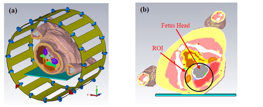
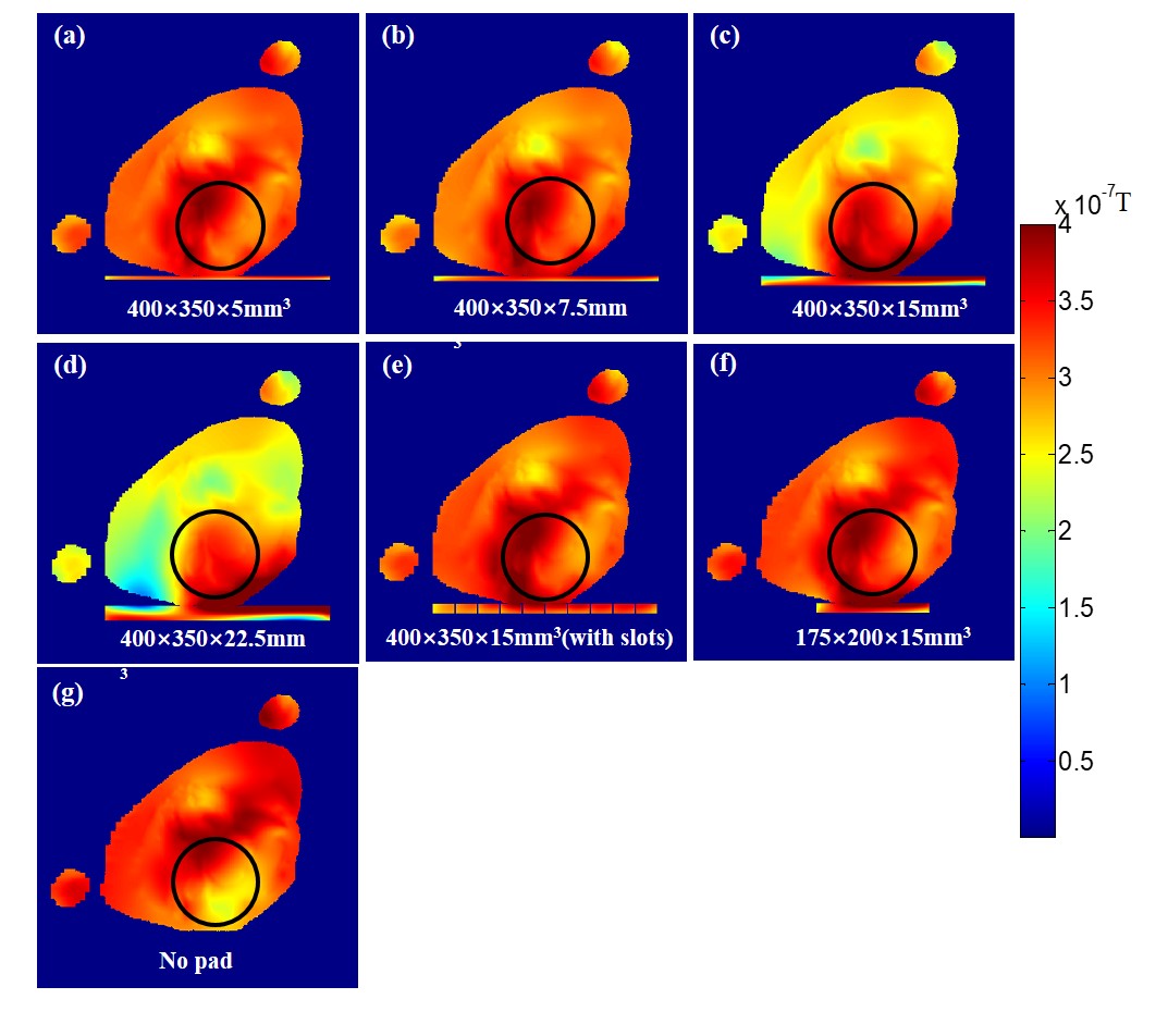
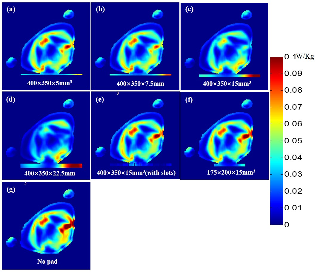
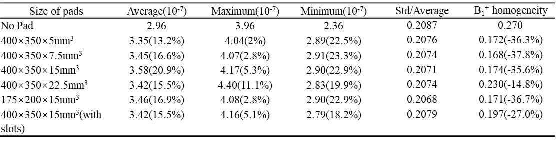
We investigated the effects of different sizes of the high dielectric pads by the numerical methods for calculating electromagnetic field2,3. The commercial CST software (Darmstadt, Germany) is used to simulate the model. Different sizes of high dielectric pads were modeled to represent a dense mixture with 4:1 barium titanate-to-water (mass/ mass ratio)4. From previous research results, we believe that the thickness and size of the high dielectric pad have the different influences on human tissue. Specifically, we have designed a pad with 3mm width slots to make the electromagnetic field permeate the pad easily. Therefore, the high dielectric pads with some different sizes as shown in Table I was placed on the sickbed in our designing. The relative permittivity and the electric conductivity of the high dielectric pad were respectively set to 220 and 0.473 S/m 4. A quadrature 16-element high-pass birdcage coil, end ring radius 312.5mm, length 470mm, tuned to 123.2MHz, simulated as a volume coil. In order to acquire the best results, a human tissue model imported from a 387mm length pregnant woman in CST was placed in the center of the birdcage in the left lateral decubitus and rotated 140 degrees from the supine position, as shown in Figure 1 (b). The two orthogonal discrete excitation ports were set on the side of end ring. The capacitors used to tune the frequency of 123.2MHz were 66pF in the birdcage coil. All the models were split to 7.5million mesh cells, and the boundary condition were open in all direction. The models were simulate using two GPU accelerator card with/without the high dielectric pad load. Time-domain solver were employed, and the steady-state accuracy limit was set to -30 dB. All the post-processing results were normalized to 1W input power and calculated by MATLAB (Mathworks, Natik, MA, USA) code. To evaluate the B1+ field in the fetus head region, select the transverse plane centered on the volume coil and the radius of the ROI black circle is 60mm as shown in Fig. 1(b). The maps of SAR were calculated using 10g average mass and IEEE/IEC62704-1 as average method. The maximum value of SAR corresponds to the whole simulated human tissue. The homogeneity of B1+ field in the ROI was calculated as:
B1+ homogeneity = ( B1+Max - B1+Min )/( 2×B1+Average )
The transmit efficiency in the ROI was calculated as:
Transmit Efficiency = B1+Average/Total SAR
Results
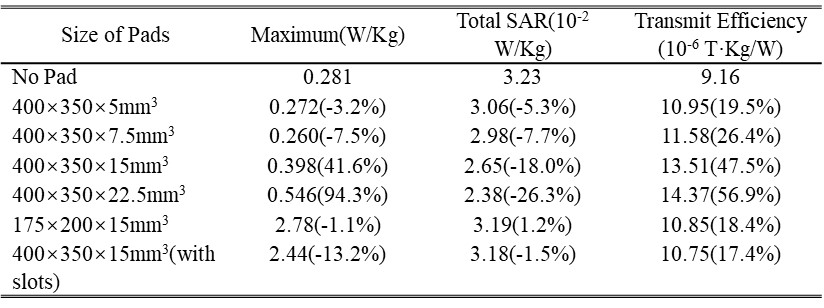
As shown in Table II, the larger and thicker the size of pad, the lower total SAR. The pad can increase the transmit efficiency nearby and uniform the B1+ field in ROI. Although thicker pads reduce the total SAR and improve the transmit efficiency, but the maximum SAR in the human body increased and the distant B1+ field were shielded at certain level. If the pads are too thin, they have limited effect on changing the distribution of the magnetic field. In this case, the pad with thickness of 400×350×7.5mm3 has the best effect that improves the transmit efficiency of 26.4% in ROI, reduces A by 37.8% which means the B1+ field is more homogeneous. It also reduces the maximum SAR and total SAR by 7.5%, 7.7% respectively. The pads with size of 175×200×15mm3 or 400×350×15mm3 with gaps have similar effect of pad with size of 400×350×7.5mm3.Discussion/Conclusion
Our results obtained from numerical simulation demonstrate the relationship between the size of high dielectric pad and its impact to imaging performance. The results could be used to guide the selection of appropriate size and thickness of dielectric pads in fetal magnetic resonance imaging in order to gain the highest performance. To avoid any possible shielding of the magnetic fields by the dielectric material, the dielectric pad should large enough to cover the region of interest and should use slots in the dielectric pad.Acknowledgements
This work is supported in part by National Natural Science Foundation of China No. 81527901, 81327801, national key R&D program no. 2016YFC0100100, national grants no. 51307171, 61571433, 61401450, 81470077, 81571669 and 2013CB733800/2013CB733803, provincial grants no. 2014B030301013, 2015B020214006 and 2014A030310200, city grant no.KQJSCX20160301143250, CYJ20140417113430589, JSGG20141020103440414,KQCX2015033117354154, internal grant no. 201314, and a Pengcheng Scholar Award.References
[1] Van Gemert, Jeroen, et al. An Efficient Methodology for the Analysis of Dielectric Shimming Materials in Magnetic Resonance Imaging. IEEE transactions on medical imaging 36.2 (2017): 666-673. [2] Luo Minmin, et al. Numerical assessment of the reduction of specific absorption rate by adding high dielectric materials for fetus MRI at 3T. Biomedical Engineering/Biomedizinische Technik 61.4 (2016): 455-461. [3] Brink, Wyger M.,et al., The effect of high-permittivity pads on specific absorption rate in radiofrequency-shimmed dual-transmit cardiovascular magnetic resonance at 3T. Journal of Cardiovascular Magnetic Resonance 17.1 (2015): 82. [4] Wyger M. et al. High permittivity pads reduce specific absorption rate, improve B1 homogeneity, and increase contrast‐to‐noise ratio for functional cardiac MRI at 3T. Magnetic resonance in medicine 71.4 (2014): 1632-1640.Figures