4180
Investigating the Relationship between CEM43 and Brain Tissue Damage with MRI1CMRR, University of Minnesota, Minneapolis, MN, United States, 2Mortimer B. Zuckerman Mind Brain Behavior Institute,Columbia University, New York, NY, United States
Synopsis
An experimental paradigm that can be used to investigate the relationship between CEM43 metric and tissue damage is presented.
Introduction
Radio-frequency (RF) heating of tissue during MRI scans is an important patient safety issue in MRI1,2,3. Current safety guidelines impose limits on specific absorption rate (SAR) and temperature4. On the other hand, tissue damage depends not only on temperature but also on exposure time. CEM43 is a metric that takes both factors into account therefore it is potentially relevant for patient safety. CEM43 and its implications for thermal tissue damage has recently been studied5 for different whole body SAR and scan duration at 1.5 T.
MRI is capable of detecting thermal damage and blood brain barrier disruption as shown in previous imaging studies that also involves High Intensity Focused Ultrasound (HIFU) . T2 weighted images reveal hyper-intense regions around the lesions, even after short periods of time after the damage is made(i.e t<30 min). The outcome of the previous imaging studies were validated with histology work in swine6.
In this work, we present an RF heating and imaging study designed to correlate CEM43 to the tissue damage (blood brain barrier disruption) in the swine brain.
Methods
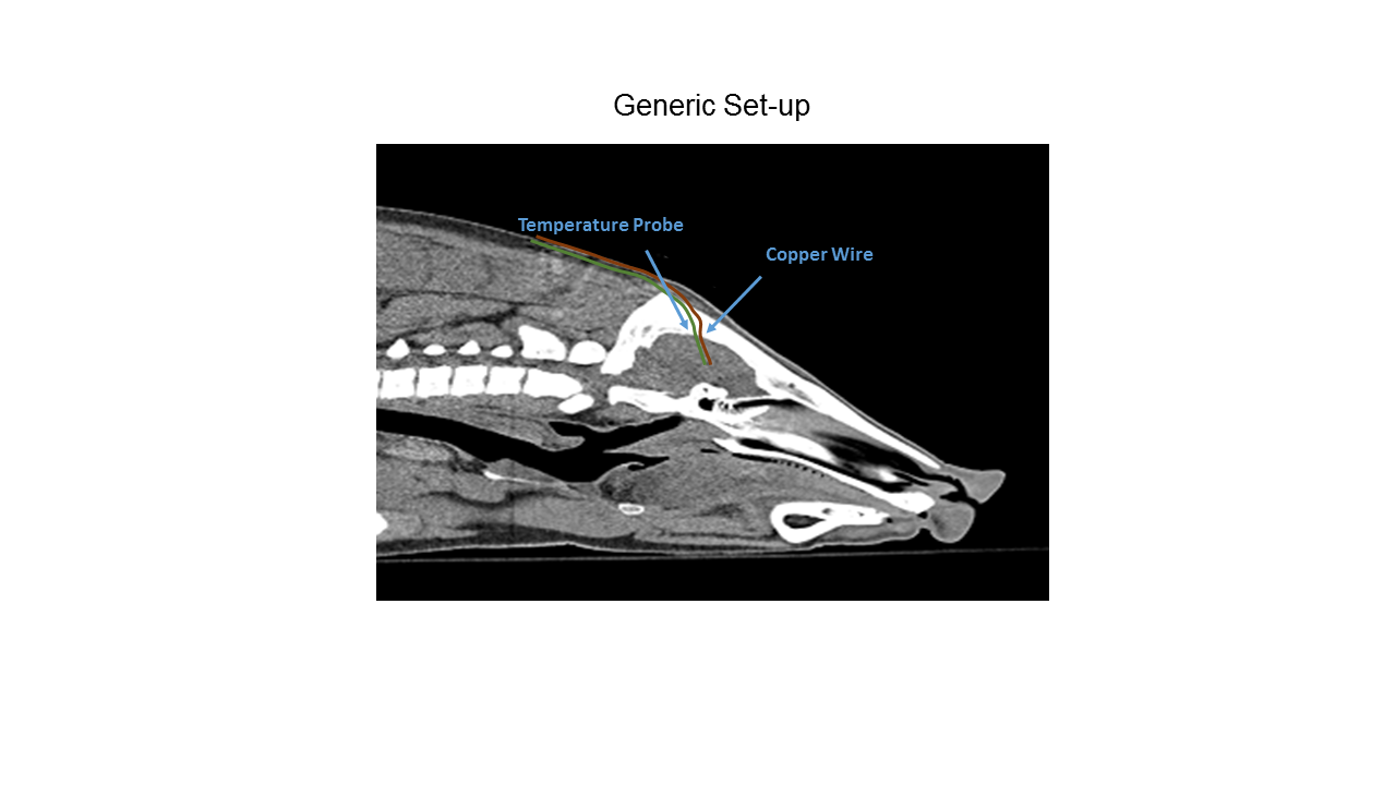
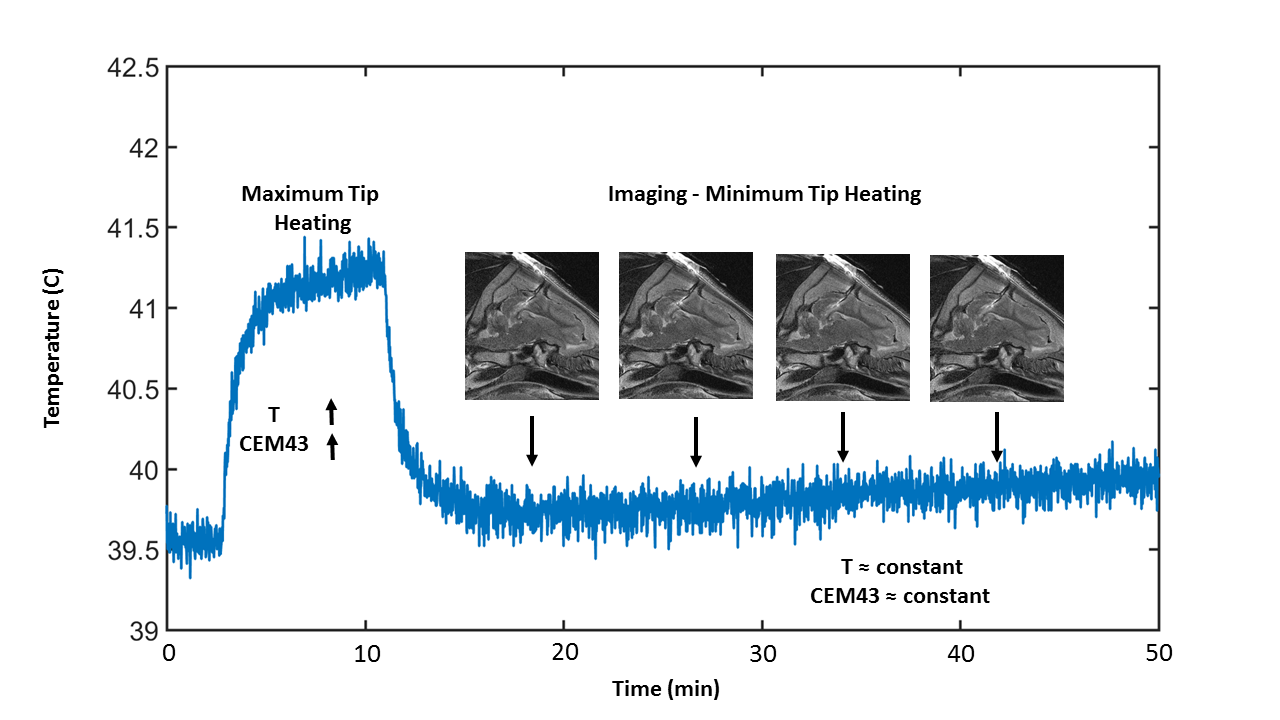
First, we prepared the animal and made sure that it is in sufficiently deep anesthesia (UMN, IACUC protocol #1706-34923A). We drilled the skull of the animal and placed a metallic wire (with a fiber-optic temperature probe attached at the tip) in the brain. We placed the animal in a 3 T system and optimized the body coil excitation to calculate minimal and maximal heating at the wire tip7. Then we performed a set of consecutive RF heating and imaging studies on the anesthetized swine. First, we used the minimal heating pattern to acquire an RF artifact free, control image around the lead using a T2-Weighted TSE sequence. Then we applied the maximum heating pattern with the same sequence with extended duration (up to 45 minutes). Imaging with maximum heating pattern increased the tip temperature and the CEM43 level (monitored real time) as expected. After each heating sequence, we applied the minimal heating pattern to acquire images of the tissue. The tip temperature remained approximately constant during the minimal heating pattern. This enabled us to asses any possible tissue damage caused by the CEM43 level reached at the end of the previous heating sequence. Once we acquired enough number of images with the minimal heating pattern, we switched back to the maximum heating pattern and started increasing the CEM43 at the tip again. This alternating paradigm continued until the end of the study.
Results
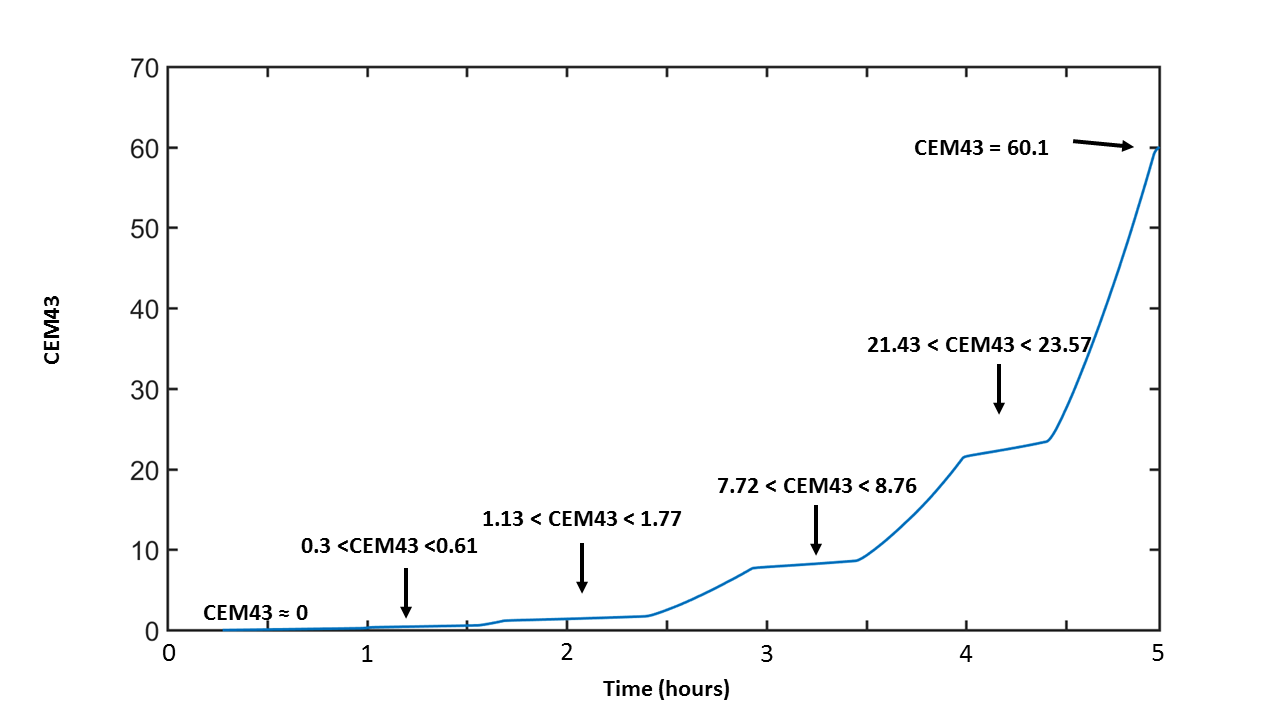
We performed five consecutive heating scenario which allowed us to reach a maximum CEM43 of 60.1 at the tip of the wire. In between the heating scenarios, we acquired RF artifact free images (every 8-10 minutes) using the minimal heating pattern. This allowed us to compare the tissue contrast around the wire to our control image (CEM43=0) during the course of the experiment. Figure 2 shows the comparison of the images acquired by the maximum and minimum heating pattern. Note that first one generated RF artifact free images while the latter generated regions with signal loss around the wire. Figure 3 shows the single step of the experimental paradigm that consists of tip heating followed by multiple image acquisition. Figure 4 and 5 shows the time variation of the temperature and CEM43 throughout the whole experiment.
Based on the heating and imaging experiments performed with a single animal, we did not encounter any significant contrast change that might indicate a tissue damage around the wire.
Discussion and Conclusion
We observed a baseline temperature increase in the probe location (39.2 oC to 41.2 oC in 4.5 hours). This was an expected consequence of aggressive RF power delivery during the course of the whole experiment.
In this work we used a copper wire (1mm diameter,18 cm length) insulated with teflon to generate heat at the tip. In order to generate more rapid heating and reach higher CEM43 levels, the dimensions/insulation of the copper wire may be optimized.
We proposed and tested an experimental paradigm that can be used to investigate CEM43 and its relation to tissue damage. We did not encounter any tissue damage in our limited number of heating experiments. However we believe that further animal studies may be useful to experimentally establish the relationship between tissue damage and CEM43 under MRI-related RF heating conditions.
Acknowledgements
This work is supported by the NIH grants P41 EB015894, K99EB021173.References
1)Van Rhoon, G.C., Samaras, T., Yarmolenko, P.S., Dewhirst, M.W., Neufeld, E. and Kuster, N., 2013. CEM43° C thermal dose thresholds: a potential guide for magnetic resonance radiofrequency exposure levels?. European radiology, 23(8), pp.2215-2227.
2)Henderson JM, Tkach J, Phillips M, Baker K, Shellock FG, Rezai AR.Permanent neurological deficit related to magnetic resonance imagingin a patient with implanted deep brain stimulation electrodes for Par-kinson’s disease: case report. Neurosurgery 2005;57:E1063.
3)Vaughan, T., DelaBarre, L., Snyder, C., Tian, J., Akgun, C., Shrivastava, D., Liu, W., Olson, C., Adriany, G., Strupp, J., Andersen, P., Gopinath, A., van de Moortele, P.-F., Garwood, M. and Ugurbil, K. (2006), 9.4T human MRI: Preliminary results. Magn. Reson. Med., 56: 1274–1282.
4)IEC 60601-2-33 International Electrotechnical Commission. International standard,. Medical equipment: particular requirements forthe safety of Magnetic resonance equipment, 3rd edition.Geneva 2010
5)Murbach, M., Neufeld, E., Capstick, M., Kainz, W., Brunner, D.O., Samaras, T., Pruessmann, K.P. and Kuster, N., 2014. Thermal Tissue Damage Model Analyzed for Different Whole‐Body SAR and Scan Durations for Standard MR Body Coils. Magnetic resonance in medicine, 71(1), pp.421-431.
6) Zvi R. Cohen, M.D., Jacob Zaubermann, M.D., Sagi Harnof, M.D., Yael Mardor, Ph.D., Dvora Nass, M.D., Eyal Zadicario, Ph.D., Arik Hananel, M.D., David Castel, D.V.M., Meir Faibel, M.D., Zvi Ram, M.D.; MAGNETIC RESONANCE IMAGING-GUIDED FOCUSED ULTRASOUND FOR THERMAL ABLATION IN THE BRAIN: A FEASIBILITY STUDY IN A SWINE MODEL, Neurosurgery, Volume 60, Issue 4, 1 April 2007, Pages 593–600
7)Eryaman, Y., Turk, E. A., Oto, C., Algin, O. and Atalar, E. (2013), Reduction of the radiofrequency heating of metallic devices using a dual-drive birdcage coil. Magn Reson Med, 69: 845–852. doi:10.1002/mrm.24316
Figures