4169
Real-time SNR enhancement in surgical region for intraoperative MRI combining a stationary and a small freely-moving RF coils1Magnetic Resonance Imaging Laboratory, Department of Bio and Brain Engineering, Korea Advanced Institute of Science and Technology, Daejeon, Republic of Korea, 2Division of MRI Research, StemLab Inc, Sungnam-si,Gyeonggi-Do, Republic of Korea
Synopsis
This study introduces a new technique to enhance SNR in a surgical region for real-time intraoperative MRI that combines a static big and an endoscopic small RF coils. Segmented imaging using the static RF coil and fast imaging using the small RF coil were performed simultaneously with a reduced FOV. The images from the static and freely-moving RF coils were updated in multiple shots and in every shot, respectively. This technique not only reduces computing time, but also improves SNR in surgical region without intensity variation, which would be beneficial in intraoperative MRI.
Introduction
Nowadays, CT-guided surgery for pituitary tumor is widely performed. However, since MRI has less harmful effects on the patient than CT, MRI is more desirable for intraoperative imaging. In order to increase accuracy of operation, high SNR or high temporal resolution imaging is more important around the surgical region rather than in the whole brain. Whole brain imaging is still needed to check brain dislocation caused by CSF leakage, but can be done with relatively low temporal resolution. This study suggests a new method for real-time intraoperative MRI (iMRI) using segmented imaging strategy and small RF coil attachable to an endoscope. Although small FOV imaging causes an aliasing artifact in images from a big RF coil, it would not be an issue in images acquired from a small RF coil due to the smaller region of high signal intensities. To improve temporal resolution, images are acquired from small coil with small FOV. At the same time, 2- or 4-shot segmented imaging is performed using head coils to resolve the aliasing artifact and reconstruct images with larger FOV. This technique enhances SNR around surgical region with high temporal resolution.Methods
Aqueous gel type phantom (PVA polymer) was scanned using 1.5T STEMLAB MRI scanner with a 7-channel head coil and a one-channel small RF coil (Fig. 3). GRE sequence was used for the dynamic segmented scan. Imaging parameter were: TR=135ms, TE=8ms, In case of 2-shot segmented imaging, matrix size=128X64 and FOV=256X128mm2, and temporal resolution of small RF coil imaging and head coil imaging were 8.8 seconds and 17.6 seconds, respectively. In case of 4-shot segmented imaging, matrix size=128X32 and FOV =256x64mm2, and temporal resolution of small RF coil imaging and head coil imaging were 4.4 seconds and 17.6 seconds, respectively (Table.1).
Each RF coil obtained small FOV k-space data for each shot because of the segmented acquisition. K-space data acquired from the small RF coil were reconstructed to images for every shot immediately, and k-space data acquired from the head coil were combined after two or four shots to reconstruct full FOV images. For example in 2-shot segmented imaging, FOV became 50% smaller in the phase‑encoding direction. Odd‑numbered phase‑encoding lines were acquired in the first shot, and even‑numbered phase encoding lines were acquired in the second shot. For small RF coil imaging, images with 50% FOV was reconstructed in every shot. For head coil imaging, images were reconstructed after merging k-spaces from the two segmented shots. After the first shot, small RF coil images were merged with previously acquired full‑FOV head coil images. After the second shot, small RF coil images were merged with updated full‑FOV head coil images. In case of 4-shot segmented imaging, FOV became 25% and phase‑encoding step became bigger so that four k-space data were combined to compose one head coil image (Fig.1)
Result and Discussion
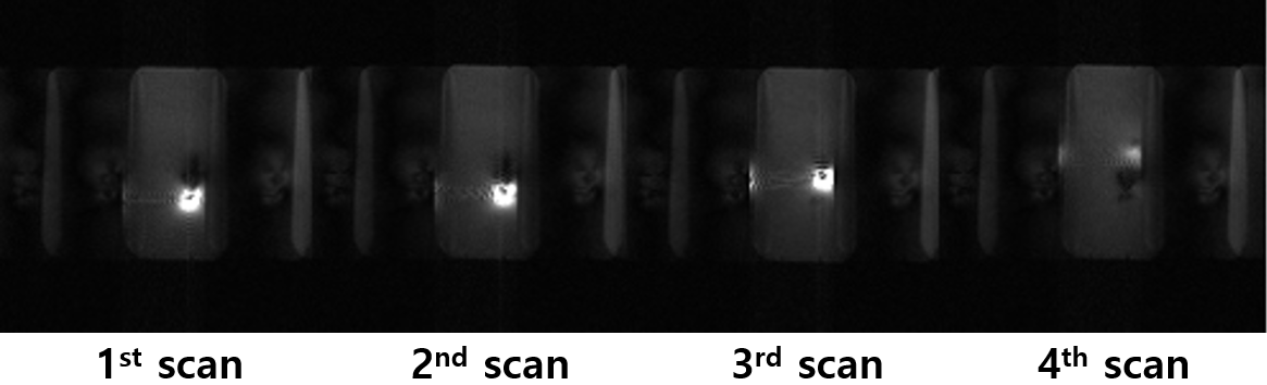
Image reconstruction was successful in both 2-shot and 4-shot cases. Figure 2 shows SNR of 2 segmented image and 4 segmented image visually increased after combining images from the two coils. Compared to the case with no small RF coil, SNR increased by 363.42% and 455.39% for 2‑segmented and 4‑segmented imaging respectively, consistent with the visual observation. Using segmented imaging sequence and small RF coil, we could update images in surgical region in real time and whole image in semi-real time. Despite the reduced FOV down to 50% or 25%, we could reconstruct images without aliasing from both the head coil (due to segmented acquisition) and the small coil (due to the small sensitivity region).
Compared to the conventional scans, the proposed 2-segmented imaging and 4-segmented imaging have better temporal resolution in the surgical region thanks to high temporal‑resolution of the moving small RF coil. This approach is applicable to imaging with endoscopic small RF coil and intraoperative MRI. Additionally, the proposed method provides SNR enhancement in the surgical region.
Conclusion
This research suggests a real-time imaging method for intraoperative MRI. The proposed method not only enhances SNR in surgical region, but also improves temporal resolution using a small RF coil and spatial coverage through segmented imaging with a big RF coil.Acknowledgements
No acknowledgement found.References
1. Yuichiro et al. IEEE 2013.
2. Scott B et al. MRM 1999; 41:375-385.
Figures
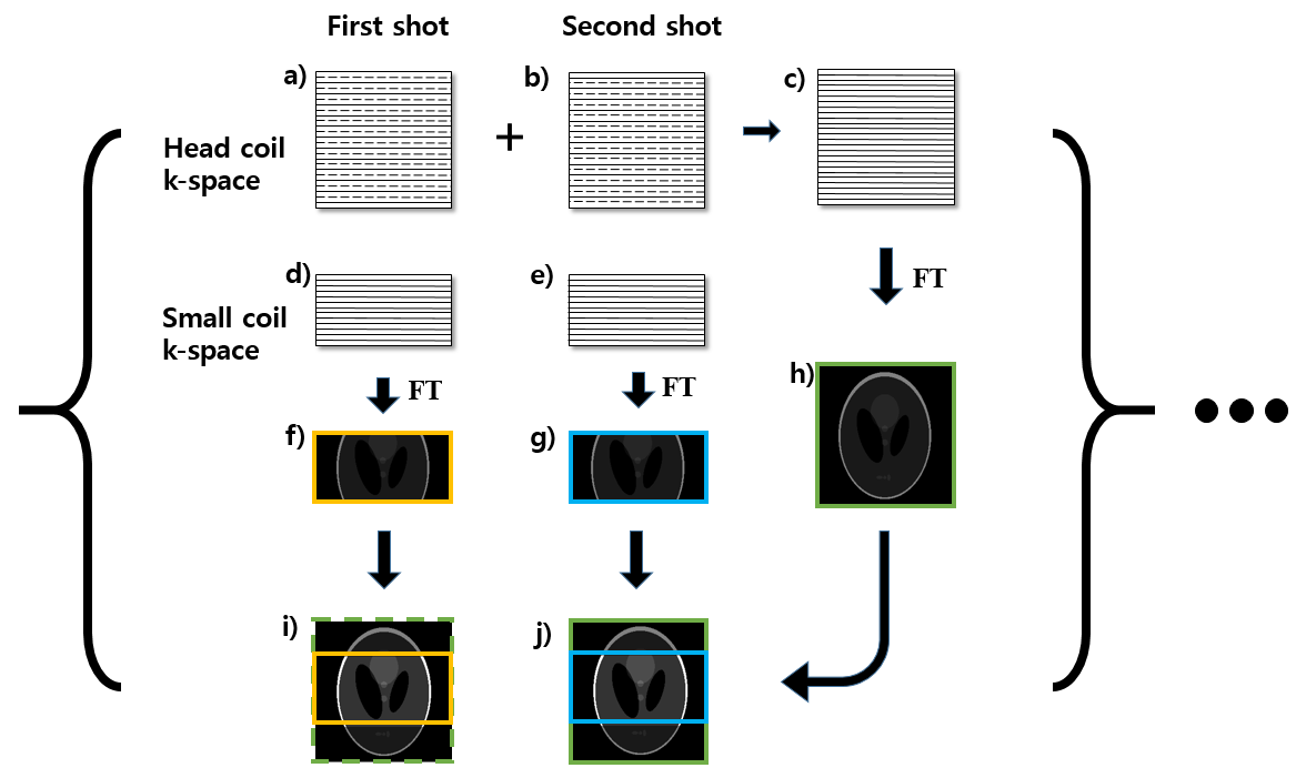
Fig 1. 2-segmented imaging reconstruction.
a) head coil k-space (even line) from first shot.
b) head coil k-space (odd line) from second shot.
c) merged head coil k-space from two shots.
d), e) small coil k-space from first and second shot.
f), g) reconstructed image from d), e).
h) reconstructed image from c).
i) merged image of f) and h) (dash line represents previously acquired image from head coil).
j) Merged image of g) and h).
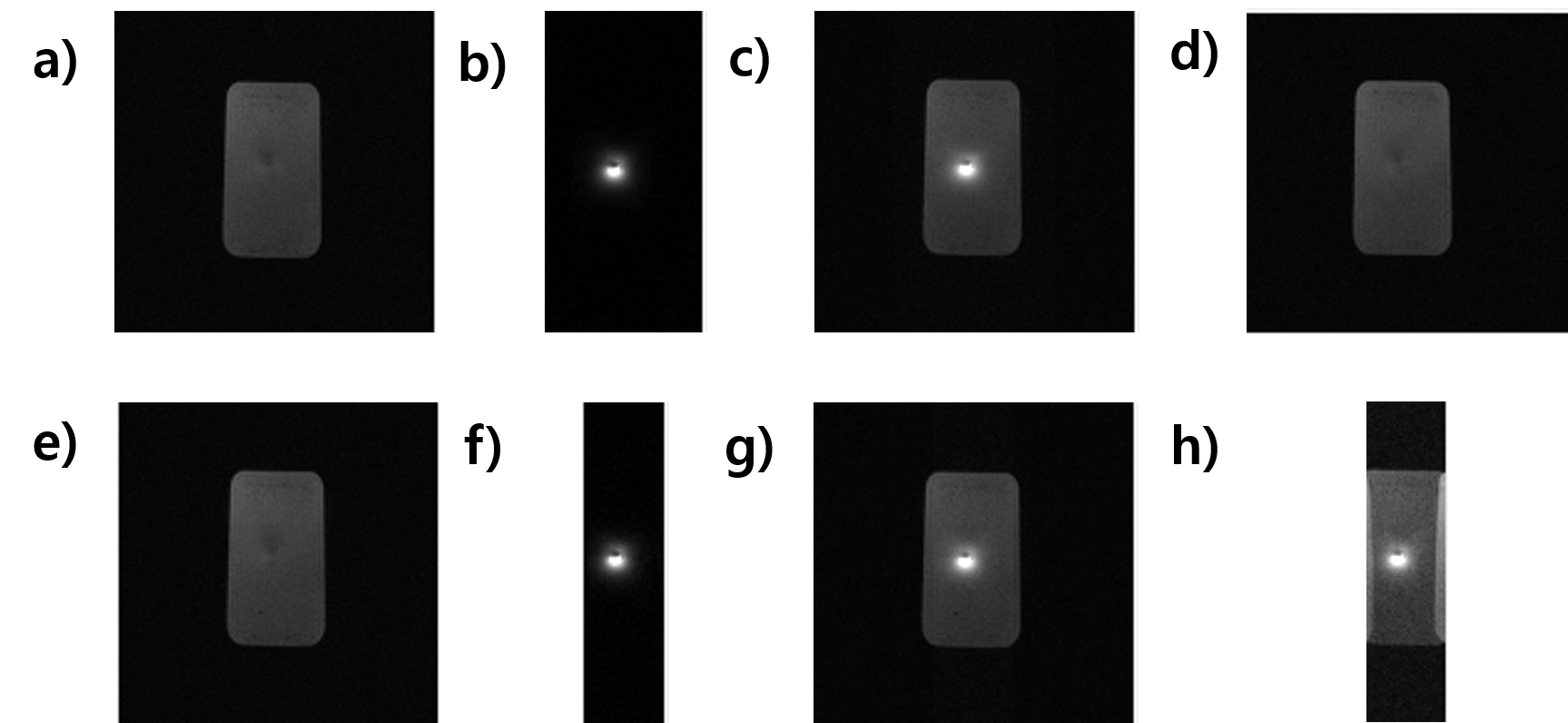
Fig 2. a),e) Head coil images from 2- and 4-shot segmented imaging.
b),f) small RF coil image from 2- and 4-shot segmented imaging.
c) merged image of a) and b).
d) image without using small RF coil and segmented imaging.
g) merged image of e) and f).
h) merged image without using segmented imaging (small FOV) – aliased.
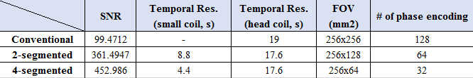
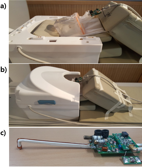
Fig 3. a), b) Aqueous gel type phantom with a small RF coil placed inside of head coil. c) Small RF coil attachable to endoscopy.