4168
Robust quantitative diffusion weighted MR on an MR-Linac system1Radiotherapy, UMC Utrecht, Utrecht, Netherlands, 2Radiology, UMC Utrecht, Utrecht, Netherlands
Synopsis
Quantitative diffusion weighted imaging is demonstrated on a clinical MR-Linac system. ADC values were measured using a standardized diffusion phantom using two different diffusion sequences, single shot EPI and single shot TSE: SPLICE. In general, accurate and repeatable ADC measurements could be acquired using both sequences. EPI showed more susceptibility and eddy current related image distortions, whereas SPLICE was geometrically more robust. Large deviations in ADC start to occur when moving further away from the isocenter, suggesting the need for gradient non-linearity correction of ADC values.
Introduction
MRI is increasingly used in radiotherapy. The integration of MRI in the radiotherapy workflow is underlined by the introduction of the MR-Linac [1], a hybrid 1.5T close-bore MR system with an integrated 7MV linac. The MR-Linac offers high quality MRI at the time of treatment. This allows for repeated quantitative MRI such as diffusion weighted imaging (DWI) to follow the response to radiotherapy over time. This requires the use of robust, reproducible DWI. Here, we assess the performance of a clinical MR-Linac system to measure ADC values using a standardized diffusion phantom and a specialized sequence robust for B0 inhomogeneities.
Methods
Imaging was performed on a 1.5T MR-Linac system (Elekta/Philips) using a specialized QIBA diffusion phantom (High Precision Devices, USA). The phantom was positioned in the isocenter using an external laser positioning system.
Two different diffusion sequences were used: single shot EPI, and single shot TSE, DW-SPLICE [2,3]. Common parameters: FOV: 220x220x125 mm3; acquired voxelsize: 1.72x1.72x4 mm3; SENSE2; TR 10000 ms; TE EPI/SPLICE: 130.98/128.546 ms; b-values EPI: 0/500/900/2000 s/mm2; SPLICE: 0/200/500/1000 s/mm2. Diffusion encoding gradients were applied along three principle axes (X,Y, Z) of the MR-Linac. To assess the influence of the modified gradient system, ADC maps were generated online for each direction separately using the vendors software. Additionally, isotropic ADC maps were generated.
Accuracy of diffusion weighted MR imaging was tested for EPI and the TSE based sequence, SPLICE, with emphasis on geometric fidelity and ADC quantification. First, the ADC values were compared with the reference values provided by the QIBA phantom. To test the precision of ADC quantification, both sequences were repeated three times to evaluate repeatability. Furthermore, the effect of gradient non-linearity on ADC was assessed by moving the phantom 120 mm along the Z-axis towards superior. ADC maps were processed offline for analysis and ROIs were drawn manually in the tubes, avoiding susceptibility artefacts. ROIs were reused for repetitions of the same sequence. Geometry was evaluated by comparison of the outer contour of the phantom, determined through thresholding.
Results
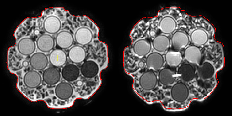
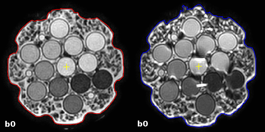
Figure 1 shows a transverse slice through the phantom and depicts b=0 s/mm2 for SPLICE and EPI. The outer contour (red) from SPLICE is propagated to EPI, which shows B0 related geometric distortions.
Further geometric evaluations show the effects of eddy currents by propagation of the outer contour of the b=0 s/mm2 for SPLICE and EPI to their respective diffusion weighted images (b=500 s/mm2 in X, Y and Z) (figure 2, animated gif). In EPI the outer contour of the phantom deforms depending on gradient orientation, whereas this effect not observed in SPLICE.
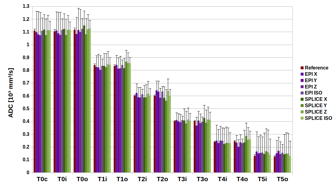
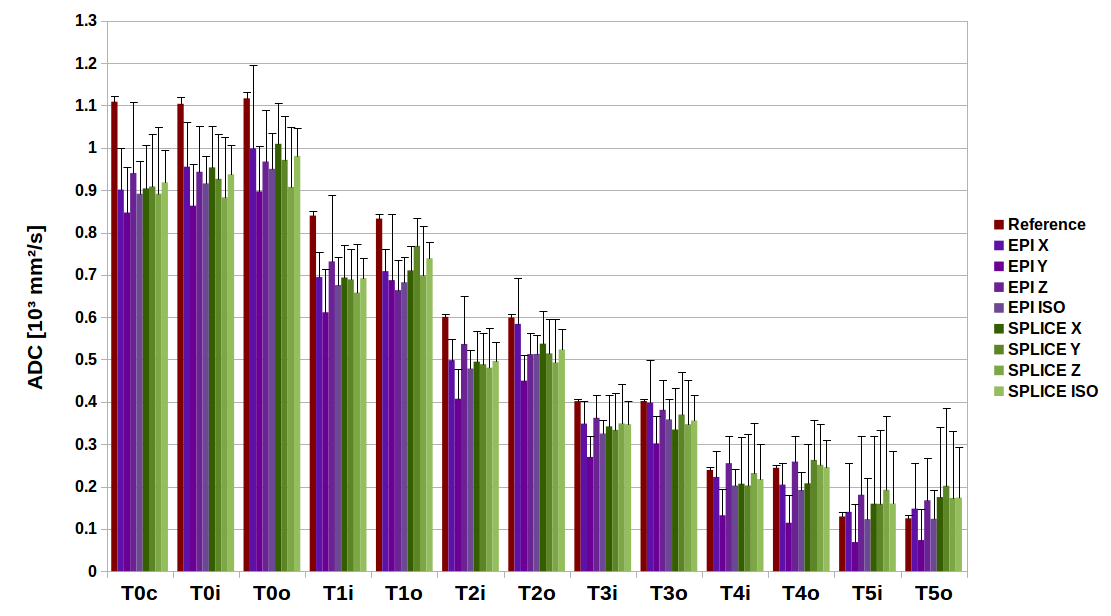
Mean ADC values for each tube of the phantom for both the EPI and SPLICE sequence and each diffusion direction separately (figure 3). No differences in the ADC values between the different directions were found (X vs Y vs Z vs ISO).
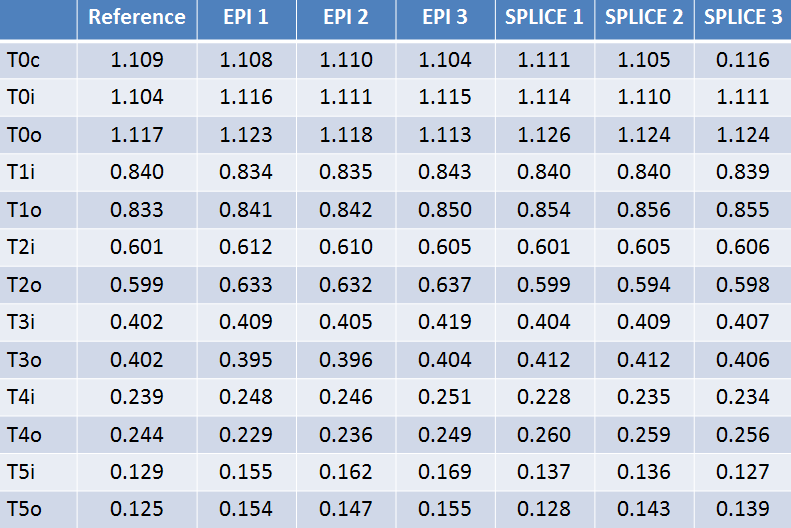
Figure 4 shows the repeated measurements of both sequences compared to their reference value. Good repeatability was achieved for both sequences over the whole range of ADC values.
No differences between the inner and outer tube of the same ADC value were observed (figure 3 and 4). Large deviations start to occur when moving further away from the isocenter as shown in figure 5.
Discussion
Diffusion weighted imaging on the MR-Linac was shown to be feasible for both EPI and TSE bases acquisition methods. The SPLICE sequence is more robust to susceptibility related image distortions as is illustrated by the accurate depiction of the phantom shape. In EPI, the phantom is deformed depending on gradient orientation. This is likely due to the varying effects of eddy currents on the spatial encoding. This effect is minimal in SPLICE. The disadvantage of SPLICE is the long acquisition time. For ADC quantifications, the eddy current variations between the different diffusion gradient directions hardly affect ADC values. This indicates good gradient performance for ADC calculation in both evaluated sequences, within the dimensions of the phantom. Furthermore, both SPLICE and EPI show excellent reproducibility over multiple acquisitions within the same scan session.
In the central region of the MR-Linac, no differences between the inner and outer tube were observed, indicating that the effects of gradient non-linearity on the measured ADC values are minimal. However, when moving further away from the isocenter, larger differences occur. This suggests the need for the implementation of correction methods [4,5] for larger FOVs.
Conclusion
Quantitative diffusion weighted imaging is demonstrated on a clinical MR-Linac system. The SPLICE sequence is more robust to susceptibility and eddy current related image distortions. Correction of ADC for gradient non-linearity is required for larger FOVs.
Acknowledgements
Financial support for this work was provided by the Dutch Cancer SocietyReferences
[1] Lagendijk JJW and Bakker CJG, MRI guided Radiotherapy, a MRI based linear accelerator. ESTRO Istanbul 2000
[2] Schick F. SPLICE: sub-second diffusion-sensitive MR imaging using a modified fast spin-echo acquisition mode. Magn Reson Med. 1997 Oct;38(4):638-44.
[3] Schakel T, Hoogduin JM, Terhaard CHJ, Philippens MEP. Technical Note: Diffusion-weighted MRI with minimal distortion in head-and-neck radiotherapy using a turbo spin echo acquisition method. Med Phys. 2017 Aug;44(8):4188-4193.
[4] Bammer R, Markl M, Barnett A, Acar B, Alley MT, Pelc NJ, Glover GH, Moseley ME. Analysis and generalized correction of the effect of spatial gradient field distortions in diffusion-weighted imaging. Magn Reson Med. 2003 Sep;50(3):560-9.
[5] Malyarenko DI, Ross BD, Chenevert TL. Analysis and correction of gradient nonlinearity bias in apparent diffusion coefficient measurements. Magn Reson Med.2014 Mar;71(3):1312-23.
Figures