4164
Non-contrast MRA Guidance in Pediatric Endovascular Interventions1Phoenix Children's Hospital, Phoenix, AZ, United States, 2Philips Healthcare, Bothell, WA, United States
Synopsis
The goal of this research project is to investigate the feasibility of using non-contrast 3D MRA imaging in the planning and guidance of catheter-based pediatric vascular disease treatment. It may result in a reduction in patient radiation exposure, contrast (Gadolinium and Iodine) usage and procedure time as compared to traditional 2D fluoroscopy guidance.
Purpose
The goal of this research project is to investigate the feasibility of using non-contrast 3D MRA imaging in the planning and guidance of catheter-based pediatric vascular disease treatment.Background and Introduction
Over the last decades, the complexity of procedures performed in Interventional Radiology (IR) labs has increased dramatically. 3D perspective of anatomy is desirable for catheter guidance and device placement because of its ability to avoid superposition over 2D fluoroscopy imaging. Currently, Interventionalists often use C-arm cone beam Computer Tomography (CT) based 3D volumetric image for catheter guidance through blood vessels during IR procedure. However, radiation exposure is always a concern in children because they are more sensitive to radiation. And the longer life expectancy may result in a larger window of opportunity for developing radiation-related cancer. Meanwhile, the contrast agent is administered to boost vessel imaging and the pediatric patient is under general anesthesia during IR and MRA procedure, there always is a risk of Iodine complications or unwanted residual intracranial Gadolinium deposition from contrast.
In the last few years, Philips IGT-Systems has developed and launched software tools such as roadmapping and VesselNavigator that allows for both the planning and the live incorporation of pre-existing 3D image data in the treatment of vascular diseases. Meanwhile, a recently developed novel MRA sequence, Relaxation-Enhanced Angiography without Contrast and Triggering (REACT), provides high quality vascularity in MRA imaging, compared with traditional Contrast-Enhanced MRA (CEMRA). It is possible to segment 3D vessel structure from REACT MRA imaging and fuse with 2D fluoroscopy imaging with VesselNavigator assistance.
Methods
Pre-selected patients are examined on a 3T Philips Ingenia scanner with REACT sequence. This technique is based on a two-echo Dixon Turbo Field Echo (TFE) sequence, with additional magnetization preparation by a non-volume-selective inversion pulse preceded by a T2-preparation module to suppress signals from static tissues, such as muscles and organs, and to enhance signal contrast between blood vessels and background. The 3D REACT images are imported to the VesselNavigator workstation. Then, the vasculature of interest pertinent to the procedure is segmented. Third, the planning of the procedure is carried out. Points of interest, such as thrombus location and branch vessel ostia, can be marked in the segmented vessels and are later displayed on the live 3D roadmap. Next, the patient on the operating table is registered to the segmented REACT dataset. Registration can be done in one of two ways: 1) 2D-3D registration, by means of projection of two images with an angulation of at least 30 degrees. 2) 3D-3D registration, by means of C-arm cone-beam CT with curvature of branch vessel as landmarks. Finally, VesselNavigator is displayed on a separate screen as a fusion of the segmented dataset and live fluoroscopy image and serve as a live 3D roadmap during the procedure. The 3D roadmap position and opacity can further be adjusted at the table at this point for increased precision. Reasons of potential fusion image inaccuracy are analyzed. The radiation exposure to patients and IR staff, procedure length and contrast usage are collected to evaluate and compare with 2D fluoroscopy guided intervention.Preliminary Results
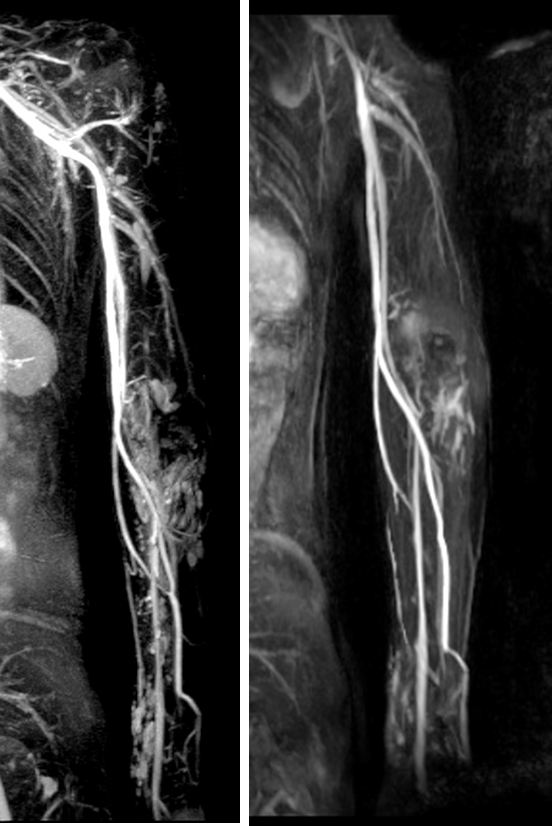
Figure 1. Long-FOV, multi-stack REACT without cardiac/respiratory triggering (left) and contrast-enhanced MRA(right) in a 12-years-old female with vascular malformation. REACT more clearly shows the detailed structure of venous malformation that extends from the level of the left shoulder down to the level of the left wrist compared with CEMRA .
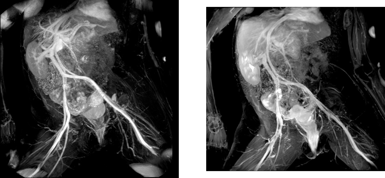
Figure 2. Respiratory-triggered REACT(left) vs. contrast-enhanced MRA(right) in a 12-years-old female with history of myelomeningocele and sever scoliosis. REACT image shows superior signal suppression from muscles and abdominal organs as well as slightly superior vessel conspicuity in the particularly in the liver and kidney.
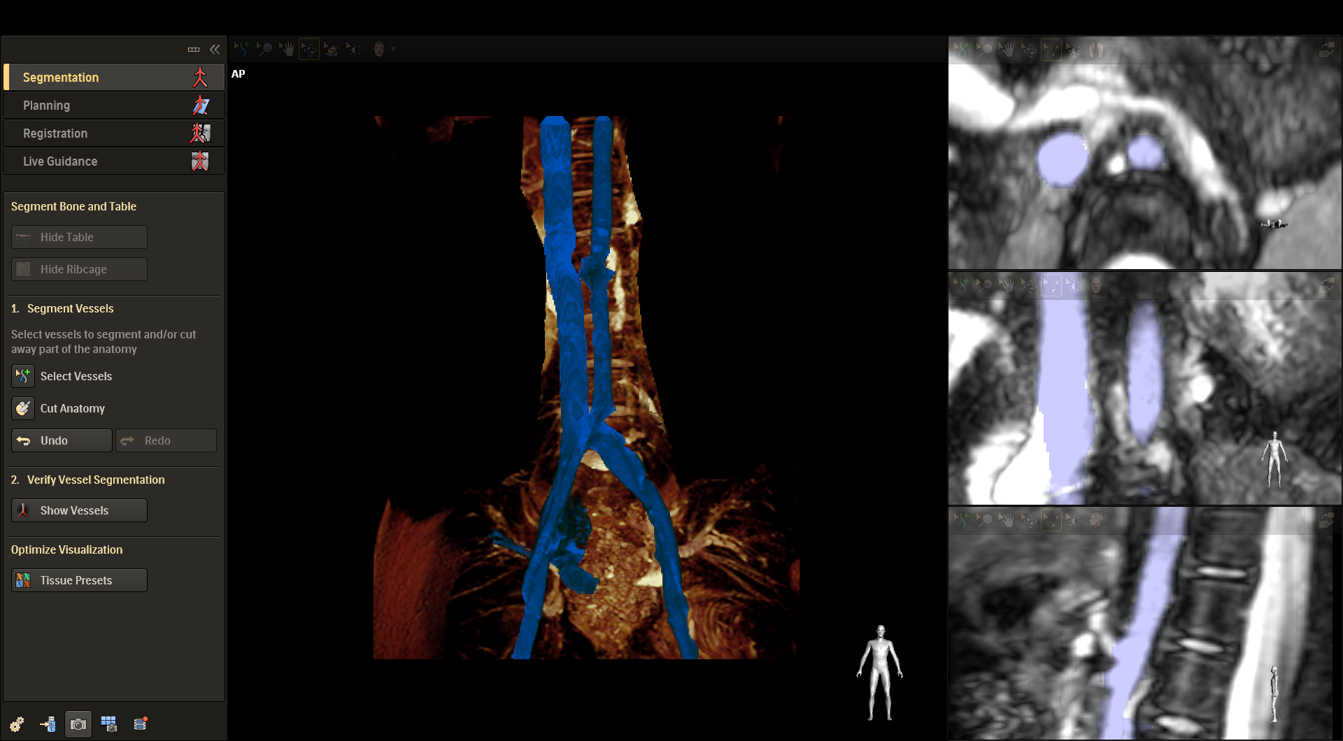
Figure 3. AP view(left) of VesselNavigator segmentation of REACT aorta and relevant side branches. Axial, Coronal and sagittal cross sections of aorta and selected side branches. .
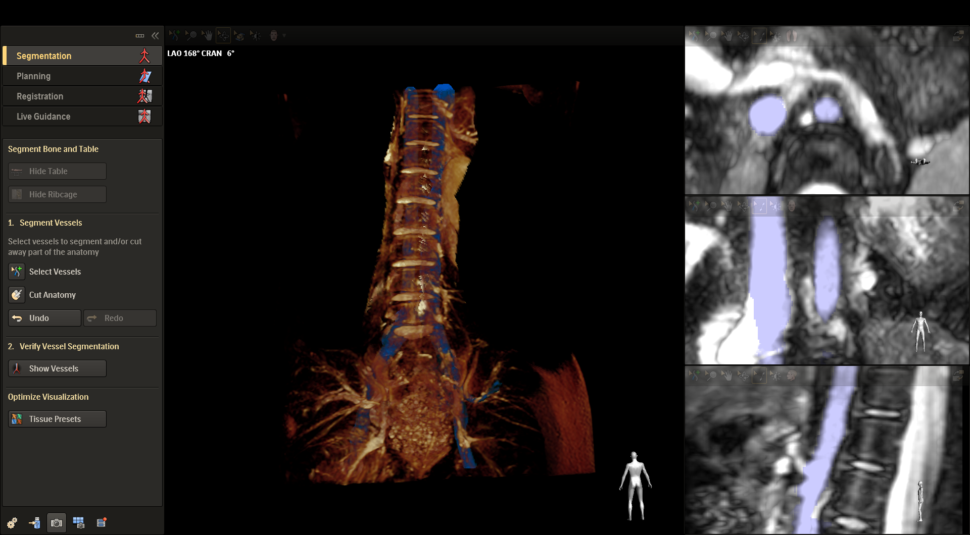
Figure 4. LAO view(left) of VesselNavigator segmentation of REACT aorta and relevant side branches. Axial, Coronal and sagittal cross sections of aorta and selected side branches.
Conclusion
Utilization of pre-intervention REACT MRA image manipulation with VesselNavigator may provide an intuitive and continuous 3D roadmap endovascular interventional procedure. This will result in a reduction in patient radiation exposure, contrast (Gadolinium and Iodine) usage and procedure time as compared to traditional 2D fluoroscopy guidance.Acknowledgements
This work is funded by Philips HealthcareReferences
Sailer A et al, “CTA with fluoroscopy image fusion guidance in endovascular complex aortic aneurysm repair”, European Society for Vascular Surgery 2014.
Stangenberg L et at, “A novel too for three-dimensional roadmapping reduce radiation exposure and contrast agent dose in complex endovascular interventions”. Society for Vascular Surgery 2015.
Figures