4149
A rapid 3D spiral readout with uniform sampling density and smooth T2* weighting1ETH Zürich, Zurich, Switzerland, 2Skope Magnetic Resonance Technologies, Zurich, Switzerland
Synopsis
A stack of disks trajectory for fast 3D acquisitions with long readouts is introduced and compared to interleaved stack of spiral and Yarn-ball trajectories. Isotropic 1.6 mm whole-brain coverage is achieved in less than a second.
Introduction
MR scanning of 3D volumes in under a second is aimed at in applications like fMRI with high temporal resolution1. 3D Fourier encoding lends itself to this task: it provides high SNR due to data acquistion from the entire volume, while being amenable to undersampling that makes optimal use of parallel imaging acceleration in all three dimensions. Unfortunately the available 3D encoding strategies require a trade-off between the following desirable characteristics, particularly at medium resolution:
1) average k-space speed close to the maximum k-space speed
2) regular undersampling in the entire 3D k-space
3) favorable PSF due to a benign k-space filter
Stacks of EPIs2 do not cover k-space as time efficiently as spirals. Cones and concentric shells3 are inefficient in that they oversample the k-space center and/or waste time by using very different readout lengths albeit TR being determined by the longest interleaf.
Recently the stack of blipped spirals was introduced, acquiring two k planes per spiral shot4–6. However, it does not allow for more k-space volume within one shot with a benign T2* weighting over the whole 3D k-space. In this work, a method to acquire an arbitrary number of adjacent planes within one shot is introduced (‘stack of disks’). It covers 3D k-space with high efficiency, uniform sampling density, and smooth T2* weighting. This trajectory is compared with an interleaved stack of spirals and the Yarn-ball trajectory7.
Methods
Hardware:
- Philips 7T Achieva system
- 32 channel head array (Nova Medical)
- 16 19F NMR field probes mounted on the head array and a dedicated MR acquisition system8
From the probe data, the actual encoding fields up to 2nd/3rd order in space were calculated.
Sequence: A GRE sequence with T2*-contrast was implemented (TE=20ms/28ms). K-space was covered by three different 3D trajectories, designed for an isotropic resolution and whole brain coverage (FOV=24x24x12cm3) applying a total undersampling factor of R=8, cf. Table 1 for acquisition parameters. Resolution is defined by the extent of a sphere in k-space; for the non-spherical trajectories, kmax was adjusted to sample the identical k-space volume.
- Stack of disks: 1.6mm resolution, 11 shots, TAQ=42ms, TR=76 ms
- Stack of disks: 3mm resolution, 6 shots, TAQ=19ms, TR=55 ms
- Interleaved stack: 3mm resolution, 6 shots, TAQ=20ms, TR=55 ms
- Yarn-ball: 3mm resolution, 6 shots, TAQ=21ms, TR = 55ms
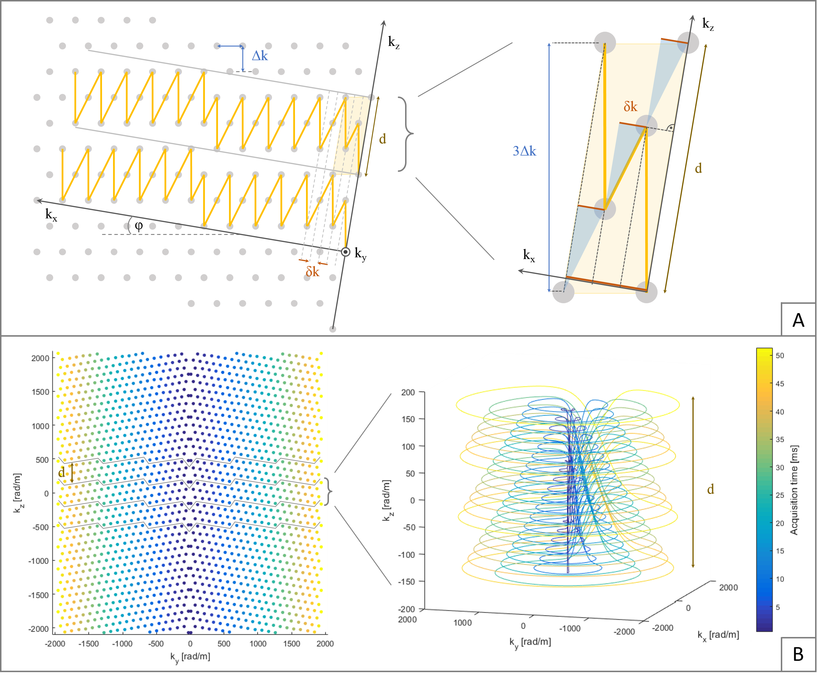
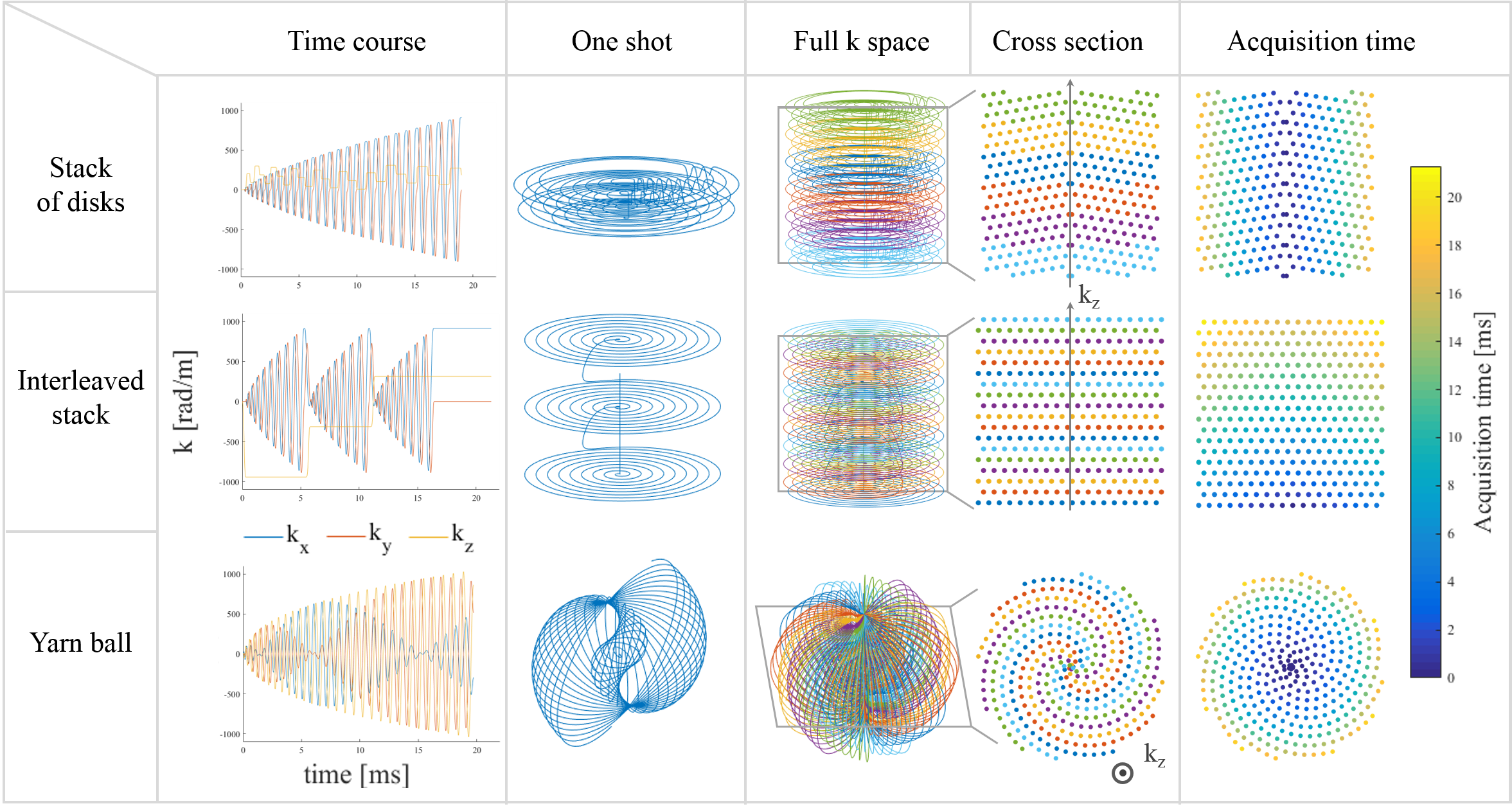
Stack of disks: The hexagonal grid6,9 formed by a conventional stack of spirals in every plane, which contains the kz axis, is tilted (Fig.1).
Interleaved stack: Each shot acquires several planes of a spiral stack, one after the other (Fig. 2). In order to obtain a smooth T2* weighting, an echo time shift has to be introduced.
Yarn-ball (7): Sampling of spherical k-space volume (Fig.2).
Image reconstruction was performed by an iterative conjugate gradient SENSE reconstruction algorithm10 extended to 3D, including multi-frequency interpolation11,12 for static off-resonance correction and based on the concurrently monitored trajectories13 of 0th and 1st spatial order. Off-resonance and coil sensitivity maps were computed from multiecho, multisclice spin-warp pre-scans (6 echoes, TE 4-9 ms, 1x1x1mm3 resolution, R=3 and 1 echo, TE=4ms, 5x5x1mm3 resolution).
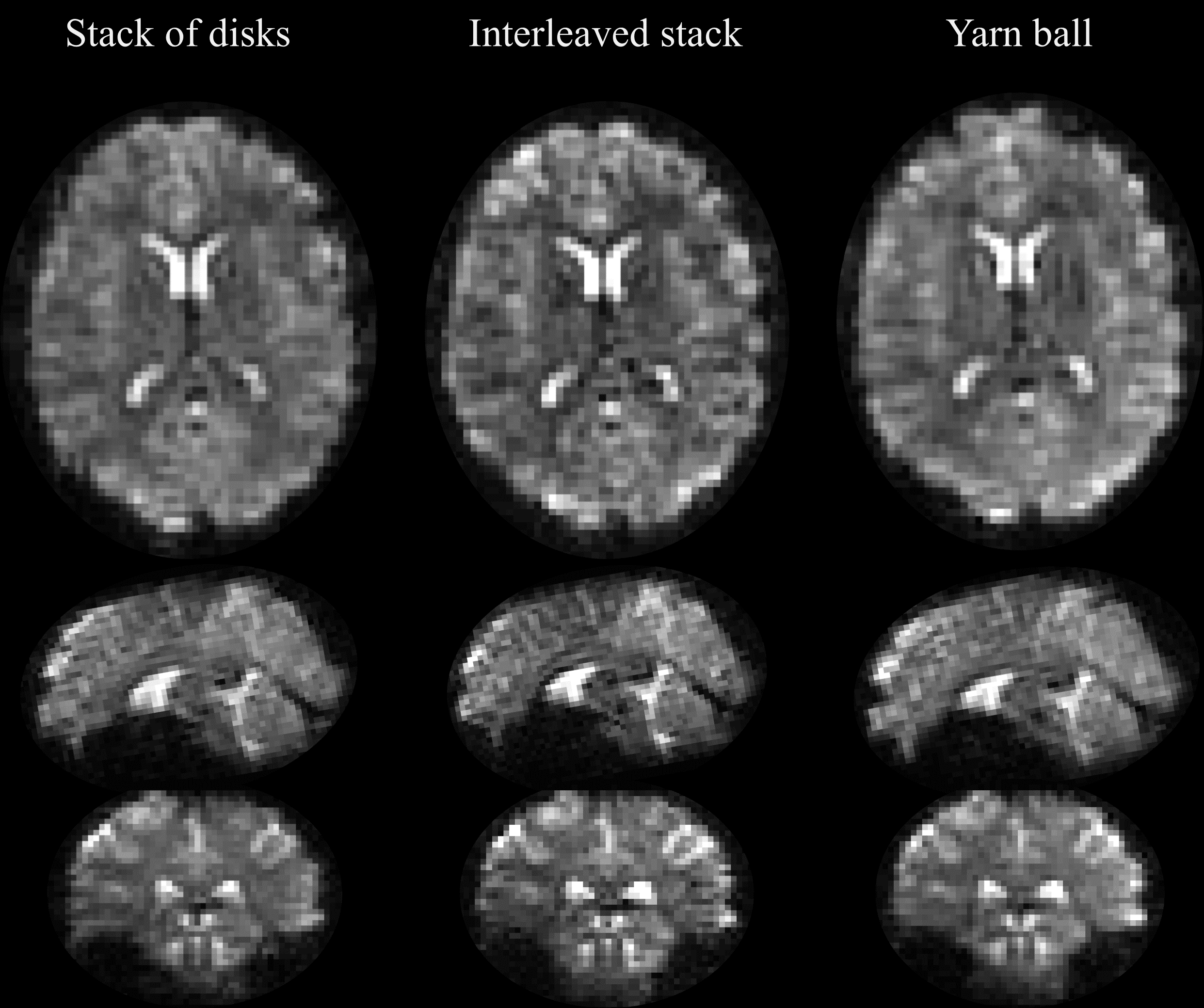
Results
See Figures 3-5.Discussion and conclusion
All of the three discussed trajectories exhibit smooth T2* weighting. However, to accomplish this goal, the interleaved stack trajectory has to introduce a costly echo time shift (here: 25% prolongation), reducing its efficiency (Fig. 3).
The Yarn-ball trajectory loses efficiency by oversampling the poles of the acquired k-space sphere in order to sample densely enough at the equator. Plus, for many shots, it approaches radial sampling in the kx-ky plane, thereby oversampling low kz values. In contrast, the stack of disks trajectory preserves a homogeneous k-space density by sampling spirals on a hexagonal grid. The strategy could equally be applied to EPI trajectories, catching however the same time loss compared to spirals as in 2D.
An advantage of the Yarn-ball trajectory is its reduced sensitivity to flow in any direction, being a potential source of artifacts. Both stack trajectories do need a prephaser gradient in kz direction. Besides, the Yarn-ball never has to slow down on its k-space path. Aiming for the same, the stack of disks trajectory leaves small undersampled areas at the site of the blips, which could be mitigated by slightly shifting the applied blips for each rotation.
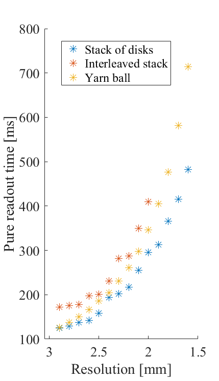
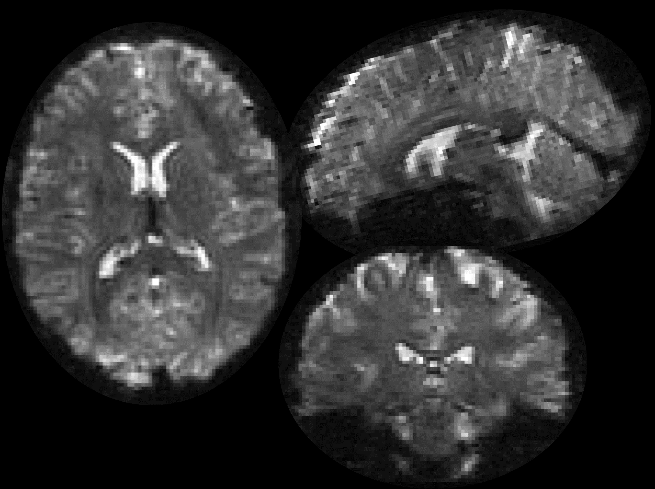
To account for off-resonance and actual gradient dynamics the reconstruction was based on the expanded signal model described above. Whole brain coverage is attained in 330ms at 3mm isotropic resolution (Fig. 4). For higher-resolved applications, Yarn-ball and interleaved stack are not competitive anymore (Fig.3). The stack of disks trajectory covers 1.6mm isotropic resolution in 840ms (Fig. 5), whereas both of the others would require more than a second.
Acknowledgements
No acknowledgement found.References
1. Feinberg DA, Moeller S, Smith SM, Auerbach E, Ramanna S, Glasser MF, Miller KL, Ugurbil K, Yacoub E. Multiplexed Echo Planar Imaging for Sub-Second Whole Brain FMRI and Fast Diffusion Imaging Valdes-Sosa PA, editor. PLoS ONE 2010;5:e15710. doi: 10.1371/journal.pone.0015710. 2. Poser BA, Koopmans PJ, Witzel T, Wald LL, Barth M. Three dimensional echo-planar imaging at 7 Tesla. NeuroImage 2010;51:261–266. doi: 10.1016/j.neuroimage.2010.01.108. 3. Irarrazabal P, Nishimura DG. Fast Three Dimensional Magnetic Resonance Imaging. Magn. Reson. Med. 1995;33:656–662. doi: 10.1002/mrm.1910330510. 4. Zahneisen B, Poser BA, Ernst T, Stenger AV. Simultaneous Multi-Slice fMRI using spiral trajectories. NeuroImage 2014;92:8–18. doi: 10.1016/j.neuroimage.2014.01.056. 5. Stenger VA, Rettenmeier C. Blipped Stack of Spirals for Fast Volumetric Functional MRI. Proc. ISMRM 2017 [Internet]. 6. Engel M, Kasper L, Pruessmann KP. Rapid 3D imaging with multiplanar spirals. Proc. ISMRM 2017 [Internet]. 7. Stobbe RW, Beaulieu C. A Looping Trajectory for Single-Shot 3D Imaging. In: Proc. Intl. Soc. Mag. Reson. Med. Vol. 19. ; 2011. 8. Dietrich BE, Brunner DO, Wilm BJ, Barmet C, Gross S, Kasper L, Haeberlin M, Schmid T, Vannesjo SJ, Pruessmann KP. A field camera for MR sequence monitoring and system analysis. Magn. Reson. Med. 2016;75:1831–1840. doi: 10.1002/mrm.25770. 9. Mersereau RM. The Processing of Hexagonally Sampled Two-Dimensional Signals. Proc. IEEE 1979;67:930–949. 10. Pruessmann KP, Weiger M, Börnert P, Boesiger P. Advances in sensitivity encoding with arbitrary k-space trajectories. Magn. Reson. Med. 2001;46:638–651. doi: 10.1002/mrm.1241. 11. Man L-C, Pauly JM, Macovski A. Multifrequency interpolation for fast off-resonance correction. Magn. Reson. Med. 1997;37:785–792. doi: 10.1002/mrm.1910370523. 12. Barmet C, Tsao J, Pruessmann KP. Sensitivity encoding and B0 inhomogeneity – A simultaneous reconstruction approach. In: Proceedings of the ISMRM. ; 2005. p. 682. 13. Barmet C, Wilm BJ, Pavan M, Katsikatsos G, Keupp J, Mens G, Pruessmann KP. Concurrent higher-order field monitoring for routine head MRI: an integrated heteronuclear setup. In: Proceedings of the 18th Annual Meeting of ISMRM, Stockholm, Sweden. ; 2010. p. 216.Figures