4148
Wind Out-In Dual-Echo Yarn-Ball with Application to Knee Imaging1Biomedical Engineering, University of Alberta, Edmonton, AB, Canada
Synopsis
3D centre-out Yarn-Ball k-space acquisition is implemented in a wind-out/wind-in dual-echo format for the first time. As with standard 3D-Radial acquisition this technique facilitates short (first ‘echo’) TE with utility for imaging tissues with short T2*. However, the advantage of centre-out Yarn-Ball is much greater k-space sampling efficiency than 3D-Radial. Smooth variation from wind-out to wind-in minimizes potential errors in k-space trajectory evolution as a result of eddy-currents. Spoiled-steady-state dual-echo 3D Yarn-Ball images with 0.7x0.7x0.7 mm3 voxels were acquired from the knee of a healthy volunteer in 6 minutes, and the difference image shows ligament and meniscus conspicuity.
Introduction
Dual-echo 3D ultra-short TE (UTE) imaging has value for imaging tissues with short T2* values [1]. However, a drawback of standard 3D-Radial acquisition is that only ½ a line of k-space is sampled (per image) with each RF excitation, and much of the centre of k-space is highly oversampled. The 3D centre-out Yarn-Ball technique has recently been developed to facilitate much greater k-space sampling efficiency [2], and here we present dual-echo Yarn-Ball imaging for the first time with application to imaging ligaments and menisci in the knee.Methods
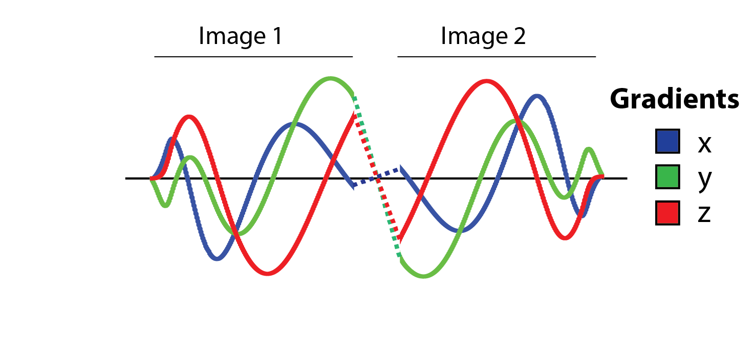
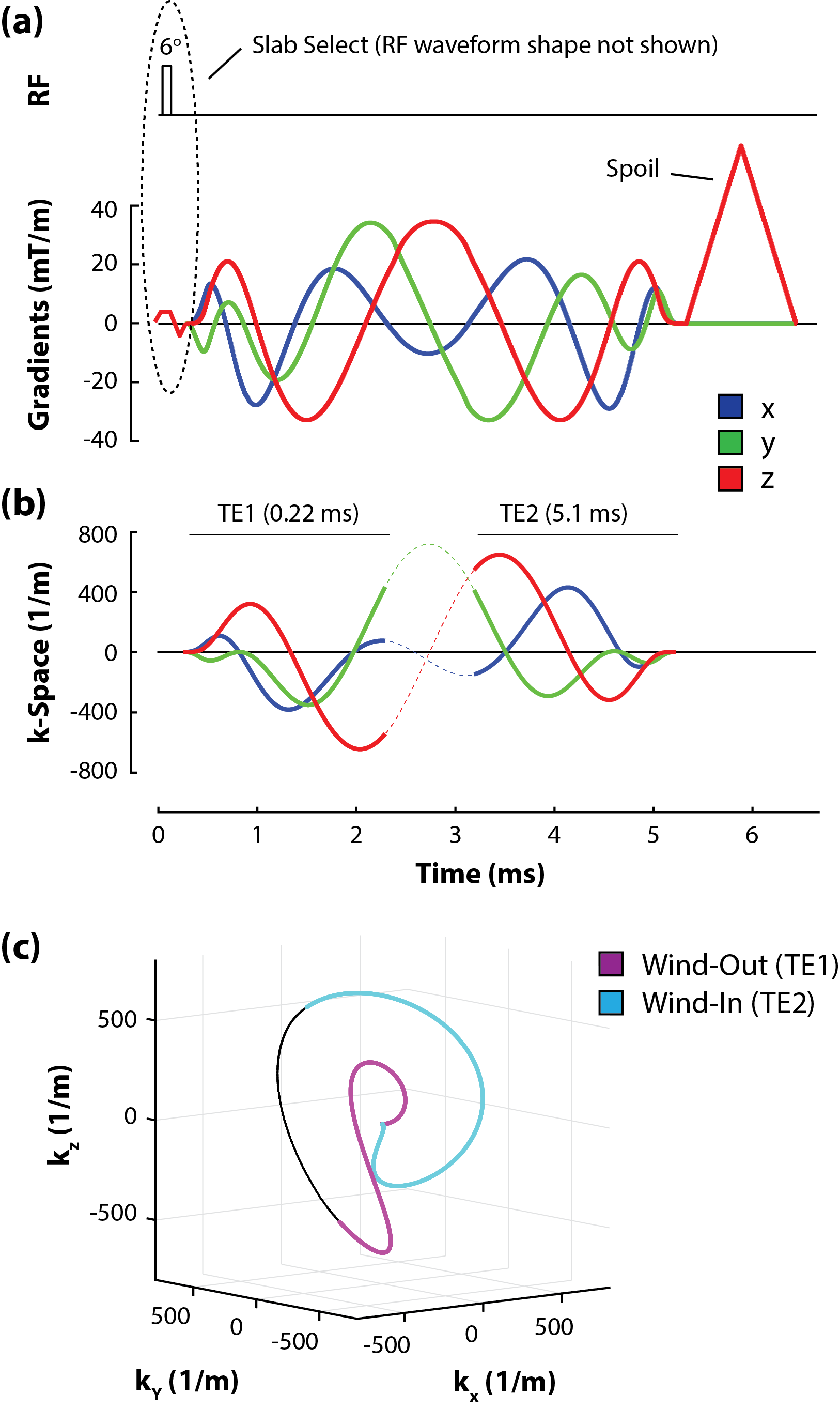
Perhaps the first approach one might consider in the implementation of dual-echo Yarn-Ball is simply to replay the gradient waveform backwards (i.e. staring from the end) with inverted polarity (Figure 1). In theory this will resample k-space in reverse (i.e. toward the middle) along the same path. However, sharp transitions within the waveform can lead to ‘unexpected’ k-space sampling delays and deviation from the intended path as the result of eddy-currents. An alternative approach is to continue the direction of winding, rather than reverse it as in Figure 1. A key aspect of the Yarn-Ball technique is that radial evolution (r′) decreases with distance from the centre of k-space as r′=c/r2. At the edge of the sampled k-space sphere (i.e. kmax) typical Yarn-Ball trajectories will be travelling near perpendicular to the radial dimension (i.e. r′ will be very small). Thus, a switch to solving r′=-c/r2 to wind back in will result in minimal radial discontinuity. To ensure sufficient r′ reduction at the point of radial direction change, the trajectory can be solved slightly past kmax with increased radial slowing. While winding out, the azimuthal and polar angles evolve as θ′=cr′ and φ′=crθ′ respectively. To maintain the same winding direction θ′=c|r′| is solved on the way back in.
Images were acquired from a healthy 19 year old female volunteer on a Siemens Prisma 3T scanner using a 4-channel flexible receive-coil wrapped around the knee. The k-space sampling readout duration (or time from the centre of k-space to kmax and visa-versa on return) was chosen to be 2 ms for this initial test. Longer readouts will result in increased signal smearing from rapid T2* decay, but will yield greater sampling efficiency within peripheral nerve stimulation constraints. Slab select excitation along the superior-inferior dimension resulted in a TE of 0.22 ms for the first image; a TE of 5.1 was chosen for the second image. The inclusion of gradient spoiling resulted in a TR of 6.5 ms and a flip-angle of 6o was chosen to maximize signal. A pictorial description of the sequence is given in Figure 2. A total of 18445 trajectories fully sampled k-space for 130 mm isotropic FoV support with 0.7x0.7x0.7 mm3 voxel volume (defined by 1/2kmax). The acquisition of 3 averages yielded a total scan time of 6 minutes. Images were created with standard gridding reconstruction.
Results
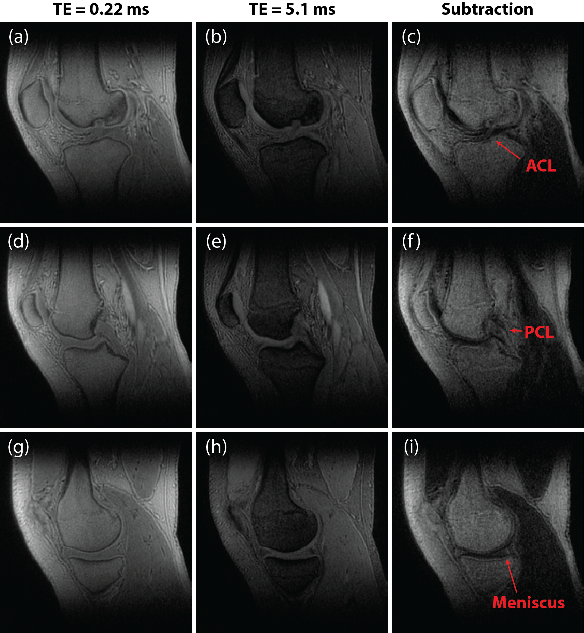
Columns 1 and 2 of Figure 3 show representative slices from both images (TE = 0.22 and TE = 5.1) of the spoiled-steady-state dual-echo Yarn-Ball sequence. Tissues with short T2* values such as bone, ligaments, tendons and menisci exhibit intensity similar in magnitude to muscle and cartilage on the TE = 0.22 image. On the TE = 5.1 image, intensity in the short T2* tissues is greatly reduced. A subtraction image (column 3 of Figure 3) yields positive short T2* contrast, with anterior and posterior cruciate ligament (ACL, PCL) as well as menisci conspicuity.Discussion
3D Yarn-Ball k-space acquisition is particularly amenable to implementation in a dual-echo format. Smooth transition to return-to-zero winding is facilitated with continuity in winding direction. For 3D-Radial imaging ~108000 trajectories are required to fully sample the same k-space matrix sphere, almost 6x more than Yarn-Ball. Future work will consider optimization of readout duration and TE separation for maximum short T2* conspicuity on difference images as well as the calculation of T2* maps. Dual-echo Yarn-Ball implementation within a steady-state free-precession context [3] will also be considered for enhanced signal to noise ratio. Other applications including B0 mapping, susceptibility mapping and shimming may also benefit from dual-echo Yarn-Ball k-space acquisition.Acknowledgements
Funding from the Canadian Institute of Health ResearchReferences
[1] Du J., et. al., Magn Reson Imaging, 29(4):470–482, 2011
[2] Stobbe R.W. and Beaulieu C., ISMRM, abstract 2442, 2015 (Toronto)
[3] Martirosian, P., et. al., Magn Reson Med, 71:294–301, 2014
Figures