4141
Improved Parallel Echo-Planar Imaging (EPI) with Ghost Removal and Distortion CorrectionYuan Zheng1, Yu Ding1, Xiaodong Ma2, and Weiguo Zhang1
1UIH America, Inc., Houston, TX, United States, 2United Imaging Healthcare, Shanghai, China
Synopsis
We have developed a procedure for reconstructing high-quality EPI images with parallel imaging, and presented both in-vitro and in-vivo results. Interleaved EPI and the PLACE technique are used to generate coil sensitivity maps (CSM) and a distortion map. These maps do not suffer from ghosting and match the distortion of the imaging data. The CSM are used in the PEC-SENSE reconstruction to generate images with significantly reduced ghosting artifacts. Geometric distortions are subsequently corrected using the distortion map. This technique can be used to improve the image quality of many EPI applications, such as diffusion imaging, when parallel imaging is involved.
Introduction
EPI is a fast imaging technique, particularly widely used in neuro imaging applications including functional and diffusion MRI. However, due to differences in system responses to readout gradients with opposing polarities, EPI images often suffer from ghosting artifacts. When EPI acquisition is used together with parallel imaging, the ghosting artifacts become more complicated. Moreover, EPI images are susceptible to off-resonance induced geometric distortions due to the low bandwidth in the phase encoding direction. We present here an approach to reconstruct ghosting-free parallel EPI images by extending the PEC-SENSE technique recently proposed by Xie et. al.1 We also demonstrate that the geometric distortions can be corrected to further improve image quality.Theory
The PEC-SENSE technique1 satisfactorily removes EPI ghosting artifacts. However, it requires high-quality coil sensitivity maps (CSM) with no ghosts and with the same distortion as the imaging acquisitions, which can itself be difficult to obtain when parallel imaging is involved. We have utilized an interleaved-EPI (iEPI) sequence to collect the CSM. The number of interleaves is set to be same as the parallel imaging acceleration factor to match the effective echo spacing, and thus the geometric distortions between CSM and imaging data. However, the iEPI image (I1) cannot be directly used as the CSM for PEC-SENSE, since it suffers from EPI ghosting artifacts itself. In order to eliminate the artifacts, and also to extract a map for distortion correction, we extended the PLACE technique proposed by Xiang et. al.2 to parallel imaging. Two more iEPI images are collected. For the second image (I2), the k-space is shifted in the ky direction by an amount (in units of k-space lines) equal to the number of interleaves. For the third image (I3), the k space is shifted in the same direction by twice as much. An artifact-free CSM can be generated by combination of I1 and I2 (or I2 and I3)2. A distortion map can be extracted from I1, I2 and I32. The reconstruction pipeline for parallel EPI (acceleration factor=2) is shown in Fig. 1. The sampling pattern and reconstruction procedure for generating coil sensitivity and distortion maps are shown in Fig. 2.Methods
All data were collected on a uMR 560 1.5 T scanner (United Imaging Healthcare, Shanghai, China) with a 16 channel head coil. A gradient-echo EPI sequence was used for this study. Three echoes with phase encoding turned off were inserted after the excitation to collect information for preliminary odd-even line corrections3,4. The calibration scans include six shots as shown in Fig. 2. For comparison, a low resolution (24 central k space lines) image is also collected for conventional GRAPPA reconstruction5. 2D images were acquired with slice thickness=3.5mm, FOV=230×230 mm, resolution= 112×112, FA=90°, TE=32 ms, BW=1910 Hz, echo spacing=0.94 ms, acceleration factor=2. The first data set was collected on a 16 cm diameter water cylinder to demonstrate the effect of ghost removal. The second data set was acquired on several closely positioned small water phantoms (expected to suffer from off-resonance due to air-water interfaces) to demonstrate distortion correction. The third data set was collected on a volunteer head. All data were first corrected using information extracted from the three echoes without phase encoding3,4. The data were then reconstructed using either GRAPPA or the proposed procedure.Results
Fig. 3 a-c show I1, I2 and I4 (as indicated in Fig. 2) of the water cylinder phantom. Although I1 and I2 have obvious ghosts, I4 is almost artifact free by combination of I1 and I2 according to the PLACE method, and is used as CSM for the PEC-SENSE reconstruction. Fig. 3 d-e compare the water cylinder images reconstructed using GRAPPA and the proposed method (without distortion correction). The latter significantly removed the ghosts. Fig. 4 shows images of the small water phantoms reconstructed by the proposed method, before and after distortion correction. Geometric distortions are largely corrected. Fig. 5 shows a high-quality parallel EPI head image generated using the proposed procedure.Conclusion
We have developed a procedure for reconstructing high-quality EPI images with parallel imaging. IEPI and PLACE are used to generate coil sensitivity and distortion maps. These maps do not suffer from ghosting and matches the distortion of imaging data. The CSM are used in the PEC-SENSE reconstruction to generate images with significantly reduced ghosting artifacts. Geometric distortions are subsequently corrected according to the distortion map. The proposed technique can be used to improve the image quality of many EPI applications, such as diffusion imaging, when parallel imaging is involved.Acknowledgements
No acknowledgement found.References
- Xie VB, Lyu M, Liu Y, Feng Y, Wu EX, Robust EPI Nyquist ghost removal by incorporating phase error correction with sensitivity encoding (PEC-SENSE), Magn Reson Med. 2017 Jun 7. doi: 10.1002/mrm.26710.
- Xiang QS, Ye FQ, Correction for geometric distortion and N/2 ghosting in EPI by phase labeling for additional coordinate encoding (PLACE), Magn Reson Med. 2007 Apr;57(4):731-41.
- Bruder H, Fischer H, Reinfelder HE, Schmitt F, Image reconstruction for echo planar imaging with nonequidistant k-space sampling. Magn Reson Med. 1992;23:311–323.
- Zhang W, Wu H, Jiang X, An image reconstruction method for the Echo-Planar Imaging sequence. CN Patent 104035059 A.
- Griswold MA, Jakob PM, Heidemann RM, Nittka M, Jellus V, Wang J, Kiefer B, Haase A, Generalized autocalibrating partially parallel acquisitions (GRAPPA), Magn Reson Med. 2002 Jun;47(6):1202-10.
Figures
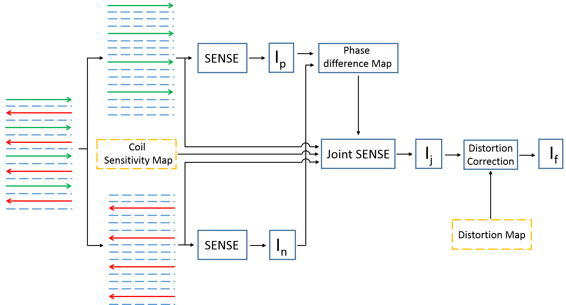
Basic image reconstruction pipeline for PEC-SENSE1 with
parallel imaging (acceleration factor = 2). Two ghost-free images from odd and
even echoes are reconstructed using SENSE. A phase error map due to the
odd-even line inconsistency is estimated from these two images. The phase error map
is then incorporated into the coil sensitivity maps and the odd and even echoes
are reconstructed together with joint SENSE. Distortion correction is applied
to generate the final image. The coil sensitivity and distortion maps
are generated as shown in Fig. 2.
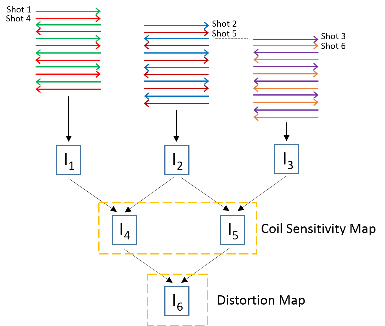
Six shots are acquired
with shifts of -2, 0, 2, -1, 1, 3 lines in the ky direction. The echo time of
shots 4, 5, 6 are shifted by half of the echo spacing with respect to shots 1,
2, 3. Shots (1, 4), (2, 5), (3, 6) are combined to generate three full k
spaces, corresponding to images I1, I2, and I3. Ghost-free images I4 and I5 are
generated by combining I1 and I2, I2 and I3. A distortion map I6 is extracted
from the phases of I4 and I5 to correct geometric distortions.
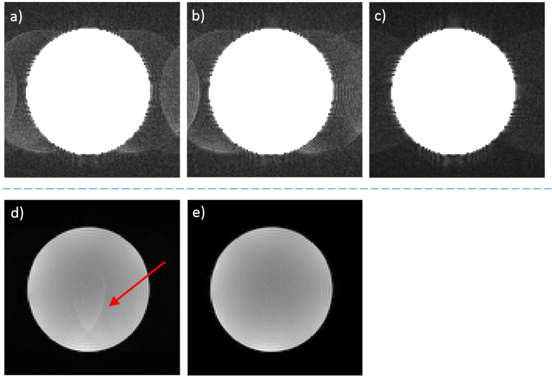
Images of the water
cylinder phantom experiment. a), b) and c) correspond to the calibration scan
images I1, I2 and the combined image I4 in Fig.2. I4 has significantly reduced
ghosts compared with I1 and I2, and is used as the CSM for
reconstructing e). Conventional GRAPPA recon of the imaging acquisitions d) leaves
obvious ghosts, while the proposed parallel PEC-SENSE approach shows no visible
ghosting artifacts.
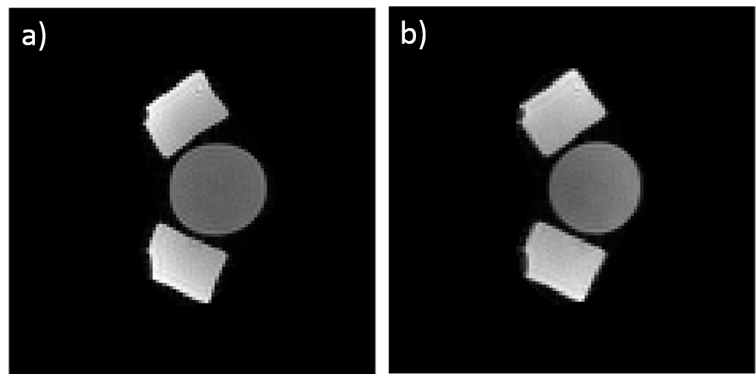
Images reconstructed
using the proposed procedure, before a) and after b) distortion
correction. The circular shape of the cylinder at the center is restored.
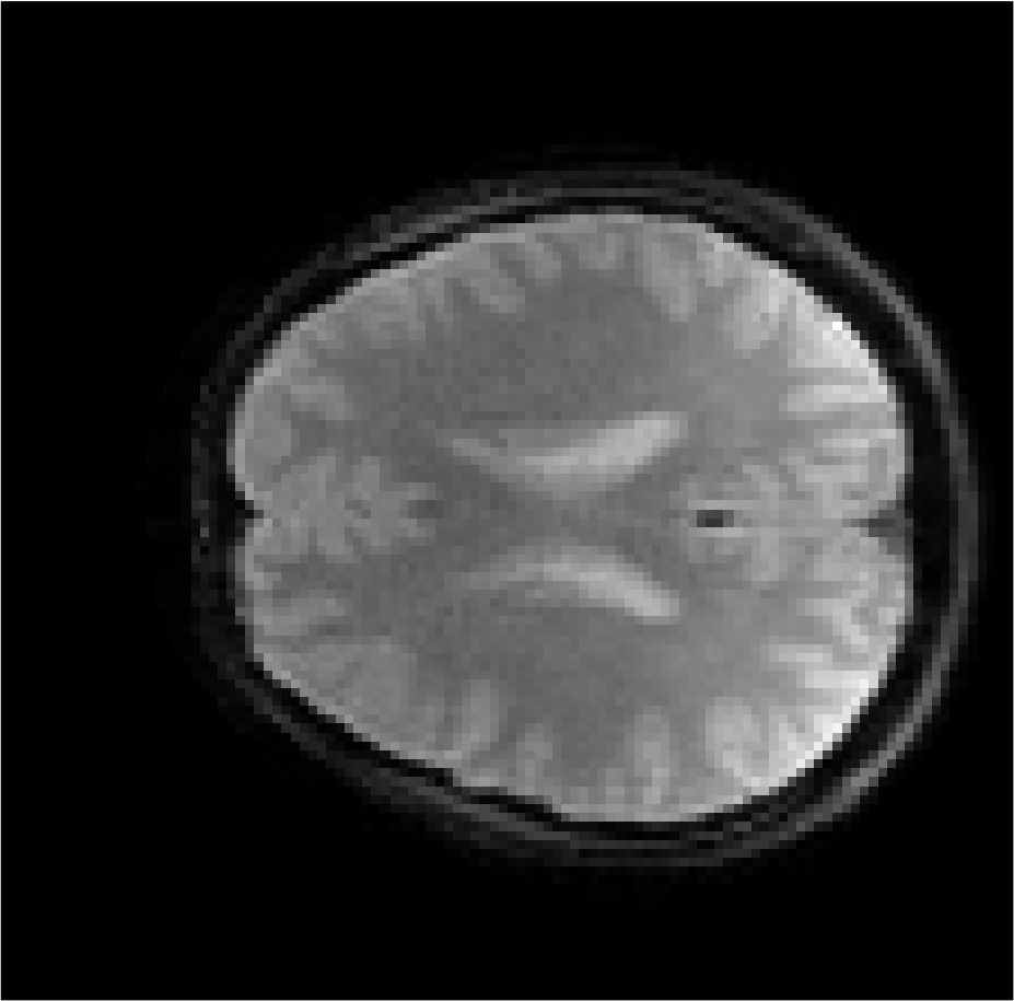
A volunteer head
image acquired with a parallel EPI sequence (acceleration factor = 2) and
reconstructed with the proposed procedure.