4128
A CAIPIRINHA-based approach to the referenceless reconstruction of high-definition SPEN images with simultaneous multislice1Chemical Physics, Weizmann Institute of Science, Rehovot, Israel
Synopsis
Spatiotemporal Encoding (SPEN) is an ultrafast imaging technique where the low-bandwidth axis is rasterized in a joint spatial/k-domain. SPEN benefits from increased robustness to inhomogeneities, folding-free reconstruction of subsampled data, and an ability to combine multiple interleaved or signal averaging scans in a referenceless fashion. SPEN’s relatively high SAR, however, complicates its volumetric uses. Here we show how this can be solved by merging a controlled aliasing for parallel imaging (CAIPIRINHA) protocol involving phase-cycling of multi-banded excitation pulses in independent scans, so as to enable a referenceless reconstruction of interleaved multislice acquisitions delivering high in-plane definition and excellent inter-slice decoupling.
INTRODUCTION
Controlled aliasing for parallel imaging (CAIPI, CAIPIRINHA)1,2, enables simultaneous multislice (SMS) excitation while reducing the noise amplification resulting from similar coil sensitivity maps for different slices. This requires specific slice-selective manipulations, which in combination with multi-coil multiplexing decreases the coupling among the images. CAIPI’s blipping of the slice-select (SS) gradient during the acquisition is particularly useful in « ultrafast » experiments, aimed at covering large volumes using echo-planar imaging (EPI)3. SPatiotemporal ENcoding (SPEN) is an alternative ultrafast technique that relies on a frequency-swept chirp pulses acting in combination with gradients to spatially rasterize spins in a sample4. SPEN provides images that are folding-free even if subsampled5; it can successfully average and interleave multiple scans even in diffusion or functional experiments, and it benefits from reduced distortions due to its use of stronger gradients than EPI4. SPEN suffers, however, from SAR values that are higher than EPI’s in proportion to the ratio between the sequences’ PE-axis bandwidths. Here we demonstrate how to alleviate these coverage complications, by invoking a CAIPIRINHA-like procedure whereby SMS 2D SPEN images are separated by coil- and phase-cycling multiscan procedures. Further, these various scans are arranged so that each 2D SPEN image is acquired in an interleaved fashion5. Suitable processing therefore achieves both an in-plane resolution improvement and improved multiband separation. Applications of this in human diffusion experiments are presented.
METHODS
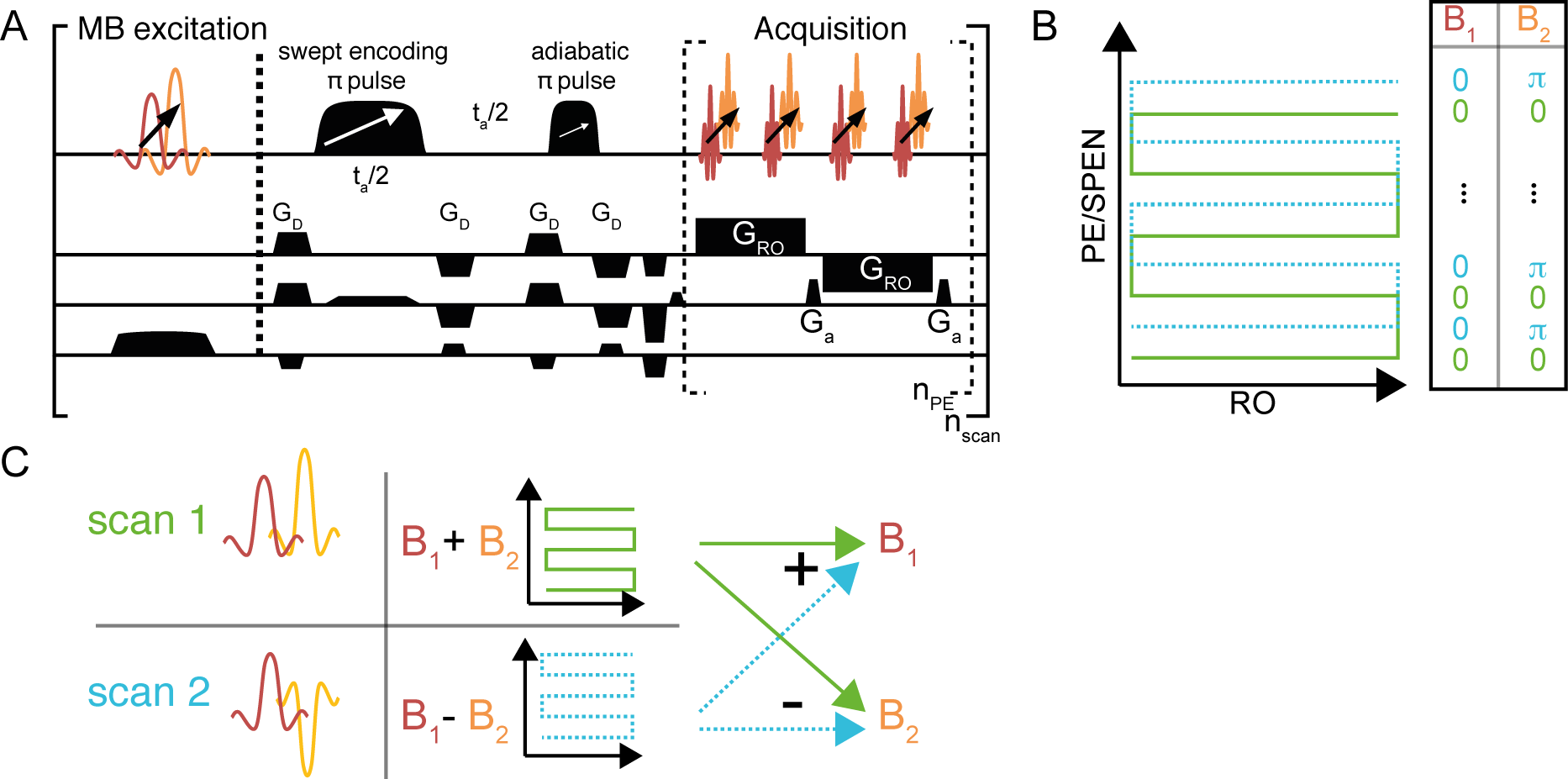
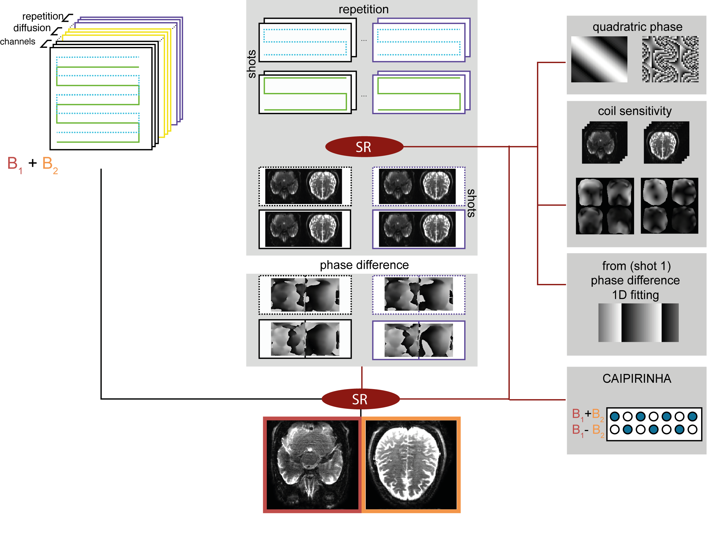
The pulse sequence for the interleaved SMS-SPEN experiment introduced here is in Fig. 1A. The different scans use an interleaved approach meant to improve resolution (Fig 1B)1. In addition, different scans change each SMS band’s phase according to a CAIPIRINHA phase-cycling scheme as shown in Fig. 1B for a two-band scenario. SPEN’s reconstruction of these subsampled (but folding-free) data then proceeds by suitable linear combinations, as shown in Fig. 1C. This leads to a robust decoupling among the different bands2, and it was preferred over its blipped CAIPI counterpart, owing to SPEN’s special nature as a mixed spatial/k-space encoding technique. Indeed, the application of CAIPI’s blips will phase-modulate the SMS bands in a conventional way, but will only move them along SPEN (PE) dimension by a fraction of the corresponding field-of-view given by the strength of SPEN’s encoding gradient. By contrast, the phase-cycling approach cleanly separates the frequency bands. It also exploits the fact that, unlike EPI, SPEN is compatible with multiscan acquisitions even for diffusivity measurements. This, however, requires that the multiscan data be suitably compatibilized among one another. To do so, the reconstruction approach shown in Figure 2 was followed. It involves calculating the phase of each band independently for every scan and diffusion weighting. These phase maps are then used in the final reconstruction, where all the scans’ data are used, using a super-resolution (SR) approach6 which factors-in the per-scan phase maps, all the channels sensitivity maps, the scan interleaving, and the CAIPIRINHA modulation. The sequence in Figure 1A was programmed on a 3T Siemens TrioTIM scanner equipped with a 32 channels head coil. SMS-SPEN was run with an encoding bandwidth of 2.4 kHz, nPE= 96, Nshots=4, CAIPIRINHA=2, encoding two bands positioned 3.3cm apart. 11 slices were thus collected over a 19x21x6cm 3D volume, with a 1x1x3mm resolution in a TR=8sec time. All human volunteers were scanned following suitable written consent.RESULTS & DISCUSSION
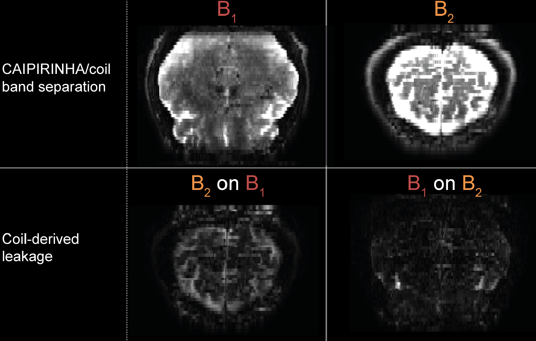
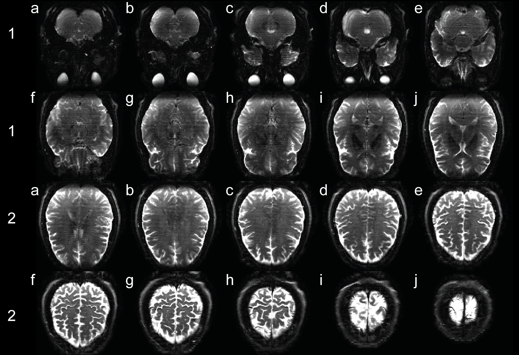
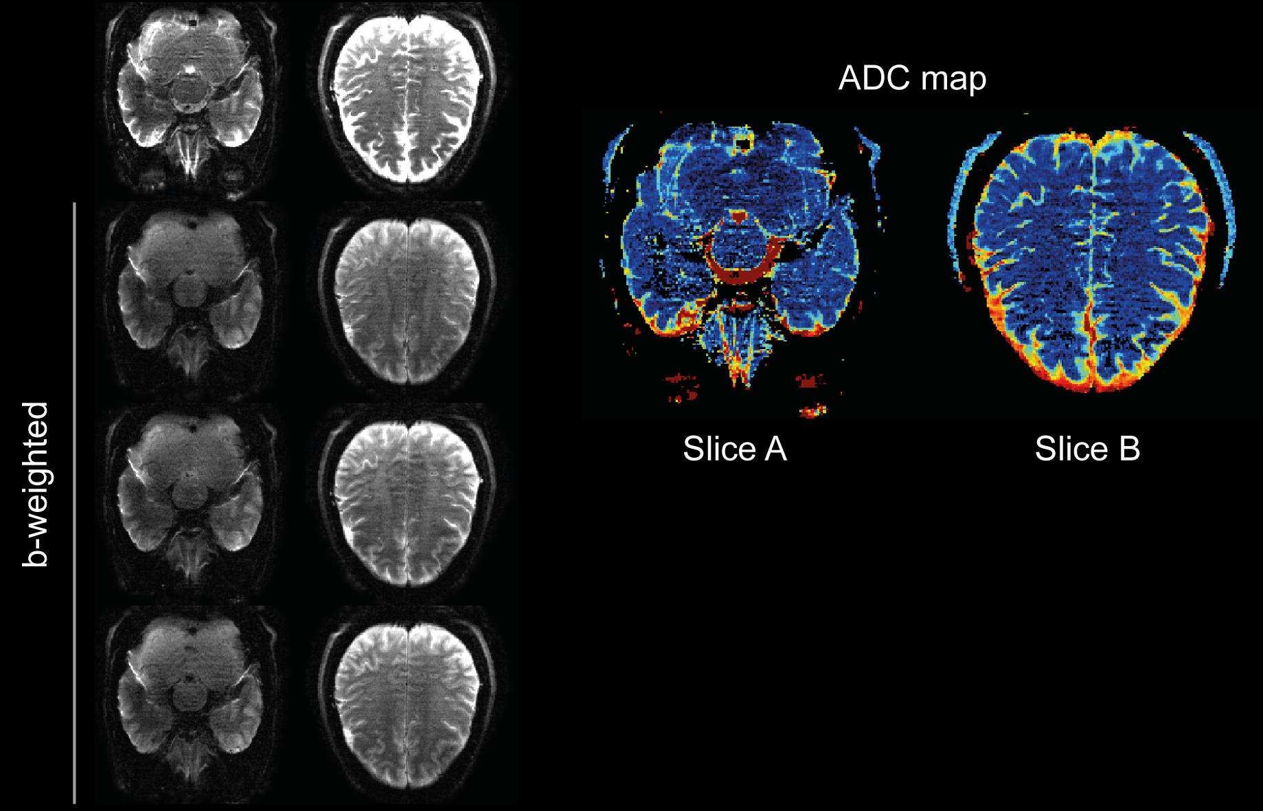
The suitability of the CAIPIRINHA-based processing for removing multislice leakage can be appreciated from Figure 3. Shown on the bottom part are experimental SPEN results arising from two bands excited as B1+B2 and B1-B2, reconstructed using only channel sensitivity information. Given the 3cm separating the bands, the separation is clearly imperfect and reflects the leakage arising in an unaided multiband scheme. Shown on the upper part are the estimations arising if the two scans B1±B2 are added/substracted. By adding this CAIPIRINHIA approach to the multishot SPEN experiments, all slices become well separated (Figure 4). Based on this success, the procedure was applied to acquire whole-brain ADC and DWI data; taking in account the phase induced by motion during b-weighted experiments (Figure 5). SPEN’s robustness is visible in areas usually distorted in EPI, particularly in tissue-air boundaries.CONCLUSION
A novel sequence and a reconstruction procedure for SMS-SPEN experiments were described, whereby use of multiple scans are combined with multiband phase cycling and interleaving, to achieve clear slice separations with increased 3D coverage and good in-plane resolution –at negligible increase in the SAR (which is dominated by the single π-sweep encoding all the slices) or g-factor related SNR penalties. Despite its multiscan nature the method was successfully applied for diffusion experiments.Acknowledgements
We are grateful to Dr. Sagit Shushan (Wolfson Medical Center) and Edna Furman-Haran (Weizmann) for assistance in the scans. SfC thanks the Feinberg Graduate School (Weizmann) and French Foreign Service for partial postdoctoral fellowships. Financial support came from the Israel Science Foundation grant 2508/17, the EU through ERC-2016-PoC grant # 751106, Minerva funding (#712277) from the Federal German Ministry for Education and Research, the Kimmel Institute for Magnetic Resonance and the generosity of the Perlman Family Foundation.References
1. Breuer, F. A., Blaimer, M., Heidemann, R. M., Mueller, M. F., Griswold, M. A., & Jakob, P. M. (2005). Controlled aliasing in parallel imaging results in higher acceleration (CAIPIRINHA) for multi‐slice imaging. Magnetic resonance in medicine, 53(3), 684-691.
2. Setsompop, K., Gagoski, B.A., Polimeni, J.R., Witzel, T., Wedeen, V.J., Wald, L.L., 2012. Blipped‐controlled aliasing in parallel imaging for simultaneous multislice echo planar imaging with reduced g‐factor penalty. Magnetic Resonance in Medicine 67, 1210–1224. doi:10.1002/mrm.23097
3. Stehling, M.K., Turner, R., Mansfield, P., 1991. Echo-Planar Imaging: Magnetic Resonance Imaging in a Fraction of Second. Science 254, 43.
4. Tal, A., Frydman, L., 2006. Spatial encoding and the single-scan acquisition of high definition MR images in inhomogeneous fields. Journal of Magnetic Resonance 182, 179–194. doi:10.1016/j.jmr.2006.06.022 [1]
5. Schmidt, R., Seginer, A., Frydman, L., 2016. Interleaved multishot imaging by spatiotemporal encoding: A fast, self-referenced method for high-definition diffusion and functional MRI: Referenceless Interleaved SPEN MRI. Magnetic Resonance in Medicine 75, 1935–1948. doi:10.1002/mrm.25742
6. Ben-Eliezer, N., Irani, M., Frydman, L., 2010. Super-resolved spatially encoded single-scan 2D MRI. Magnetic Resonance in Medicine 63, 1594–1600. doi:10.1002/mrm.22377
Figures