4123
Characterization and suppression of stripe artifact in velocity-selective magnetization-prepared unenhanced MR angiography1Ewha Womans University, Seoul, Republic of Korea, 2Case Western Reserve University, Cleveland, OH, United States, 3Johns Hopkins University, Baltimore, MD, United States
Synopsis
While velocity-selective magnetization-prepared MR angiography (VS-MRA) has shown great promise for diverse arterial territories, artifactual stripes often occur in resultant angiograms. We investigate the formation of the stripe artifact using extended phase graph analysis and show that the stripes contain not only the fundamental frequency determined by the area of VS unipolar gradient but higher-order components with multiples of the fundamental frequency. Based on this finding, we propose and test efficient strategies for suppression of the artifact in a phantom and healthy human subjects.
Introduction
Velocity-selective magnetization-prepared MR angiography (VS-MRA) enables 3D-encoded angiograms using a single acquisition without contrast agents, and has shown great promise for various arterial territories1-5. The VS magnetization preparation should ideally have velocity only selectivity, but can involve erroneous spatial selectivity which appears as stripes repeated in the direction of the VS gradient6. In this study, we analyze the generation of the tripe artifact using extended phase graph analysis and propose strategies for suppression of the artifact.
Methods
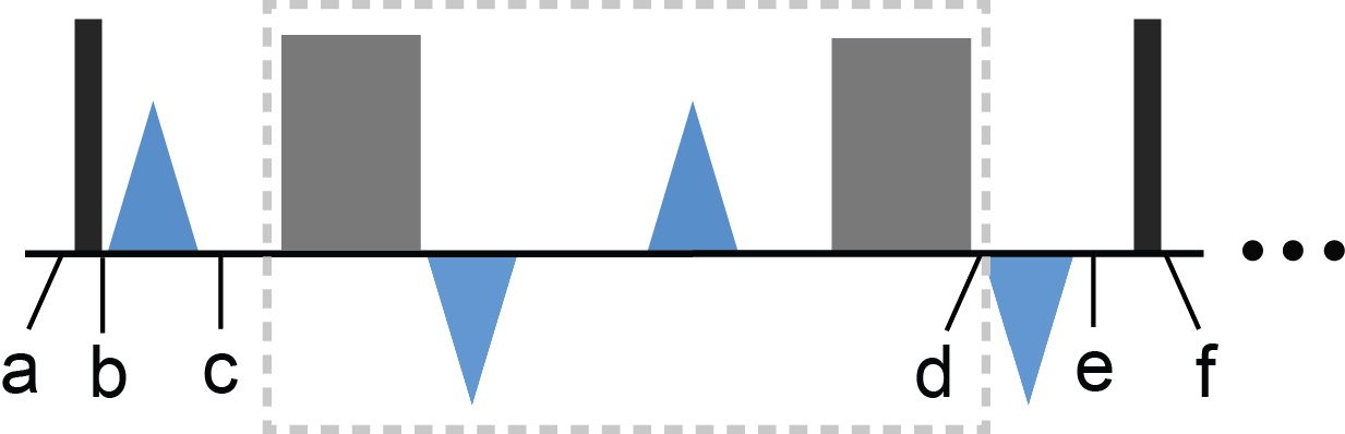
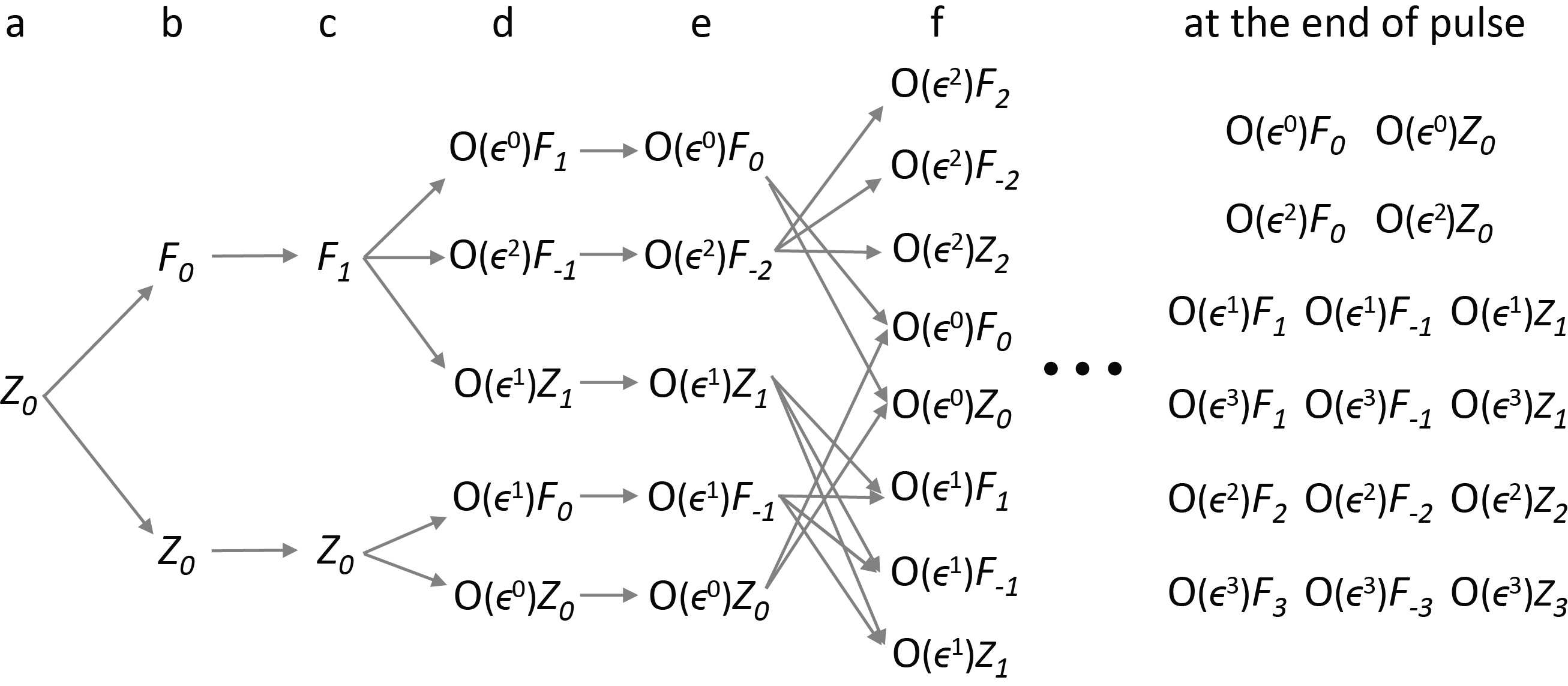
Extended phase graph analysis: VS magnetization preparation concatenates multiple VS excitation steps each of which consists of a hard RF pulse, a unipolar gradient, a refocusing module, and a reversed unipolar gradient (Fig. 1)2. We analyze the spatial frequencies of the stripe artifact by using the extended phase graph formalism which decomposes spin ensemble within a voxel into a basis of transverse (Fn) and longitudinal (Zn) states (n is the number of cycles across a voxel)7-10. Figure 2 illustrates the evolution of the ensemble states during the first VS excitation step by concatenating the effects of hard RF pulses, unipolar gradients and imperfect refocusing pulses which induce excitation of tiny flip angle ϵ. After amplitude orders lower than ϵ3 are ignored, six independent longitudinal states are derived at the end of the VS preparation pulse, containing Z1, Z2, and Z3 states.
Strategies to suppress stripe artifact: With typical area of unipolar gradient > 5 mT∙msec /m which corresponds to the spatial period of λ < 4.8 mm, and typical spatial resolution of ~1 mm, the stripe artifact will contain up to 2nd order harmonics (λ and λ/2) since λ/3 < 1.5 mm < 2*spatial resolution = 2 mm. We propose to alternately apply four VS preparation pulses which have excitation profiles spatially shifted by 0, λ/2, λ/4, and 3λ/4, respectively. The underlying rationale is that a portion of the spatial fluctuation made by each VS pulse is preserved until the next VS preparation due to incomplete Mz recovery so that the 4-phase cycled spatial modulations affect the fluctuation amplitude in a destructive manner. Since the quality of stripe suppression will decrease with increasing Mz recovery between VS prepared acquisitions, we also propose to acquire the kx-ky plane at kz = 0 four times for the four VS preparations and to average the resultant 4 kx-ky data sets. Since each kz partition is covered by segmented acquisitions over 2 cardiac cycles, the scan time is increased by (4-1)×2 = 6 cardiac.
Results
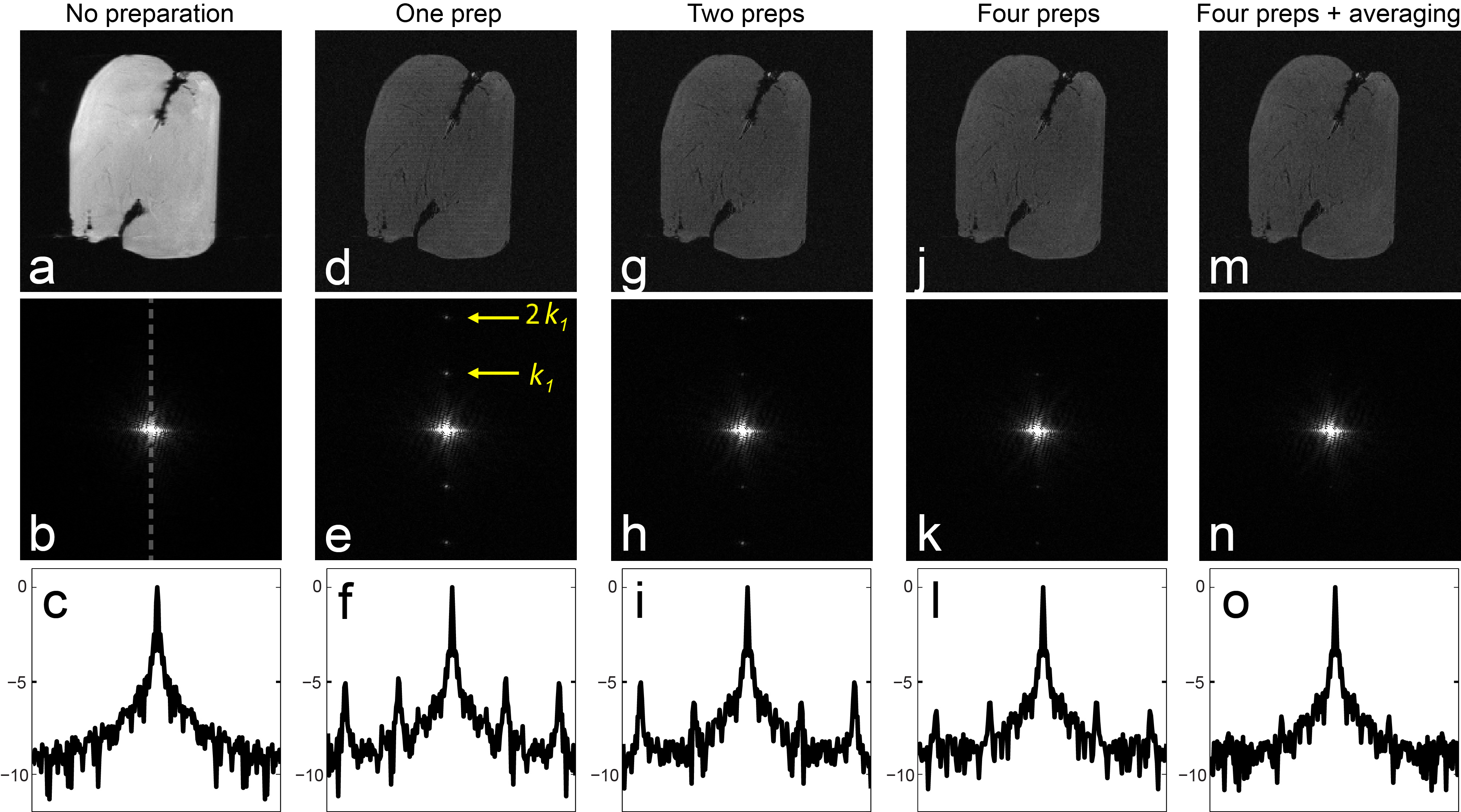
All experiments were performed on a 1.5 T clinical MR scanner (Avanto; Siemens Medical Solutions). Figure 3 shows the results of scanning a chicken breast phantom using various schemes of VS preparations. The image obtained by applying a single VS pulse (Fig. 3d) shows stripe artifact, which appears, in the corresponding k-space, as erroneous peaks at the locations of kx = k1 (1.9 cm-1) and 2k1 (3.8 cm-1) (Figs. 3e and 3f), where k1 = fundamental frequency ≡ λ-1. When the two VS preparations with spatial shifting of 0 and λ/2 were alternately used, the peak at kx = k1 was significantly reduced but the 2k1 harmonics remained nearly the same (Figs. 3g, 3h and 3i). With alternate application of the four VS preparations with 0- λ/2- λ/4 - 3λ/4 shifting, the 2k1 harmonics were significantly reduced as well. With the k-space averaging at kz = 0, the spurious k-space peaks are further suppressed and no longer visible in k-space (Figs. 3n and 3o).
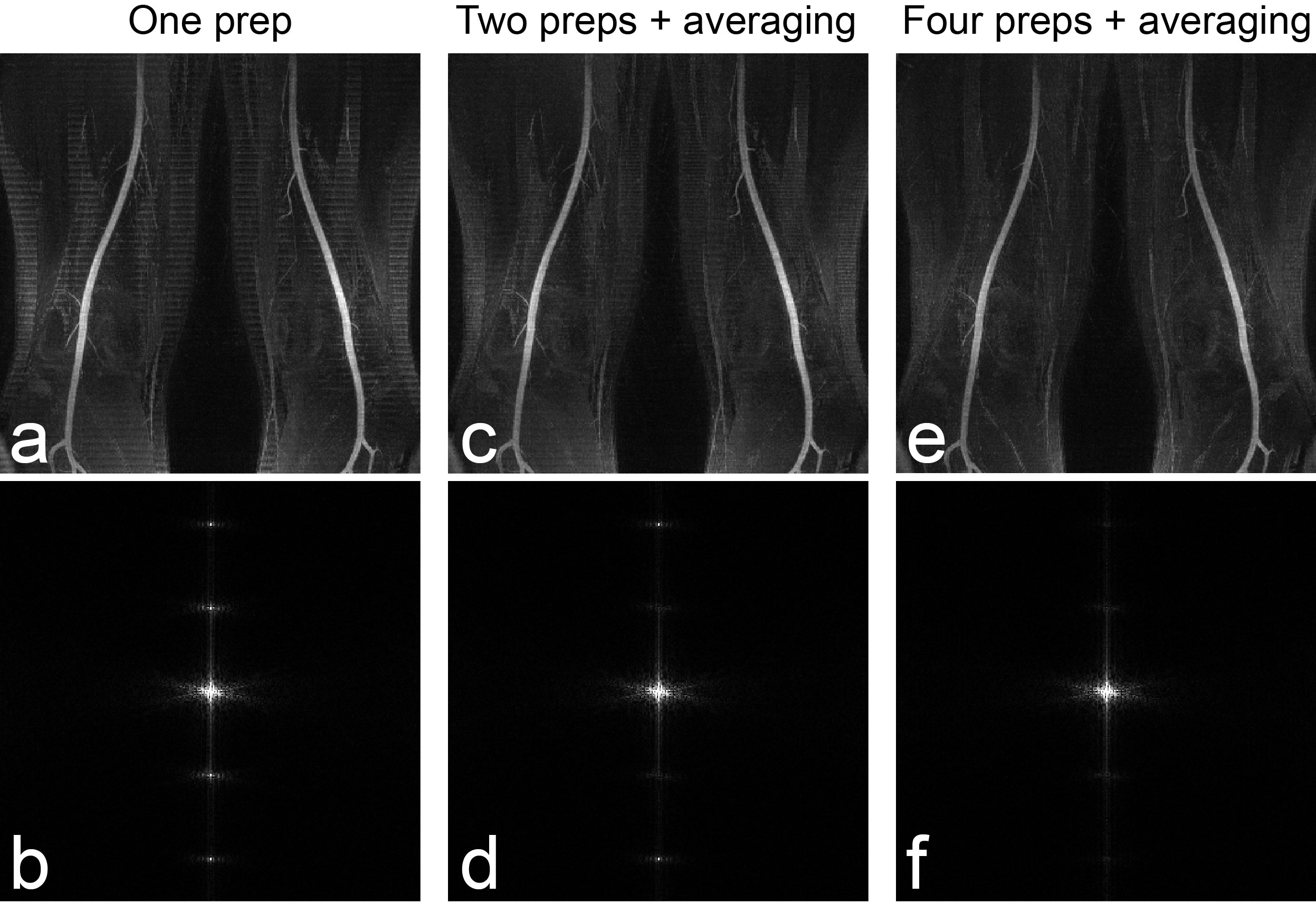
Figure 4 shows the results of VS-MRA in the thigh of a healthy subject. The alternate application of 0-λ/2 cycled VS preparations well suppressed the k harmonics but barely affected the 2k1 harmonics, which resulted in reduced, but still noticeable stripe artifact. When the 0- λ/2- λ/4 - 3λ/4 cycled VS preparations were applied alternately with the averaging at kz = 0, the stripes in image domain and the peaks at kx = 2k1 in k-space were further suppressed (Figs. 5e and 5f).
Discussion
Extended phase graph analysis has shown that the stripe artifact associated with VS magnetization prepared MRA contains high-order harmonics as well as the fundamental frequency that is determined by each unipolar VS gradient. With approximation of the stripes as superposition of the first and second order harmonics, the artifact can be suppressed by alternate application of four VS preparation pulses whose excitation profiles are spatially shifted by quarter the fundamental period of the stripes. The suppression of the artifact can be further enhanced through additional k-space averaging with only marginal increase in scan time (6 cardiac cycles).Acknowledgements
This work has been supported by NIH R01 HL135500 and Ewha Womans University Research Grant of 2017.References
1. Shin T, Worters PW, Hu BS, Nishimura DG. Non-contrast-enhanced renal and abdominal MR angiography using velocity-selective inversion preparation. Magn Reson Med 2013;69:1268-1275.
2. Shin T, Hu BS, Nishimura DG. Off-resonance-robust velocity-selective magnetization preparation for non-contrast-enhanced peripheral MR angiography. Magn Reson Med 2013;70:1229-1240.
3. Qin Q, Shin T, Schar M, Guo H, Chen H, Qiao Y. Velocity-selective magnetization-prepared non-contrast-enhanced cerebral MR angiography at 3T. Magn Reson Med 2015;75(3):1232-1241.
4. Watson DB, Grasu B, Menon R, Pensy R, Crawford RS, Shin T. Novel, non-gadolinium-enhanced magnetic resonance imaging technique of pedal artery aneurysms. J vASc Surg Cases & Innov Tech 2017;3:87-89.
5. Li W, Xu F, Schär M, Liu J, Shin T, Zhao Y, van Zijl PCM, Wasserman BA, Qiao Y, Qin Q. Whole-brain arteriography and venography: Using improved velocity-selective saturation pulse trains. Magn Reson Med 2017; Epub ahead of print (doi: 10.1002/mrm.26864).
6. Shin T, Qin Q, Park J-Y, Crawford RS, Rajagopalan S. Identification and reduction of image artifacts in non-contrast-enhanced velocity-selective peripheral angiography at 3T. Magn Reson Med 2016;76(2):466-477.
7. Hennig J. Echoes - how to generate, recognize, use or avoid them in MR-imaging sequences. Part I. Concepts Magn Reson 1991;3:125-143.
8. Hennig J. Echoes - how to generate, recognize, use or avoid them in MR-imaging sequences. Part II. Concepts Magn Reson 1991;3:179-192.
9. Scheffler K. A pictorial description of steady-states in rapid magnetic resonance imaging. Concepts Magn Reson 1999;11:291-304.
10. Weigel M, Schwenk S, Kiselev VG, Scheffler K, Hennig J. Extended phase graphs with anisotropic diffusion. J Magn Reson 2010;205:276-285.
Figures