4111
Atlas-based breathing motion correction for dynamic lung XeMRI1Department of Engineering Science, University of Oxford, Oxford, United Kingdom, 2Department of Oncology, University of Oxford, Oxford, United Kingdom, 3Department of Radiology, University of Oxford, Oxford, United Kingdom, 4Department of Biomedical Engineering, King's College London, London, United Kingdom
Synopsis
In this work we propose a framework to compensate for the breathing motion for dynamic lung XeMRI (DXeV), aligning images from different breathing phases and therefore increasing the accuracy of the ventilation analysis process. We build a lung atlas, to delineate a plausible shape of the lungs in the ventilation images, which is further used to co-register all ventilation volumes in the sequence to the reference lung atlas. After applying the proposed breathing motion correction method, the tidal breathing motion has been largely compensated and all masks of ventilation volumes correspond to each other spatially.
Introduction
Hyperpolarised 129Xe MRI (XeMRI) is a non-invasive functional imaging method, which enables assessment of regional lung function. Such imaging techniques enable the evaluation of lung ventilation in static images acquired within a single breath-hold. The recently proposed Dynamic Hyperpolarized 129Xe Ventilation (DXeV)1 imaging technique extends these capabilities to capture ventilation volumes during the breathing thus creating a temporal sequence.
During breathing, the lung volume increases and decreases as the organ inflates and deflates. This breathing motion impacts on spatial correspondence between ventilation volumes of DXeV and therefore it impedes the temporal analysis of the localized ventilation. This work proposes a framework to compensate for the breathing motion for dynamic lung XeMRI, aligning images from different breathing phases and therefore making the ventilation analysis process more accurate.
While motion compensation can be achieved through registration of lung masks, extracting such masks directly from image intensities is a challenge, since different lung regions receive inhaled gas at different times, or not at all if they are a part of a ventilation defect. Alternatives include laborious manual annotation or an additional acquisition of structural data.
Methods
We propose to build a lung atlas, to delineate a plausible shape of the lungs in the ventilation images. We further use it to co-register all ventilation volumes in the sequence to the reference lung atlas.
Atlas construction and fitting: For building the lung atlas we used 10 CT volumes form Dir-Lab dataset2, as no structural imaging data was available for the DXeV dataset. We segment the lungs from CT volumes and perform their joint registration using a Demons-based group-wise image registration method3. Subsequently, we fit the atlas to one of the ventilation volumes of the DXeV using affine image registration and further use it as a reference volume. We choose the peak inhalation ventilation volume as it provides the best definition of lung boundaries and because by representing the largest lung volume, it naturally includes the area of the lungs in all other frames. On the chosen ventilation volume we performed a thresholding followed by morphological operations to segment an initial shape of the lungs to which we fitted the atlas.
Breathing motion correction: Since the atlas was fitted to the peak inhalation volume, the remaining ventilation volumes of the lungs in the sequence should be localised inside of it. Subsequently, we perform a similar segmentation step, this time restricting the region of interest to the inside of the atlas. At this stage we compensate for the difference between the segmented lungs for each ventilation volume from the sequence and the atlas using an affine image registration method.
Results
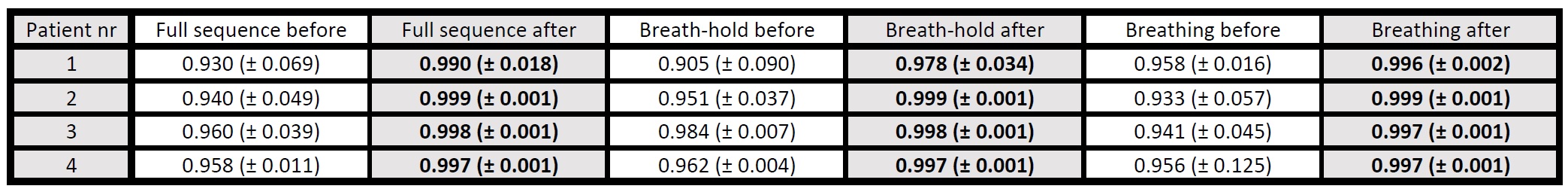
We have tested our method on a dataset of four DXeV sequences of healthy patients, each consisting of 30 volumetric ventilation images from the data presented in 1. The tidal breathing pattern presented in Fig.1 a) is mostly compensated by our method as shown in Fig.1 b), where all volumes of the lungs have visually similar shape. The results from Tab. 1 show that the proposed method results in close to 1 (average of 0.996) Dice overlap between the ventilation volumes and the reference volume.
In Fig. 2 we show the influence of the motion correction for landmarks placed inside the lungs. The reduction in the intensity of the ventilation images corresponding to the loss of the polarization of the gas in the lungs is expected to be at an approximately constant rate. However, for the lower parts of the lungs (blue and red curves) noticeable variations in the curves shapes can be observed, especially for the ventilation volumes pointed by arrows, which vary from the expected shape of the curves. The motion correction for the upper parts of the lungs (green and magenta curves) results in almost no change in the intensity distribution, as little motion is observed there.
Discussion
As shown in Fig.1 b) the proposed method compensates for the breathing motion in the dynamic sequence. The sudden changes in blue and red curves pointed by arrows in Fig. 2 b) and c), do not correspond to loss of the gas polarization or any other physical property but are caused by misalignment of the subsequent ventilation volumes in the sequence.Conclusions
In this work we have presented a framework for breathing motion correction in dynamic XeMRI. The presented results show that the analysis of the DXeV data without breathing motion correction might be prone to the spatial misalignment of the subsequent ventilation volumes and that the proposed breathing motion correction method shows a potential to compensate for such motion.Acknowledgements
A.S., B.P., J.A.S., and V.G. gratefully acknowledge funding from the CRUK and EPSRC Cancer Imaging Centre at Oxford (CICO), C5255/A16466.References
[1] Doganay Ozkan, et al. “Fast dynamic ventilation MRI of hyperpolarized 129Xe using spiral imaging”, Magnetic Resonance in Medicine, 2017. [2] Castillo Richard, et al. 2009. A framework for evaluation of deformable image registration spatial accuracy using large landmark point sets. Phys Med Biol 2009; 54; 1849-1870. [3] Papiez Bartlomiej W., et al. "Facial expression recognition using diffeomorphic image registration framework." Mathematical Methodologies in Pattern Recognition and Machine Learning. Springer, New York, NY, 2013. 179-194.Figures