4104
Self-Calibrated Soft Gating for Respiratory Resolved 3D+Time Lung ZTE1Global MR Applications and Workflow, GE Healthcare, Menlo Park, CA, United States, 2Global MR Applications and Workflow, GE Healthcare, Waukesha, WI, United States, 3Global MR Applications and Workflow, GE Healthcare, Munich, Germany
Synopsis
3D + Time lung ZTE reconstruction can provide combined anatomical imaging and motion extraction for RTP and PET/MR-type applications. In this work, we propose a soft-gating method for non-Cartesian imaging, via a solver preconditioner, to improve the sharpness and SNR of 4D ZTE. No a priori knowledge is used for the soft gating weights, as they are self-calibrated based on coil-image bin to bin similarities. The proposed soft gating method provides a SNR gain while maintaining resolution.
Introduction
The potential benefits of a high-quality 3D+Time ZTE reconstruction method are multiple: efficient quiescent lung imaging, respiratory-resolved MRAC
for motion compensated PETMR reconstruction1, and high-fidelity motion calibration for RTP. Previously2, we proposed a
motion compensated reconstruction method for lung ZTE.
A respiratory resolved reconstruction was
required to estimate the motion between the different respiratory phases. This step was performed using retrospective gating (RG). Conventional RG uses a hard-thresholding which
fully accepts data within a given acceptance windows, ignoring intra-window motion corruption, and completely rejects the rest of the data, discarding their potential
for improving reconstruction. Respiratory soft-gating (SG) has already proven its benefit in the context of Cartesian imaging3,4. Here we propose to adapt the SG
method to a non-Cartesian framework, via a solver preconditioner, to improve the sharpness and SNR of 4D lung ZTE. Introduction
Methods
Methods
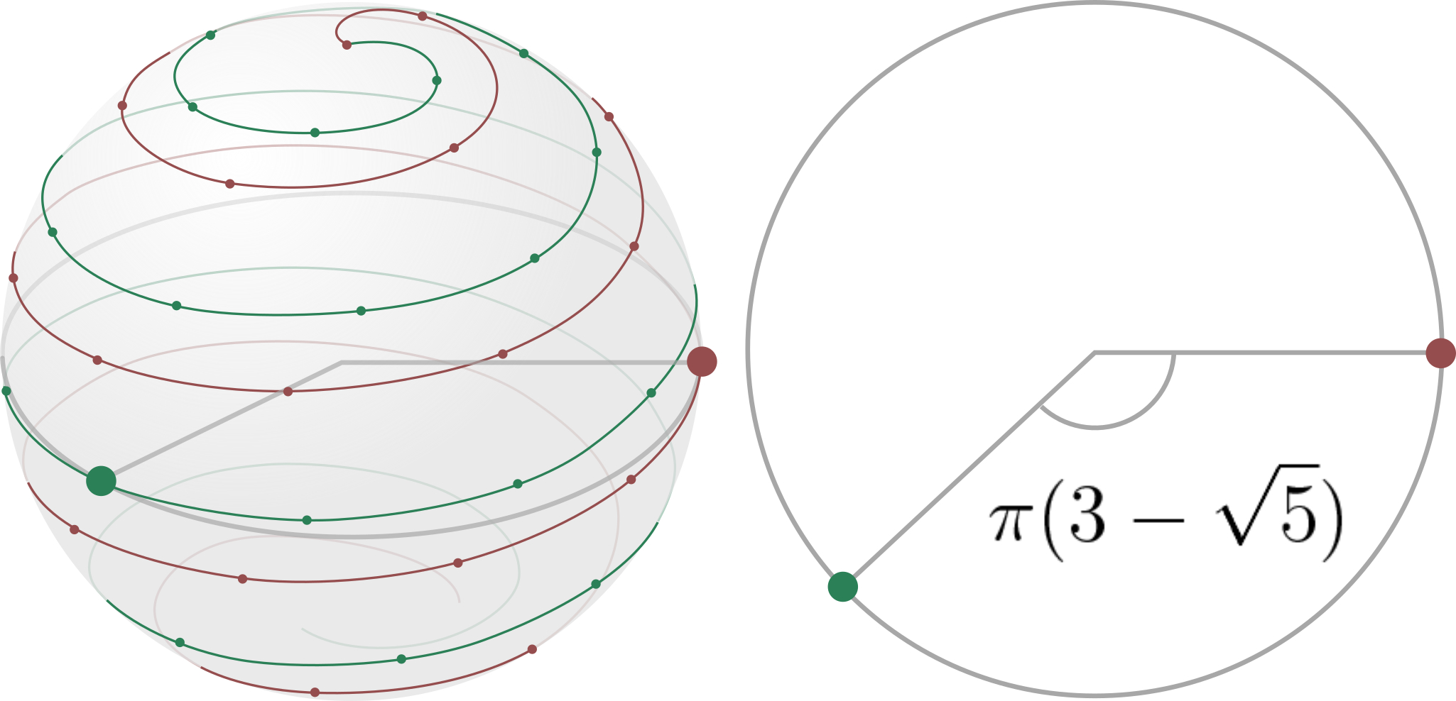
Acquisition - Two volunteers were scanned on 3T (MR750w, GE Healthcare). On each volunteer, 2 datasets were acquired with the RUFIS sequence5: in free-breathing (FB, scan time: 3min10s) and in breath-hold (BH, 26s), with a 1.1mm isotropic resolution. The trajectory (Fig.1) was optimized for retrospective gating: it is now split into multiple segments, each segment covering the k-space periphery homogeneously with a spiral. In addition, the segments were interleaved with a golden angle rotation around the z-axis such that the reconstruction of an image with any arbitrary selection of segments always corresponds to a quasi-homogeneous sampling of the k- space periphery.
Encoding operator and preconditioner – Conventionality, the reconstruction consists in solving the following inverse problem: 𝑠=𝐸𝜌, with 𝑠 the rawdata, 𝜌 the unknown image, and 𝐸=𝐺𝐹𝜎 the encoding operator composed of 𝜎 the coil sensitivities, 𝐹 the Fourier transform, and 𝐺 the gridding operator. In the case of the retrospectively gated reconstruction of the bin 𝜌𝑖, a hard sampling operator 𝛿𝑖 is added in the encoding operator 𝐸𝑖=𝛿𝑖𝐺𝐹𝜎=𝐺𝑖𝐹𝜎. Solving this problem usually requires the addition of a density correction filter to the back-projection operator 𝐸𝑖𝐻=𝜎𝐻𝐹𝐻𝐺𝑖𝐻d𝑖, where d𝑖 depends on 𝛿𝑖. This density correction can be interpreted as a preconditioner of the problem. One can note that it is equivalent to apply the density correction d𝑖 in the non-Cartesian domain or 𝐷𝑖 in the Cartesian domain after 𝐺𝑖𝐻. In the Cartesian domain, the density correction can now be precisely calibrated as follow: 𝐷𝑖=1/(𝐺𝐻𝛿𝑖𝐽), with 𝐽 the unitary matrix reflecting 𝑠.
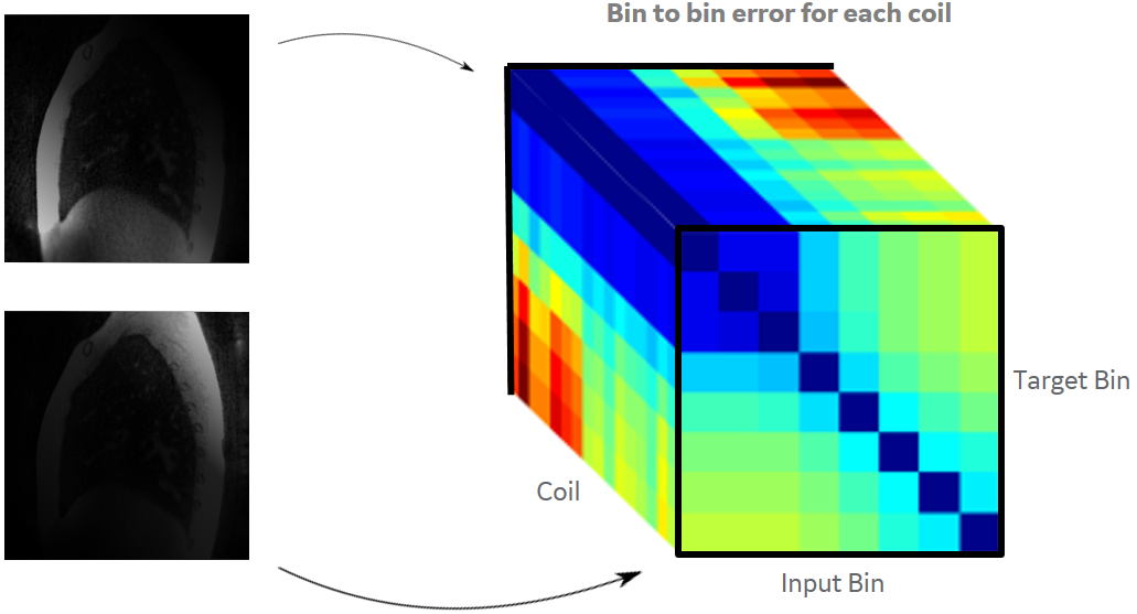
Self-calibrated soft gating – The goal of SG is to describe more accurately how well the acquired data correspond to the image to be reconstructed. For instance, data acquired in inspiration are more likely to corrupt the reconstruction of an expiration frame. In this case, a lower weight should be associated. Also, data from a posterior coil far away from the diaphragm, are less likely to be affected by motion. In this case, a higher weight could be associated. Those weights 𝑤𝑖,𝑗 can be calibrated from the data themselves by evaluating the variation of each coil image between the different respiratory phases 𝜌𝑖=1..𝑛𝐵𝑖𝑛,𝑗=1..𝑛𝐶𝑜𝑖𝑙 (Fig.2). For the 𝜌𝑖,𝑗 estimation, a fast RG relying on a belt signal and a filtered back-projection was used. The weight 𝑤𝑖,𝑗 were fitted to be inversely proportional to the error ‖𝜌𝑟𝑒𝑠𝑝,𝑗−𝜌𝑖,𝑗‖ for a data acquired in phase 𝑟𝑒𝑠𝑝. In this framework, the preconditioner becomes d𝑖,𝑗𝑊𝑖,𝑗 with the corresponding Cartesian density correction 𝐷𝑖,𝑗=1/(𝐺𝐻𝑊𝑖,𝑗𝐽).
Solver – The final respiratory resolved volumes 𝜌𝑖 were reconstructed using a primal-dual optimizer with a second-order generalized total variation (TGV)6. In addition to the SG images, the RG images, the image using the FB data without motion management, and the BH image were also reconstructed with TGV for comparison.
Results
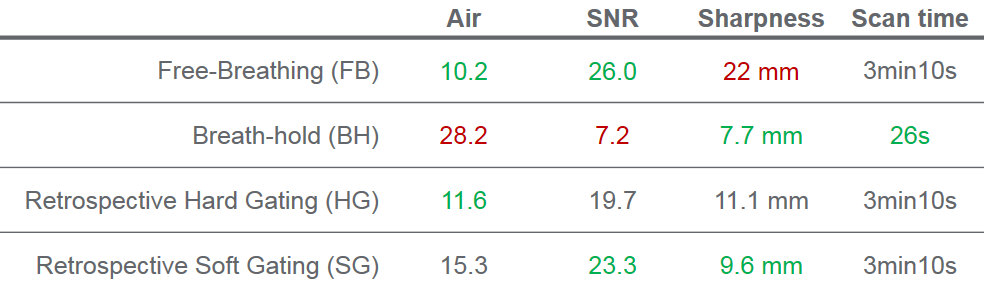
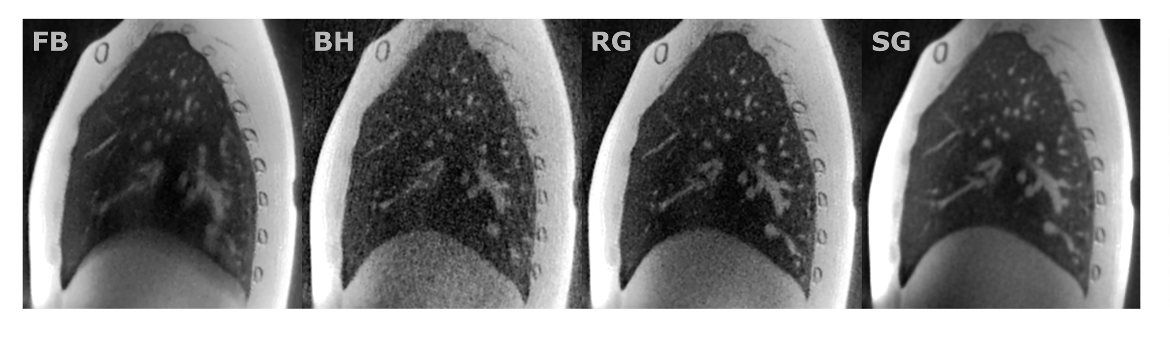
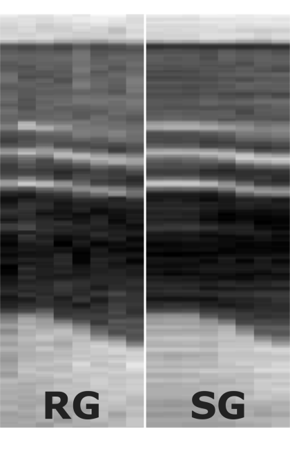
One sagittal slice of the 4 reconstructed volumes are presented in Fig.4. A vertical pencil-beam through the different respiratory phases of the RG and SG reconstruction are presented in Fig.5. In SG, the moving structures are sharper than in RG, especially for the central respiratory phases corresponding to a rapid motion (Fig.3). All the respiratory phases have a higher SNR in SG compared to RG.Discussion
Self-calibrated soft gating make the most of all the data by considering data correlation across respiratory bins and coil-specific motion. The reduction of the intra-bin inconsistency improves the sharpness and the usage of a larger amount of consistent data improves the SNR. The interesting aspect of this SG approach is that no a priori knowledge is used for the soft gating weights, since they are entirely based on the self-calibrated coil image bin to bin similarities. Also, if the quiescent phase is required alone, the reconstruction of that single phase only is needed.Acknowledgements
No acknowledgement found.References
1. Yang et al., ISMRM 2017
2. Menini et al., ISMRM 2015
3. Cheng YJ et al., JMRI 2014
4. Lai et al., ISMRM, 2017
5. Madio et al., MRM 1995
6. Knoll et al., MRM 2011
Figures