4089
Combined prospective motion correction and retrospective B0 correction for EPI using optical tracking and predictive B0 maps.1Sir Peter Mansfield Imaging Centre, School of Physics and Astronomy, University of Nottingham, Nottingham, United Kingdom, 2Philips Healthcare, Nottingham, United Kingdom
Synopsis
We demonstrate a combination of prospective motion correction and retrospective B0 unwarping using predictive field maps to improve the registration of EPI images. Prospective motion correction is performed using an optical tracking camera and custom scanner code. Field maps were generated using a multilinear fit of subject orientation using data gathered at known orientations. Results show a higher correlation when registering EPI data to a high-resolution anatomical, post-correction.
Purpose
Prospective motion correction can increase image quality by locking the image geometry to the subject’s head [1]. Volume-by-volume correction is particularly useful during fMRI acquisitions in which acquisition time is relatively short, so that inter- rather than intra- scan movement effects dominate. Prospective motion correction maintains the phase encoding direction relative to the head, allowing straightforward correction of the image warping which is prevalent in EPI. However, susceptibility induced field variation within the head is also dependent on head orientation and cannot be corrected for prospectively yet. Here, we use prospective motion correction using an optical tracking camera to correct for head motion during the scan and perform additional B0 unwarping using predicted field maps for various head movementsMethods
EPI data were acquired at 5 different head positions with prospective motion correction applied to align all volumes. Motion tracking was performed using an optical tracking camera (Metria Innovation, Milwaukee, WI) with a MPT marker fixed to a bite-bar. Prospective motion correction was performed using code for geometry adjustment running on the scanner computer. Data were acquired on a Philips 7T Achieva scanner (Philips Healthcare, Netherlands) with TE/TR=26/2000ms; FOV=210x210x45 mm3, RL phase encoding and SENSE factor 2. Subjects were asked to hold 5 poses; resting, half nod, half shake, full nod and full shake position for 5 volume acquisitions. B0 maps were also acquired independently at 10 different head positions using a dual-echo sequence with TE/ΔTE = 5.6/1.0ms; TR 25ms and a FOV of 256x160x192 mm3. The change of head position was measured using the MPT system. Magnitude image data acquired during each field mapping experiment were registered to the resting position in MATLAB and the resulting transform was applied to the corresponding B0 maps. Each voxel of the B0 map was fitted as a function of pitch, roll, yaw and a constant, using a multilinear fit [2]. A predicted B0 map for each position in the EPI scan was then created. The distorted EPI images were corrected using the predicted B0 map with FSL [3] [4]. Both the corrected and uncorrected EPI images were then registered using a rigid body transform to the anatomical image and the correlation coefficient was measured.Results
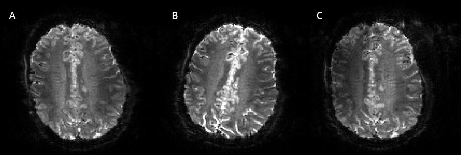
Figure 1 shows the EPI data acquired with prospective motion correction applied. A delay in the PMC geometry update allows for the volume directly after movement to be imaged before the geometry update is applied.
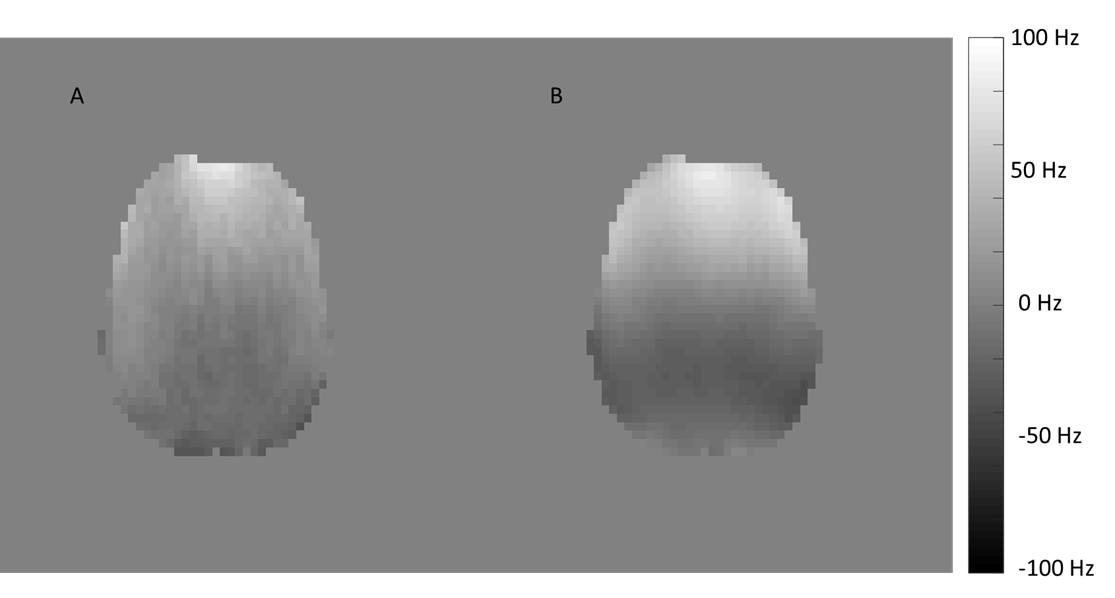
A B0 map acquired in the half nod position is shown in Figure 2 along with the predicted field map. A total of 10 B0 maps were acquired over a range of approximately 15o of nod, 10o of shake and 10o head roll.
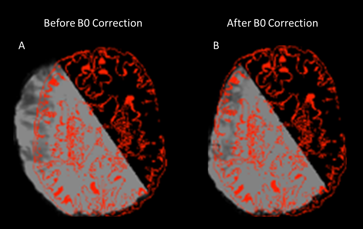
Figure 3 shows the improved registration to the high-resolution anatomical after B0 unwarping using boundary based registration.
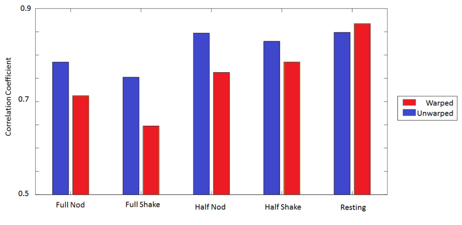
A graph of the correlation coefficients between the distorted and undistorted EPI images to the anatomical are shown in Figure 4.
Discussion
Figure 1 demonstrates the prospective motion correction for a large head shake. Signal loss in Figure 1(C) is most likely due to the change in coil sensitivity profile affecting SENSE reconstruction. Brain asymmetry as a result of B0 inhomogeneity within the head is also shown. The predictive field map matches closely with the true field map in the half nod position, with typical differences in the order of 10Hz. B0 unwarping shown in Figure 3 increases the accuracy of the registration of the anatomical to the EPI as measured by the correlation coefficient. This was shown to be the case in all positions with the exception of the resting position where there was a small decrease in correlation (Figure 4). As the size of the movement increases the correlation between the EPI and anatomical decreases probably due to the larger warping due to gradient non linearity, however predictive field maps are still able to offer a notable improvement. It is likely that for large movements the linear approximation between measured angle and actual B0 begins to break down. With further analysis it may be possible to extend the viable range of the predictive field maps by characterising fields at high angles of rotation and to predict the minimum number of required field maps.Conclusion
We have demonstrated the ability to restore image quality of EPI data using a combination of prospective motion correction and B0 unwarping. Only a limited number of B0 maps at the start of the scanning session are required to create predictive field maps, which in turn allows for correction of fMRI data corrupted by motion without increasing fMRI scan time.Acknowledgements
This work was supported by funding from the Engineering and Physical Sciences Research Council (EPSRC) and Medical Research Council (MRC) [grant number EP/L016052/1].References
[1] J. Maclaren, B. S. Armstrong, R. T. Barrows, K. Danishad, T. Ernst, C. L. Foster, K. Gumus, M. Herbst, I. Y. Kadashevich and T. P. Kusik, “Measurement and correction of microscopic head motion during magnetic resonance imaging of the brain,” PLoS One, vol. 7, no. 11, p. e48088, 2012.
[2] A. Sulikowska, Motion correction in high-field MRI, PhD Thesis: University of Nottingham, 2016.
[3] M. Woolrich, S. Jbabdi, B. Patenaude, M. Chappell, S. Makni, T. Behrens, C. Beckmann, M. Jenkinson and S. Smith, “Bayesian Analysis of Neuroimaging Data in FSL,” NeuroImage, vol. 45, no. 1, pp. S173 - S186, 2009.
[4] P. Jezzard and R. S. Balaban, “Correction for Geometric Distortion in Echo Planar Images from b0 Field Variations,” Magnetic resonance in medicine, vol. 34, no. 1, pp. 65 - 73, 1995.
Figures