4087
Revealing sub-voxel motions of brain tissue using phase-based amplified MRI (aMRI)1Radiology, Stanford University, Stanford, CA, United States, 2Stevens Institute of Technology, Hoboken, NJ, United States, 3Department of Anatomy and Medical Imaging, University of Auckland, Auckland, New Zealand
Synopsis
Problem:Amplified Magnetic Resonance Imaging (aMRI) was recently introduced as a new brain motion detection and visualization method. In this work we strive to improve aMRI by incorporating a phase-based motion amplification algorithm.Methods:Phase-based aMRI was developed, validate using digital phantom simulations and compared with EVM-based aMRI in healthy volunteers at 3T. Data were also acquired on a patient with Chiari I malformation, and displacement maps were produced using free form deformation (FFD) of the aMRI output.
Results:Phantom simulations showed that phase-based aMRI has a linear dependence of amplified displacement on true displacement. Amplification was independent of temporal frequency, phantom size, Rician noise, and partial volume effect, but had a slight dependence on phantom shape. Phase-based aMRI supported larger amplification factors than EVM-based aMRI, and was less sensitive to noise and artifacts. Abnormal biomechanics were seen on FFD maps of the Chiari I malformation patient.Conclusion: Phase-based aMRI can be used for quantitative analysis of minute changes in brain motion, and may reveal subtle physiological variations of the brain due to pathology. Preliminary data shows the potential of phase-based aMRI to assess abnormal biomechanics in Chiari I malformation.
Introduction:Amplified Magnetic Resonance Imaging (aMRI) was introduced as a new brain motion detection and visualization method1. The original aMRI approach used a video-processing algorithm, Eulerian Video Magnification (EVM)2 , to amplify cardio-ballistic motion in retrospectively cardiac-gated MRI data. We introduced a more advanced aMRI method based on phase-based video motion processing3. Here we compare EVM-based and phase-based aMRI; validate phase-based aMRI using digital phantoms; and show that the new method can be quantified to enable accurate displacement measurements. Using the free form deformation (FFD) image registration algorithm4,5 ,normalized average displacements maps are produced from Chiari I malformation pediatric patients to demonstrate the efficacy of phase-based aMRI in detecting brain motion abnormalities.
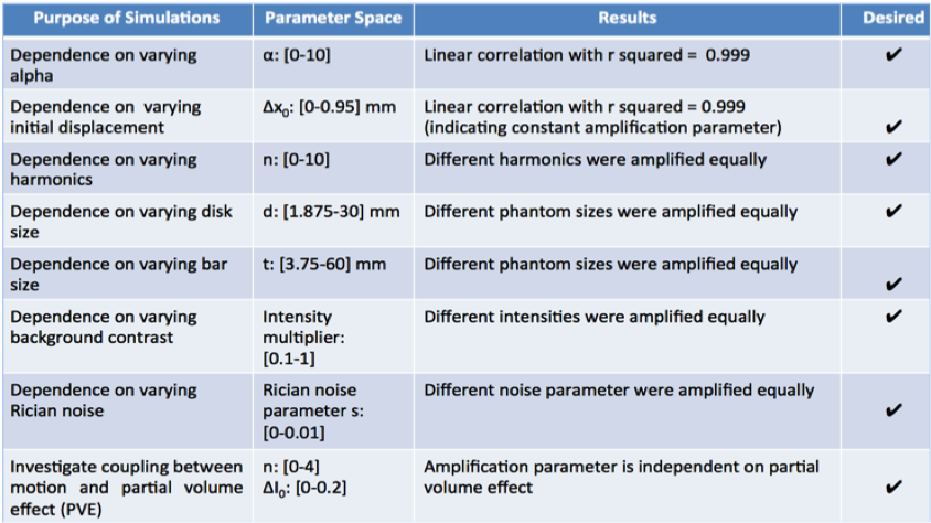
Method:Image acquisition:Scans were performed on volunteers at 3T (GE MR750) using an 8-channel head coil. A mid-sagittal slice of a normal brain (adult volunteer 29yr/F) was acquired using a 2D cardiac-gated bSSFP sequence (cine MRI), using the following parameters: matrix size=2242, flip angle=45°, TR/TE=3.6/1.3ms; FOV=22mm2; slice-thickness=4mm, acceleration factor 2, 6 views/segment, scan time ~1min, and retrospective re-binning to 150 cardiac phases/frames. Data were also acquired on 2 patients during clinical diagnostic scans: one with abnormal anatomy (4Y male), clinical symptoms of headaches and cerebellar ataxia, and obstructive features at the craniocervical junction, including peg-like, low-lying cerebellar tonsils ( below the foramen magnum), associated with Chiari I malformation and basilar invagination; and one control with normal structural MRI (3Y male).Motion amplification:With cine MRI images as input, phase-based aMRI decomposes the images into scale and orientation components using a linear steerable pyramid3. The local phase is computed over time for each component, and is then temporally bandpass-filtered to extract motion at different spatial scales and orientations. Filtered phases are multiplied by an amplification parameter and added to the original phase components, and then reconstructed, resulting in a movie with amplification of motion within the desirable frequency range. Normalized temporal variance maps, noise maps, and displacements maps were produced. Phantom Simulations:Two Gaussian-filtered 2D digital phantoms, a vertical bar (mimicking CSF around the midbrain and spinal cord) with width d and a disc (mimicking the cerebellum) with radius r, were simulated in MATLAB6. The phantom moved over time with 1D sinusoidal displacement. Rician noise was added, along with sinusoidal intensity variations to emulate partial volume effect (PVE). Amplified displacement was measured using a custom algorithm6 ,and overall amplification factor calculated as peak amplified displacement divided by the original displacement. The dependence of amplification on original displacement, harmonic number, phantom size, noise, and PVE was investigated.
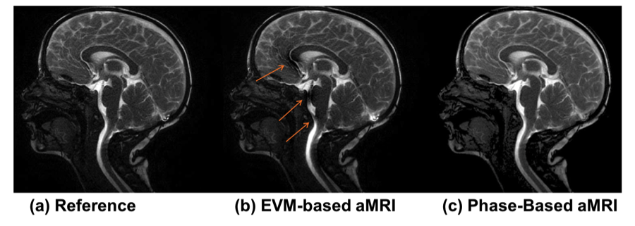
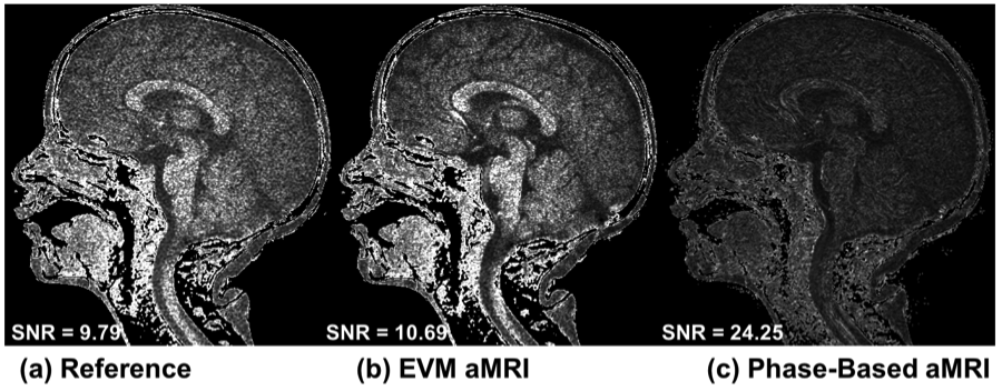
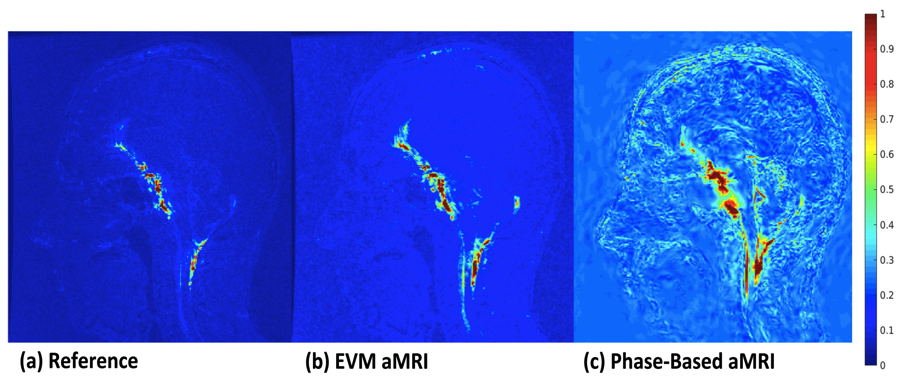
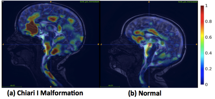
Results & Discussion:Phantom Simulations:As shown in Figure 1, phase-based aMRI demonstrated several ideal behaviors including: 1) linear dependence of amplified displacement on true displacement (i.e. constant amplification), 2) independence of amplification on temporal frequency, phantom size, Rician noise, and partial volume effect. We also observed non-ideal, slight dependence of amplification on phantom shape (standard deviation of the amplification factor was ~5% of the mean). Phase-based versus EVM-based aMRI:Phase-based aMRI was found to be less sensitive to artifacts compared to EVM-based aMRI, showing superior image quality, with an overall reduction in shading over the cerebral cortex and spinal cord, and fewer CSF flow artifacts (Figure 2). In addition, it was found to be less sensitive to noise , as shown in Figure 3. While movement of the brain was barely perceptible in the reference data, it was significantly amplified in both the EVM-based and phase-based aMRI output data, especially near the brainstem, cerebellum and spinal cord. However, phase-based aMRI revealed microscopic displacement that was not seen in EVM-based aMRI (Figure 4). Patient data:For both the patient and normal control, the input cine images displayed near-imperceptible motion, while the aMRI output showed amplified motion, particularly near the midbrain, frontal lobe, spinal cord, and cerebellum (https://web.stanford.edu/~iterem/Chiari_Vr_Normal.mov). Compared with the normal patient, the aMRI output from the Chiari I Malformation patient showed increased caudal midbrain tissue displacement, and downward displacement at the level of the brainstem and craniocervical junction. FFD maps produced from the aMRI data for both patients (Figure 5) revealed considerable differences in displacement in regions of the spinal cord, midbrain, and cerebellum.
Conclusion:This study presents a new phase-based aMRI method that enables more accurate quantification of motion, with fewer noise and artifacts and than EVM-based aMRI. Phase-based aMRI amplifies minute changes in brain motion and may provides a means of revealing subtle physiological variations of the brain due to pathology. Preliminary data shows the potential of phased-based aMRI to assess abnormal biomechanics in patients with Chiari I malformation. Application of aMRI to clinical in vivo assessment of brain biomechanical properties has the potential for a wide-range of clinical implications, such as brain injury, hydrocephalus, and other conditions associated with abnormal intracranial pressure.
Acknowledgements
Grant support and other assistance: NIH (1R21HD083803-01), Stanford-Philips Research Agreement, Stanford Interdisciplinary Graduate Fellowship, Center of Advanced MR Technology at Stanford (P41 EB015891), and the Lucas Foundation. Thanks to the LPCH technologists for their help with patient studies. The authors are grateful to Michael Iv, Tej Azad, Gerald Grant, and Samuel Cheshier for helpful clinical guidance. Thanks also to Julian Maclaren, Mahdi Salmani Rahimi, and Rachelle Bitton for helpful discussions.References
1. Holdsworth SJ, Salmani Rahimi M, Ni WW, Zaharchuk G, Moseley ME. “Amplified Magnetic Resonance Imaging (aMRI)”. Magn Reson Med; 75(6):2245-54.
2. Wu H, Rubinstein M, Shih E, Guttag J, Durand F, Freeman WT. “Eulerian video magnification for revealing subtle changes in the world”. ACM Trans Graph 2012;31:4.
3. Wadhwa N, Rubinstein M, Durand F, William T. “Phase-Based Motion processing”. ACM Trans Graph, 2014.
4. Rueckert D, Sonoda LI, Hayes C; Hill DLG, Leach MO, Hawkes DJ. “Nonrigid registration using free-form deformations: Application to breast MR images”. IEEE Transactions on Medical Imaging. 1999;18(8):712–721.
5. Modat M, Ridgway GR, Taylor ZA, Lehmann M, Barnes J, Hawkes DJ, Fox NC, Ourselin S. “Fast free-form deformation using graphics processing units”. Computer Methods And Programs In Biomedicine. 2010; 98(3): 278–284.
6. Ni W, Goubran M, Zaharchuk G, Moseley ME, Yeom K,Holdsworth SJ. “From Visualization to Quantification: Calibrating Motion Magnification by Amplified Magnetic Resonance Imaging”. In: 25th Annual Meeting of the ISMRM. Honolulu, Hawaii; 2017:777.
Figures