4059
Experience with 7 Tesla MRI of human subjects with passive implants and tattoos: an update1Erwin L. Hahn Institute for MRI, University Duisburg-Essen, Essen, Germany, 2Hamm-Lippstadt University of Applied Sciences, Hamm, Germany, 3Medical Physics in Radiology, German Cancer Research Center (DKFZ), Heidelberg, Germany, 4High Field and Hybrid MR Imaging, University Hospital Essen, Essen, Germany
Synopsis
In this retrospective study, questionnaires and screening forms were analyzed to identify all subjects with implants and/or tattoos cleared for imaging at our passively shielded 7T whole-body MR system. Over the past 11 years, 496 out of 2370 healthy volunteers and volunteers with known pathologies had implants and/or tattoos and underwent a 7T MR examination. None of the subjects reported any discomfort related to heating or force during or after imaging. No findings regarding safety occurred and all examinations could be safely performed.
Introduction
With a recently obtained CE label and a 510(k) approval by the FDA, the latest generation of 7T MR systems is currently emerging as a diagnostic modality for neuroradiology and musculoskeletal imaging. In addition, the number of clinically oriented research studies at 7T has increased considerably over the last decade 1. On the other hand, many subjects present with implanted medical devices (IMD), but almost none has been labeled MR conditional up to 7T magnetic field strength 1. Many 7T research centers conservatively exclude all subjects with implants or tattoos, regardless of type or location. The German Ultra-high field (UHF) network GUFI has recently published a recommendation for the inclusion of subjects with passive IMDs, stating that safe imaging at UHF can be performed if the IMD is 3T MR conditional and outside an RF coil-dependent safety area during the 7T exam 2. This study presents an update of our 2015 reported experience in imaging patients and healthy volunteers with implants and tattoos at 7T 3.Methods
Questionnaires and screening forms from May 2014 to September 2017 were analyzed retrospectively to identify all subjects with implants and/or tattoos cleared for 7T imaging at our institution (Magnetom 7T, type 900 passively shielded, Siemens Healthcare, Germany). These datasets were added to the results published earlier in 2015 3 to report now over a full period of 11 years (October 2006 – September 2017).
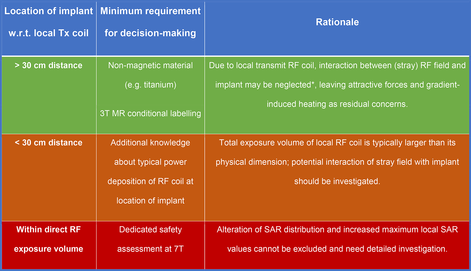
A panel of three physicists and engineers with extensive experience in MRI and RF interference cleared all subjects/implants for 7T on a case-by-case basis. The types of implants (material, dimension, geometry) were carefully examined as well as their location with respect to the exposure volume of the local RF transmit coil of each study. The decision-making process was basically divided into three categories which are explained in Figure 1. Here, a distance of 30 cm between an implant and the physical extent of the local RF coil was used to define the categories, as this distance yielded almost no stray RF fields at the location of the implant for the local RF coil. Simplified rules were applied solely for dental implants (retainer wires, bridges and crowns, inlays and pivot teeth) and tattoos (drawn within European Union after year 2000), where clearance from the safety panel was not mandatory to scan the subject. Most of the subjects were imaged at 1.5T or 3T prior to the 7T examination.
Results
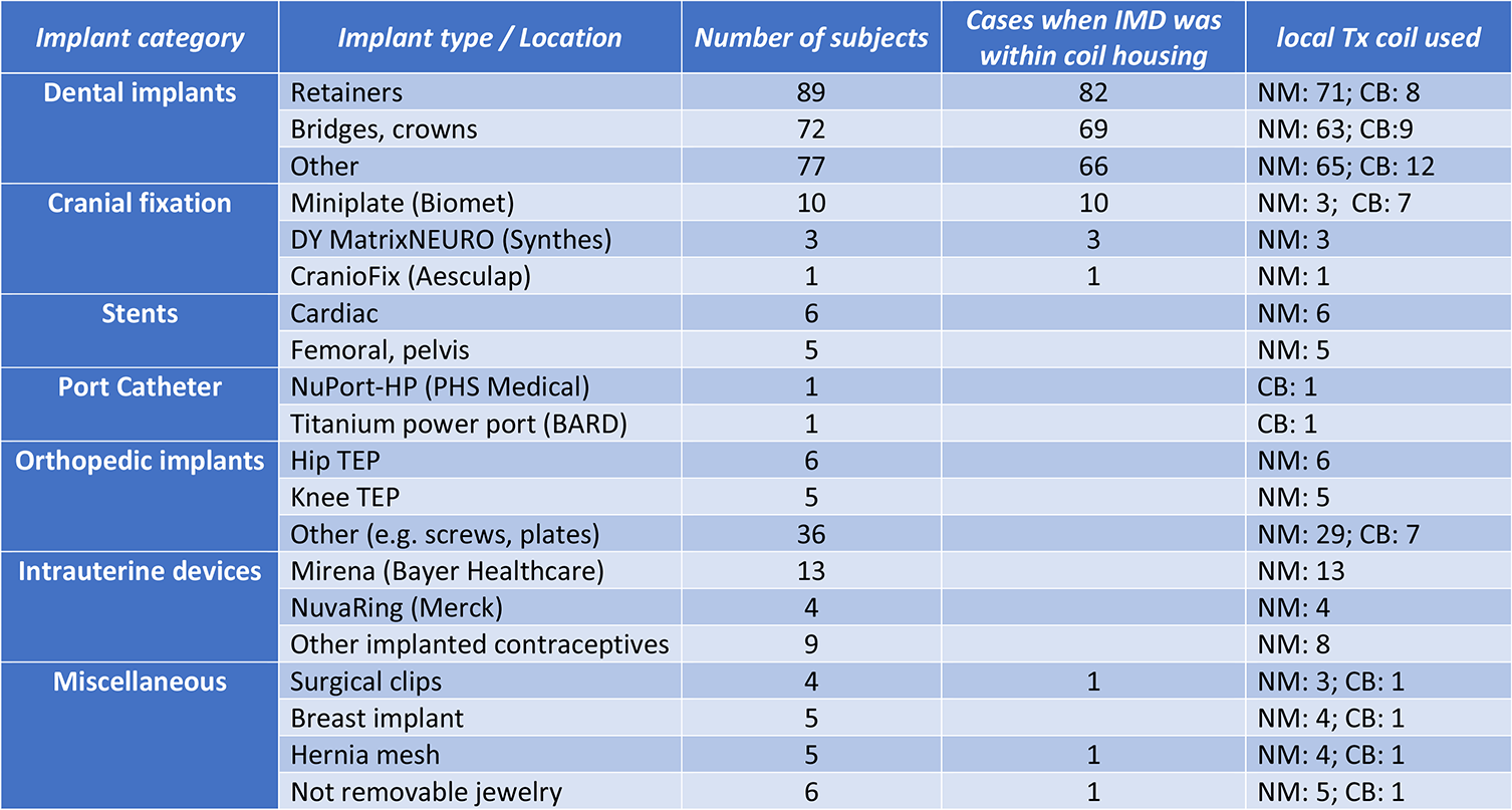
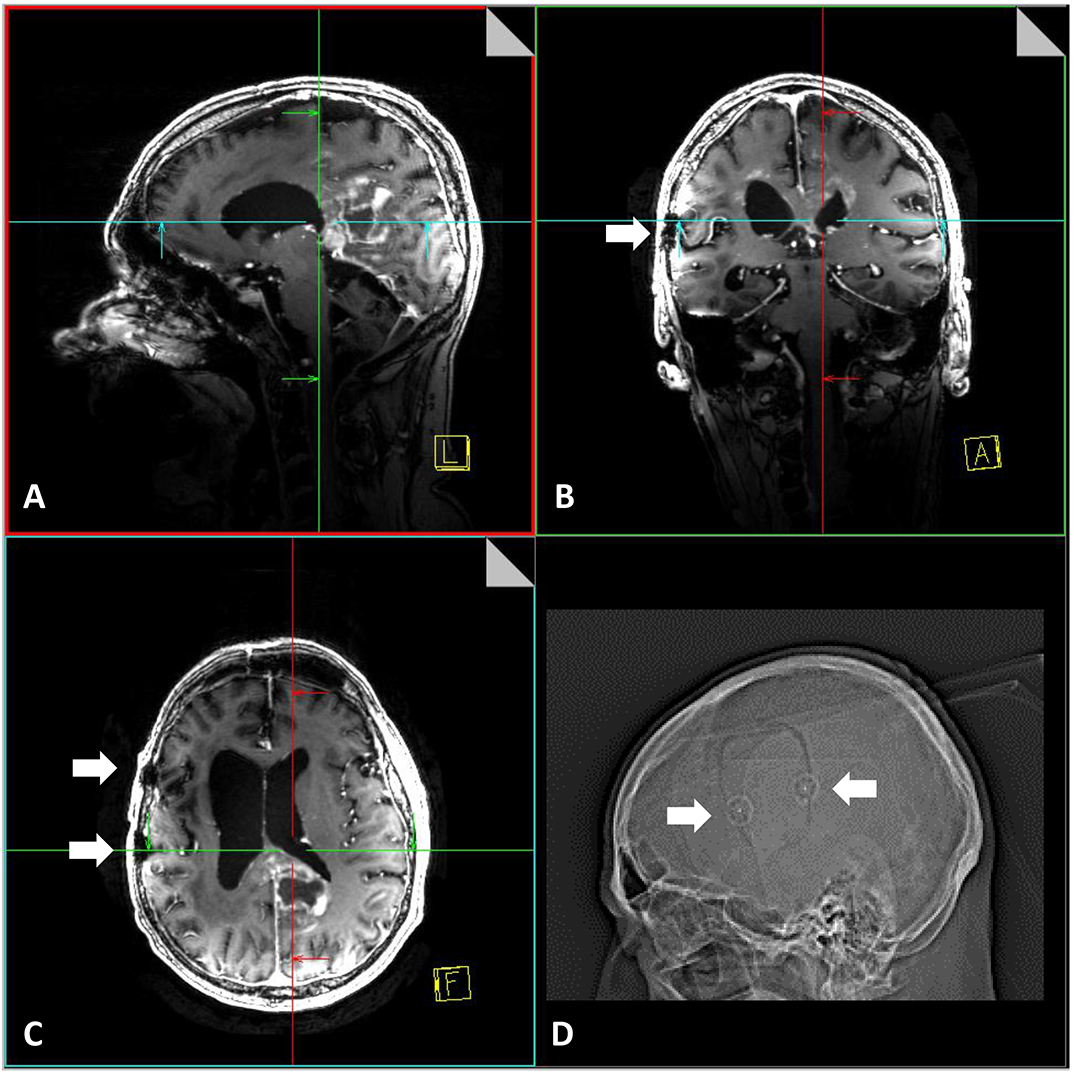
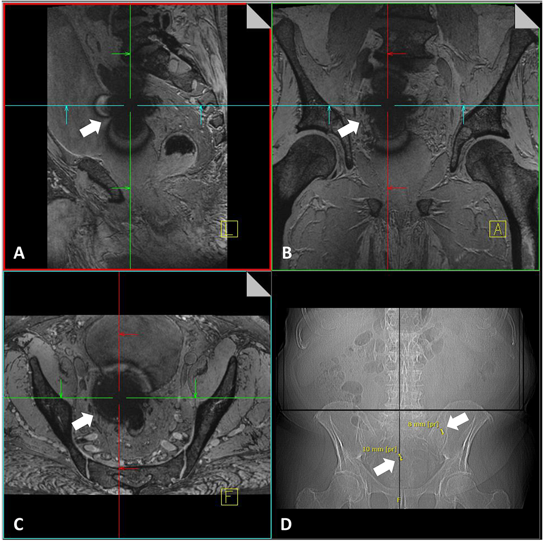
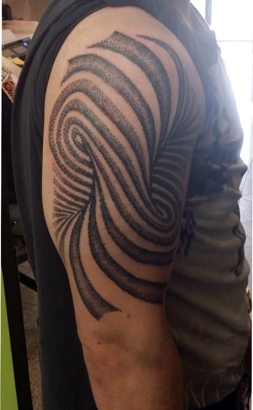
In total, 496 out of 2370 healthy volunteers and volunteers with known pathologies had implants and/or tattoos and underwent a 7T MR examination. Of the 496 subjects, 184 presented with one or several tattoos and 273 reported one or several implants; 39 subjects had both tattoos and implants. None of the subjects reported any discomfort related to heating or force during or after imaging. Please refer to Figure 2 for more details. For the subjects with implants, the majority had dental implants (230). Thereof, imaging was performed in 82 subjects with retainer wires within a head coil. Cranial osteosynthesis plates were present within a head coil in 14 subjects (Figure 3) 7,8. In 11 subjects scheduled for head imaging, total endoprostheses were reported for the hip joint (6) and knee joint (5). In 1 subject with surgical clips within a flexible body coil (Figure 4) and in 3 subjects with multiple cardiac stents 9 in close proximity to the head coil, clearance for imaging was restricted to low-SAR sequences only. Figure 5 shows an example of a large tattoo.Discussion
Since 7T examinations are not yet medically indicated and since there is only limited information available about the safety of implants at 7T, imaging should only be performed in carefully selected volunteers and after acquirement and evaluation of substantial information about the implants. The region of interest of the scan should be compared with the exact location of the implant. This is particularly relevant at 7T since only local transmit coils are used and RF heating is more probable in the exposure volume of the RF transmit coil than elsewhere. At our institution, a case-by-case decision was made for all subjects with implants, depending on size, geometry, and location of the implant with respect to the RF coil. However, for dental implants and tattoos a safe history of use has led to a more straightforward inclusion criteria. Our experience indicates that the exclusion of all subjects with implants from 7T examinations is not compulsory, and the examination of certain subjects with implants can be performed after careful evaluation of implant type and location.Acknowledgements
No acknowledgement found.References
1. Kraff O, et al., MRI at 7 Tesla and above: demonstrated and potential capabilities. JMRI 2015.
2. GUFI recommendation on “Approval of subjects for measurements at ultra-high-field MRI”, version 01/21.01.2016, http://www.mr-gufi.de/index.php/en/documents
3. Noureddine Y, et al., Experience with magnetic resonance imaging of human subjects with passive implants and tattoos at 7 T: a retrospective study. MAGMA 2015.
4. Orzada S, et al., 8-Channel Transmit/receive Head Coil for 7T Human Imaging Using Intrinsically Decoupled Strip Line Elements with Meanders. Proc. ISMRM, 2009, abstract 3010.
5. Orzada S, et al., A Flexible 8-Channel Transmit/receive Body Coil for 7 T Human Imaging. Proc. ISMRM, 2009, abstract 2999.
6. Rietsch S, et al., An 8-channel transceiver 7-channel receive RF coil setup for high SNR ultrahigh-field MRI of the shoulder at 7T. Med Phys 2017.
7. Kraff O, et al., MR safety assessment of potential RF heating from cranial fixation plates at 7 T. Med Phys. 2013.
8. Chen B, et al., Cranial fixation plates in cerebral magnetic resonance imaging: a 3 and 7 Tesla in vivo image quality study. MAGMA. 2016.
9. Winter L, et al., On the RF heating of coronary stents at 7.0 Tesla MRI. MRM 2015.
Figures