4052
Development of an 8-channel head array for MRI guided monkey ultrasound stimulation1Lauterbur Imaging Research Center, Shenzhen Institutes of Advanced Technology, Chinese Academy of Sciences, Shenzhen, China, 2Shenzhen Key Laboratory for MRI, Shenzhen, China, 3High-Field Magnetic Resonance Brain Imaging Key Laboratory of Sichuan Province, School of Life Science and Technology, University of Electronic Science and Technology of China, Chengdu, China, 4Department of Radiology and Biomedical Imaging, University of California San Francisco, San Francisco, CA, United States, 5UCSF/UC Berkeley Joint Graduate Group in Bioengineering, San Francisco, CA, United States
Synopsis
For flexible operating the ultrasound probe in a monkey model, all three instruments, stereotaxic instrument, ultrasound probe and monkey RF coil, must match to each other. In this study, an 8-channel monkey coil was custom-designed to fit the specific stereotaxic instrument, ultrasound probe, and the rhesus monkey’s head in order to get a high quality images and efficient ultrasound operation. Compared to the commercial small flexible coil array, the custom-designed monkey coil provides higher SNR in all three orientations, higher spatial resolution in vivo, and better parallel imaging capability.
Introduction
With the emergence of ultrasonic neuromodulation, the issues on the localization of ultrasound focus and the brain responses to stimulation have attracted more attentions in this field.[1,2,3] MRI sequences, including acoustic radiation force imaging (ARFI) and BOLD fMRI facilitated the accurate and real-time target localization as well as the whole-brain scanning of brain reaction in in-vivo animal studies, including non-human primate experiments.[4] In this work, an 8-channel receive monkey coil equipped with a stereotaxic module for ultrasound transducer was developed. High SNR and spatial resolution in both phantom and in-invo studies were obtained.Methods
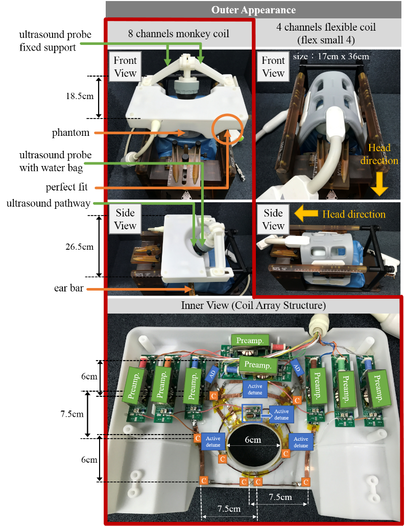
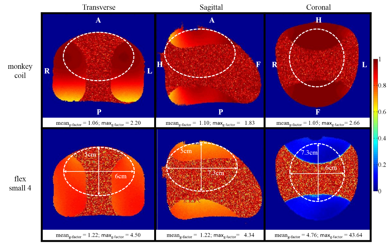
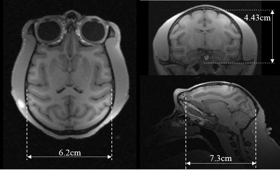
The customized ultrasound guided monkey RF coil (monkey coil) was made to fit both MRI stereotaxic instrument (KOPF 1430M) and a single-array ultrasound transducer with 55mm diameter and 60mm length (water bag included), as shown in Figure 1. The monkey coil meets the need for perfect fitting on the stereotaxic instrument, and ensures the ultrasound transducer could be steadily placed on the monkey coil. The ultrasound probe fixed support allows the probe to rotate and further more focus on different spot. In order to avoid interfere the pathway of ultrasound, a 6cm whole was cut at the bottom of ultrasound probe. All structures were printed by 3D printing using photosensitive resin. The monkey coil was made by 8 coil elements, and was arranged in three rows with number 2,3,3 in order from head to feet direction. Each coil was made with size 6.5×7.5cm2 in average, and has one active detune board and two capacitance boards included. Three boards separate the coil into three equal length and was combined by 6mm thick copper sheet. All coils were tuned on 123.2 MHz, and were well-decoupled by using over-lapping area and low noise impedance preamplifier. Phantom study: Siemens 3T Tim Trio system was used for all on-system studies, and the 4 channels small flexible coil (flex small 4) was used for comparison. A customized phantom was made to mimic the shape and size of a rhesus monkey’s head, and was design as total 432cm2 for content volume. The phantom was also printed by photosensitive resin using 3D printer and was filled with (3.75g NISO4×6H2O + 5g NACL)/1000g H2O. As for simulating the monkey coil experience, phantom was placed at the same spot of fitting monkey’s head. For fairly evaluate the performance of coil, experience was hold on without the ultrasound probe. To acquire the signal, gradient echo(GRE) sequence was used for all three orientations with following parameters: TR/TE= 300/10ms, slice thickness = 3mm, FOV = 110×110mm2, acquisition matrix = 256×256, flip angle = 60 degree, bandwidth = 130 Hz/pixel, number of average = 1. Noise on the other hand, was acquired with the same sequence by setting the power to zero. Data analysis was calculated by using MATLAB. A method called root sum of squares (sos) from reference[5] was used for SNR maps calculation. Inverse g-factor maps were acquired with acceleration factor=2. The accelerate direction was set from right to left on transverse orientation, from anterior to posterior on sagittal orientation and from head to feet on coronal orientation. In-vivo study: T1-weighted 3D MPRAGE sequence was scanned on a 5-year-old male rhesus monkey (weighted 5.5kg) for anatomic images with following parameters: TR/TE/TI = 2100ms /2.8 ms/1100 ms, FOV = 160×160×88 mm3, matrix = 320×320×176, isotropic resolution 0.5×0.5×0.5mm3, flip angle = 8 °, bandwidth = 200 Hz/pixel, echo spacing = 8.3 ms, number of average = 1.Results
Figure 2 shows the 2D SNR maps and 1D SNR profiles of both monkey coil and flex small 4 on three orientations. The white-dash-lines corresponds the position of SNR curves from each orientation, and the brain zone was measured from the rhesus monkey in the in-vivo study. Based on the SNR profiles, monkey coil improves 53% of SNR in transverse orientation, 149% in sagittal orientation, and 237% in coronal orientation. The inverse g-factor maps were shown in Figure 3. The mean and maximum of g-factor value of the brain zone were indicated at the bottom of related image. The results show that the monkey coil has a better accelerate ability than flex small 4. From Figure 4, monkey coil is capable to provide high and clear spatial resolution (0.5×0.5×0.5mm3) in in-vivo study.Conclusion/Discussion
The custom-designed monkey coil not only has a user-friendly outfit, could well fit on the specific stereotaxic instrument and perfect match with ultrasound transducer, but also provide fine performance, includes: high SNR 2D images, high spatial resolution (0.5×0.5×0.5mm3), and better parallel imaging capability.Acknowledgements
This work was supported in part by NSFC under Grant No. 61571433,81527901,81527901, 81327801and 81627901. Guangdong Province grants 2014A030313691, 2015B020214006 and 2014B030301013. Youth Innovation Promotion Association of CAS No. 2017415, city grants KQJSCX20160301143250 and JCYJ20170413161314734 and a Pengcheng Scholar Award.References
[1] Ryan Alkins, et al., Cancer Research 2013, 73(6): 1892-1899.
[2] Xuegang Xin, et al., Magn. Reson. Eng.2010, 37B(4): 237-244.
[3] Xiao Chen, et al., An ultrasound compatible rat RF array for MRI guided high intensity focused ultrasound. 24th ISMRM, 2016, 1751
[4] McDannold N, et al. MRI‐guided focused ultrasound surgery in the brain: Tests in a primate model[J]. Magnetic resonance in medicine, 2003, 49(6): 1188-1191.
[5] B. Keil et al., JMR 2013, 229, 75-89.
Figures