4025

Exploring Glutamate and Glutamine Metabolism Across Breast Cancer Subtypes using High-resolution Proton MRS Combined with Molecular Approaches
Caitlin M Tressler1, Vinay Ayyappan2, and Kristine Glunde2

1Russell H. Morgan Department of Radiology, Johns Hopkins University School of Medicine, Baltimore, MD, United States, 2Johns Hopkins University School of Medicine, Baltimore, MD, United States

Synopsis

We have studied glutamine metabolism in a panel of breast cancer cell types to examine the utilization of glutamine as an energy source in different subtypes of breast cancer. We utilized high-resolution 1H MRS detection of glutamine and glutamate from cell extracts to determine up regulation of glutamine metabolism as a whole followed by quantitative RT-PCR to begin to establish the critical pathways involved in altered glutamine metabolism within malignant cell types. Altered glutamine metabolite signatures may be used to identify malignancy within cells and key enzymes in glutamine metabolism may prove to be potential therapeutic targets.

Purpose

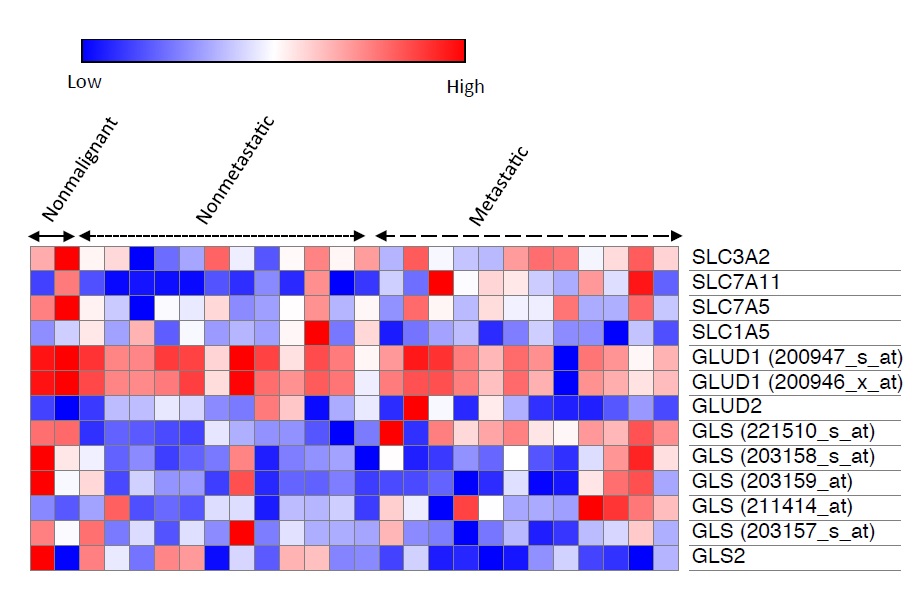
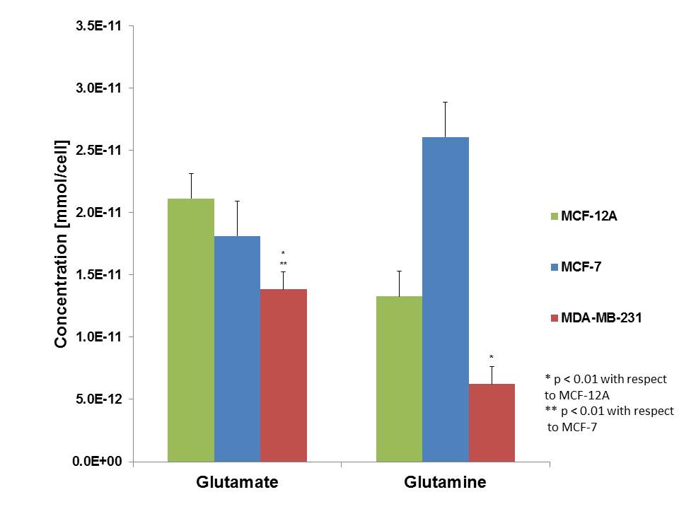
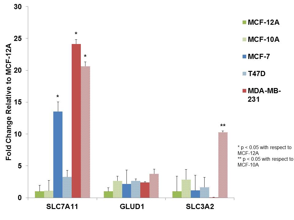
Glutamine, a non-essential amino acid, has been demonstrated as a major energy source across various cancers.1 Many types of tumors have demonstrated increased uptake of glutamine which serves as a carbon and nitrogen source as well as for shuttling essential amino acids into tumor tissue. This so-called “glutamine addiction” has been demonstrated across multiple cancer types including breast cancer, however, the mechanism of increased glutamine uptake has not been rigorously explored across the various subtypes, including nonmetastatic and metastatic, as compared to non-malignant cell lines.2 Cellular glutamine and glutamate levels can be monitored utilizing 1H MRS, and are detected at 2.34 and 2.45 ppm respectively, as previously demonstrated.3 In this study, we have investigated the glutamine profiles across multiple breast cancer cell lines via 1H MRS and explored the mechanism of glutamine uptake via computational methods and quantitative RT-PCR.

Methods

Using the NIH's GEO2R tool and the Broad Institute's Morpheus tool, we analyzed 2 nonmalignant, 11 nonmetastatic, and 14 metastatic breast cancer cell lines from a publicly available Affymetrix microarray dataset GSE-69017 to investigate gene expression of enzymes and transporters relating to glutamine metabolism that are the basis of glutamine consumption in breast cancer cell lines. Glutamine and glutamate levels were measured via high-resolution (HR) 1H MRS from cell extracts across multiple cell lines, including nonmalignant (MCF-12A), nonmetastatic estrogen/progesterone receptor positive (MCF-7), and triple-negative metastatic (MDA-MB-231), and quantified using previously established methods.3 We also measured mRNA expression levels of genes relevant to glutamine metabolism is breast cancer cells by quantitative RT-PCR to identify over- or under-expression of these enzymes/transporters. For all studies, unpaired, two-sample t-tests were used, with p-value < 0.05 considered significant.

Results

Computational analysis of the Affymetrix microarray dataset containing 2 nonmalignant, 11 nonmetastatic, and 14 metatastatic cell lines indicated statistically significant differences in the expression of glutamate transporter SLC7A11, and the two glutaminase genes GLS and GLS2 between metastatic and nonmalignant cell lines (Fig. 1). 1H MRS data of glutamine profiles demonstrated a significant decrease in both glutamine and glutamate in more aggressive, metastatic cells (MDA-MB-231), while less aggressive and nonmetastatic cell lines indicated lower levels of glutamate but increased levels of glutamine relative to nonmalignant cell lines (Fig. 2).3 We have begun qRT-PCR experiments to explore changes in gene expression level across a panel of breast cancer cell lines (Fig. 3). These data show that in all malignant cell lines, the glutamate transporter SLC7A11 is overexpressed, with higher levels of expression in more aggressive cell lines (MDA-MB-231/468), which is consistent with increased glutamate/glutamine metabolism in malignant cells.

Discussion

We are exploring the use of glutamine by differentially aggressive breast cancer cell lines to determine the correlation of glutamate/glutamine metabolism with breast cancer aggressiveness. Our data demonstrates a substantial decrease in glutamine and glutamate in highly aggressive cells (MDA-MB-231). It is possible that more aggressive breast cancer cell types are utilizing glutamine as a source of energy, thereby depleting cellular glutamine. Our initial findings indicate that an overexpression of the glutamate transporter SLC7A11 may be at least partially responsible for increased glutamate/glutamine turnover in aggressive triple-negative breast cancer cells as compared to less aggressive ER/PR-positive breast cancer cells. Increased expression levels of the glutaminase genes GLS and GLS2 in triple-negative versus ER/PR-positive cell lines may also contribute to their altered glutamate and glutamine levels.

Conclusions

Glutamine/glutamate metabolism may play an important role in the aggressive nature of breast cancer, through the utilization of glutamine as a carbon and nitrogen source. Furthermore, glutamate/glutamine metabolism may be stimulated by the overexpression of glutamate transporters in malignant cells. Altered glutamate/glutamine metabolite signatures may be used to identify malignancy within cells and key enzymes in glutamate/glutamine metabolism may prove to be potential therapeutic targets. Studies in our lab are ongoing to further unravel the altered glutamine/glutamate metabolism in relation to breast cancer aggressiveness.

Acknowledgements

We thank all members of the Division of Cancer Imaging Research in The Russell H. Morgan Department of Radiology and Radiological Science for their help and support.

References

1. Souba, WW. Glutamine and Cancer. Annals of Surgery, 1993. 218(6): 715-728.

2. Wise, DR., Thompson, CB. Glutamine Addiction: A New Therapeutic Target in Cancer. Trends Biochem. Sci., 2010. 35(8):247-433.

3. Chan KW, Jiang L, Cheng M, Wijnen JP, Liu G, Huang P, van Zijl PC, McMahon MT, Glunde K. CEST-MRI detects metabolite levels altered by breast cancer cell aggressiveness and chemotherapy response. NMR Biomed. 2016 Jun;29(6):806-16.

Figures

Figure 1: Analysis of 2 nonmalignant, 11 nonmetastatic, and 14 metastatic breast cancer cell lines among a publicly available Affymetrix microarray dataset (GSE-69017) demonstrating gene expression of glutamine uptake and metabolism enzymes. The expression values were extracted using the NIH's GEO2R tool, and the heatmap was generated via the Broad Institute's Morpheus tool. SLC7A11 and GLS (221510_s_at) both show increased expression correlated with aggressiveness, while GLS2 shows lower expression levels in more aggressive cell lines.

Figure 2: High-resolution 1H MRS quantification of glutamine and glutamate from water-soluble cell extracts across three different cell types (n = 3), nonmalignant (MCF-12A), weakly metastatic (MCF-7), and highly metastatic (MDA-MB-231). Highly metastatic cells show a marked decrease in both glutamine and glutamate while weakly metastatic cells show a slight decrease in glutamate levels and an increase in glutamine levels, relative to nonmalignant cells.

Figure 3: Quantification of qRT-PCR experiments across a panel of breast cancer cell lines including nonmalignant (MCF-10A, MCF-12A), weakly metastatic (MCF7, T47D), and highly metastatic (MDA-MB-231, MDA-MB-468). All cell lines represent one technical replicate (n = 3) and error bars indicate standard error.

Proc. Intl. Soc. Mag. Reson. Med. 26 (2018)
4025