4011
Comparative NMR urine metabolomics of whole body or partial body radiation exposure: Stepping towards delivery of potent marker for radiation exposure1NMR Research Centre, Institute of Nuclear Medicine and Allied Sciences, Delhi, India, 2Division of Radioprotective Drug Development Research, Institute of Nuclear Medicine and Allied Sciences, Delhi, India
Synopsis
Stepping forward towards the quest of high throughput radiation marker, the present study looks for a common signature on comparison of NMR based urine metabolomics post whole body or partial irradiation. Different group of animals were exposed to 10 Gy whole body or partial radiation. Irradiated group were clustered apart from controls; showed similar pattern on radiation exposure irrespective of whole body or cranial and identified taurine, citrate as common metabolites in whole body and partial radiation groups. The similar pattern observed post partial or whole body irradiation further confirm these metabolites as potent markers for radiation exposure.
Introduction
In all potential radiation hazards; nuclear accidents, wartime causalities, and even inadvertently during experiments in research laboratories, vast human population could likely to encounter whole body or significant partial body radiation exposure. Recent research has been more focused on identification of markers for radiation injury for whole body radiation exposure and few metabolite markers have been identified in recent studies1. However, estimation of sensitivity and extent of radiation specificity is still being continued worldwide. It is agreeable that the response of organism to radiation injury differs based on type of exposure (partial or whole body) and radiation dose; it may be anticipated that partial and whole body radiation exposure may have some common signature. In tune of this anticipation, the present study was conducted to look for comparative changes in mouse urine on exposure to whole body radiation or partial radiation to different regions of body using NMR metabolomics approach and to look for similar observations if any between partial and whole body radiation exposureMethodology
The study was carried out on C57 male mice (8-10 weeks old). Three group of animals (n=5 in each group) were irradiated partially to different regions; thoracic, abdominal and hind limb regions through Tele Co60 irradiation facility with a dose rate of 0.659Gy/min with SSD of 80 cm and FOV of 20x2 cm for a total single dose of 10Gy. During partial radiation exposure the body of the animals were lead shielded except for the region to be exposed. Animals were anaesthetised during partial irradiation. Another group of the animals (n=7) was whole body irradiated for a dose of 10Gy at a dose rate of 1.096Gy/min. Further, two groups of animals with n=5 in each group was kept as sham controls for whole body and partial irradiation. Urine samples were collected at 24h post irradiation and placed at -80 C. 350μl of centrifuged urine sample was added to 250μl of deuterated phosphate buffer (pH=7.4) containing 1 mM TSP for NMR spectral acquisition on. 600 MHz NMR spectrometer at 298K using 1D gradient NOESYPR pulse sequence. 64 scans were acquired with a relaxation delay of 5s, flip angle of 90° and spectral width 10ppm. All data sets were preprocessed and normalised using mean-centering and pareto-scaling. Following data normalization, multivariate analysis was carried out using metaboanalyst software (http://www.metaboanalyst.ca/Metaboanalyst.jsp).Results
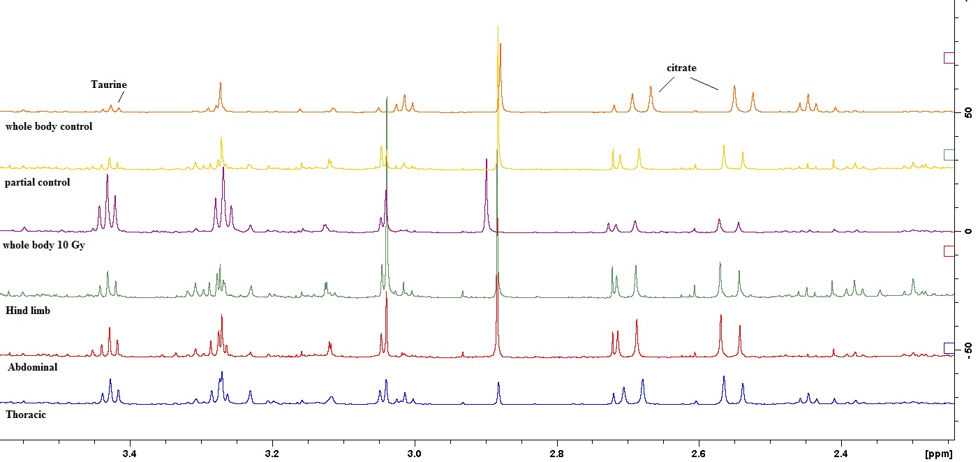
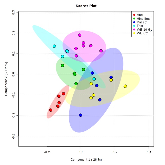
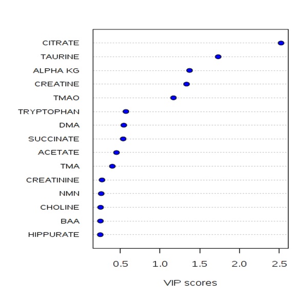
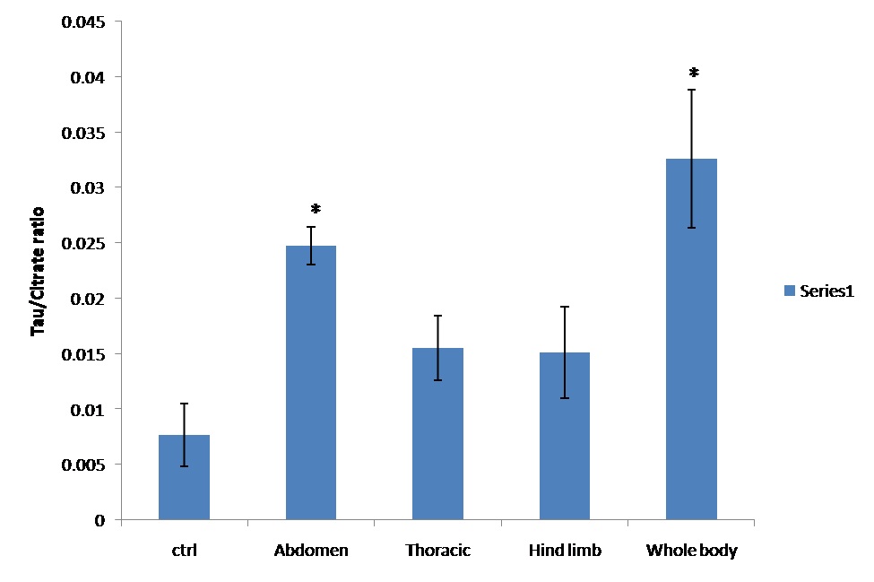
The competency of 1H NMR based metabolomics approach to differentiate partially radiated or whole body irradiated from controls was assessed using multivariate analysis in our study. Visual inspection showed distinct spectral phenotypes, which were readily observed between 1H NMR spectra of controls and irradiated groups (Figure 1). Partial irradiation groups showed similar changes as reported in earlier study on whole body radiation exposure2,3. Following data post processing, PCA and PLS-DA was performed to evaluate the overall intergroup separation. PLS-DA score plot displayed clear demarcation between whole body irradiated and control group (Figure 2). No difference was observed between two sham control groups. In fact, partial radiation group was clearly segregating from controls and whole body radiation group; however, partial overlapping of partial radiation group thoracic and hind limb was observed. VIP score >1 based on PLS-DA analysis identified similar metabolites namely, taurine, citrate, trimethyl amine oxide (TMAO), creatine, α-ketoglutarate as observed in our earlier study on whole body radiation exposure (Figure. 3). Relative integrals based taurine/citrate ratio graph(Figure 4) further ascertain radiation induced difference at metabolite levels in both the types of radiation exposure (partial or whole body).Discussion
Overall, irradiated group were clearly clustered apart from controls showing some similar changes occurring on radiation exposure irrespective of whole body or cranial. However, extent of injury and level of changes may be different in two types of radiation exposure. But pattern of metabolic changes were similar. This study further strengthens our point of observation with taurine being the potent metabolic marker of radiation exposure. Taurine has earlier been identified as most sensitive marker for radiation based on ROC analysis4. The similar pattern observed post partial or whole body irradiation further confirm, revalidate and raise a gain of confidence to consider them as potent markers for radiation exposureConclusion
The study output let us steps ahead further in our ultimate aim of delivering long awaited high throughput marker for radiation exposure. Still lot of studies are required to carry out for validation and dose response. However, the studies would be helpful in triage for mass screening of radiation exposed and not exposed population during radiological accidentAcknowledgements
The present work is supported by Defence Research and Development Organisation (DRDO), Ministry of Defencce, India
References
1. Pannkuk EL, Fornace AJ, Laiakis E. Metabolomics applications in radiation biodosimetry:exploring radiation effects through small molecules. Intl J Radiat Biol 2017. DOI: 10.1080/09553002.2016.1269218
2. Chen C, BrennerDJ, Brown TR, .Identification of urinary biomarkers from x irradiated mice using NMR spectroscopy. Radiat Res, 2011;175:622-630
3. Khan AR, Rana P, Tyagi R et al, NMR Spectroscopy Based Metabolic Profiling of Urine and Serum for Investigation of Physiological Perturbations during Radiation Sickness. Metabolomics, 2011;7: 583-592
4. Rana P, Tyagi R, Watve A. Specificity and sensitivity of early predictive urinary metabolic biomarker of radiation injury: A 1H NMR based metabolomic study. Proc of ISMRM annual meeting, May 2016, Singapore
Figures