4005
Effects of Fraction of Inhaled Oxygen on Hyperpolarized 129Xe MR Signals Acquired from the Rat Brain In Vivo1Translational Medicine, Hospital for Sick Children, Toronto, ON, Canada, 2Medical Biophysics, University of Toronto, Toronto, ON, Canada, 3Medical Imaging, University of Toronto, Toronto, ON, Canada
Synopsis
A theoretical model was developed to describe the dependence of the signal strength of 129Xe dissolved in the rat brain on the fraction of inhaled oxygen (FiO2). To validate the model, 129Xe MR signals were measured in the rat brain in vivo for four different values of FiO2 using a continuous ventilation scheme. The measured 129Xe RBC signal showed a strong dependence on FiO2 in good agreement with the trends predicted by the theoretical model. These results underscore the need to optimize and control FiO2 when using 129Xe signals to probe brain function.
Introduction
129Xe MRI has gained interest as a tool to probe for brain function such as cerebral blood flow (CBF)1,2. The purpose of this work was to develop a theoretical model for the dependence of hyperpolarized 129Xe signal from red blood cells (RBCs) in the brain on the fraction of inhaled oxygen (FiO2). The model is validated using 129Xe RBC signals acquired in vivo from rat brain following inhalation breathing paradigms which vary FiO2.Method
Theoretical Model
We incorporated previously presented equations for the relationships between oxygen concentration and T1 of 129Xe in the lungs3 and T1 of 129Xe in the blood4 with a time-dependent theoretical signal model for 129Xe in the brain based on the Kety-Schmidt equations5. In addition, we modeled the effects of oxygen saturation on the T2* of RBCs in the brain6 to develop a comprehensive model for the dependence of 129Xe signal in the brain on FiO2.
MR Spectroscopy
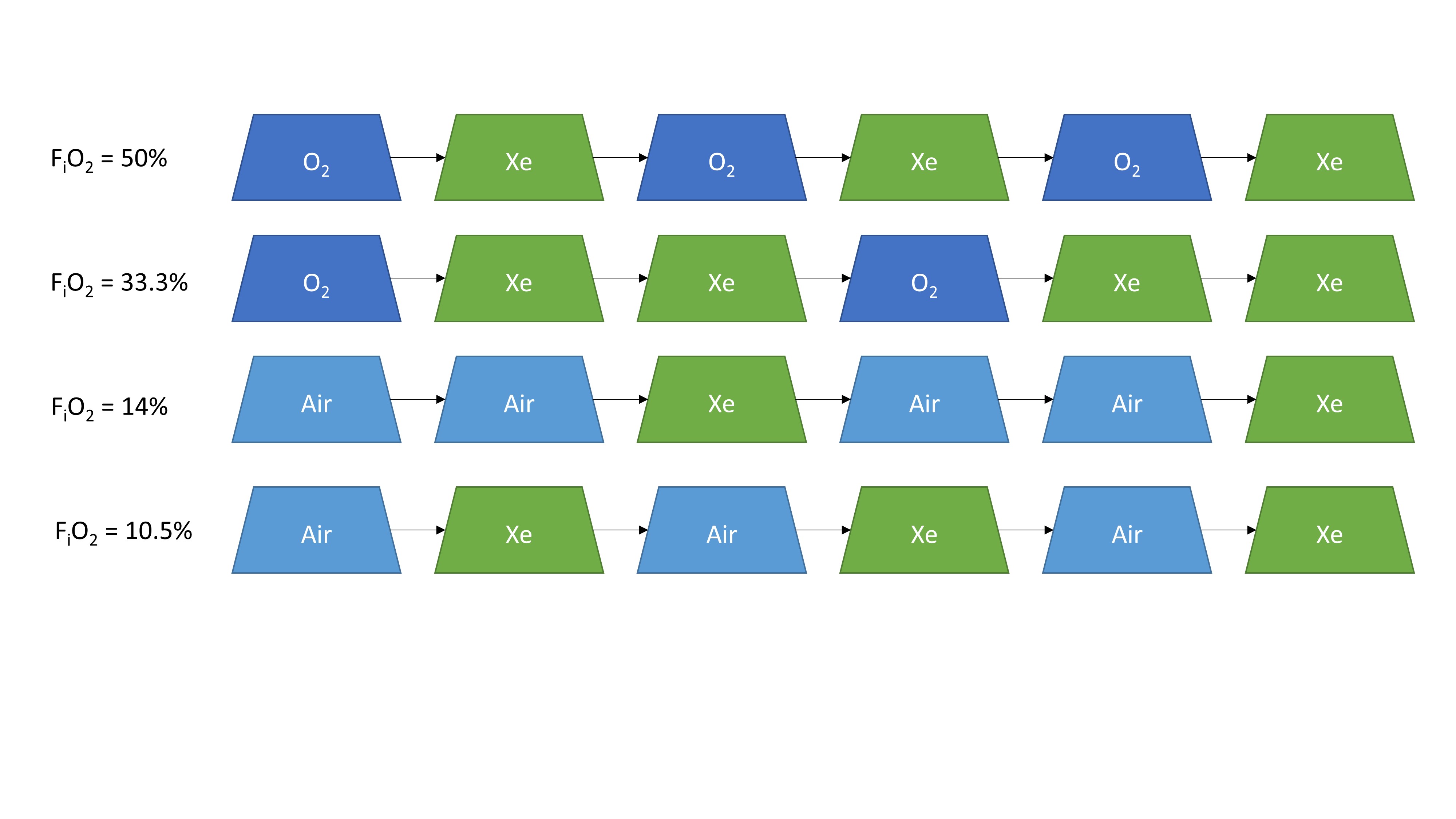
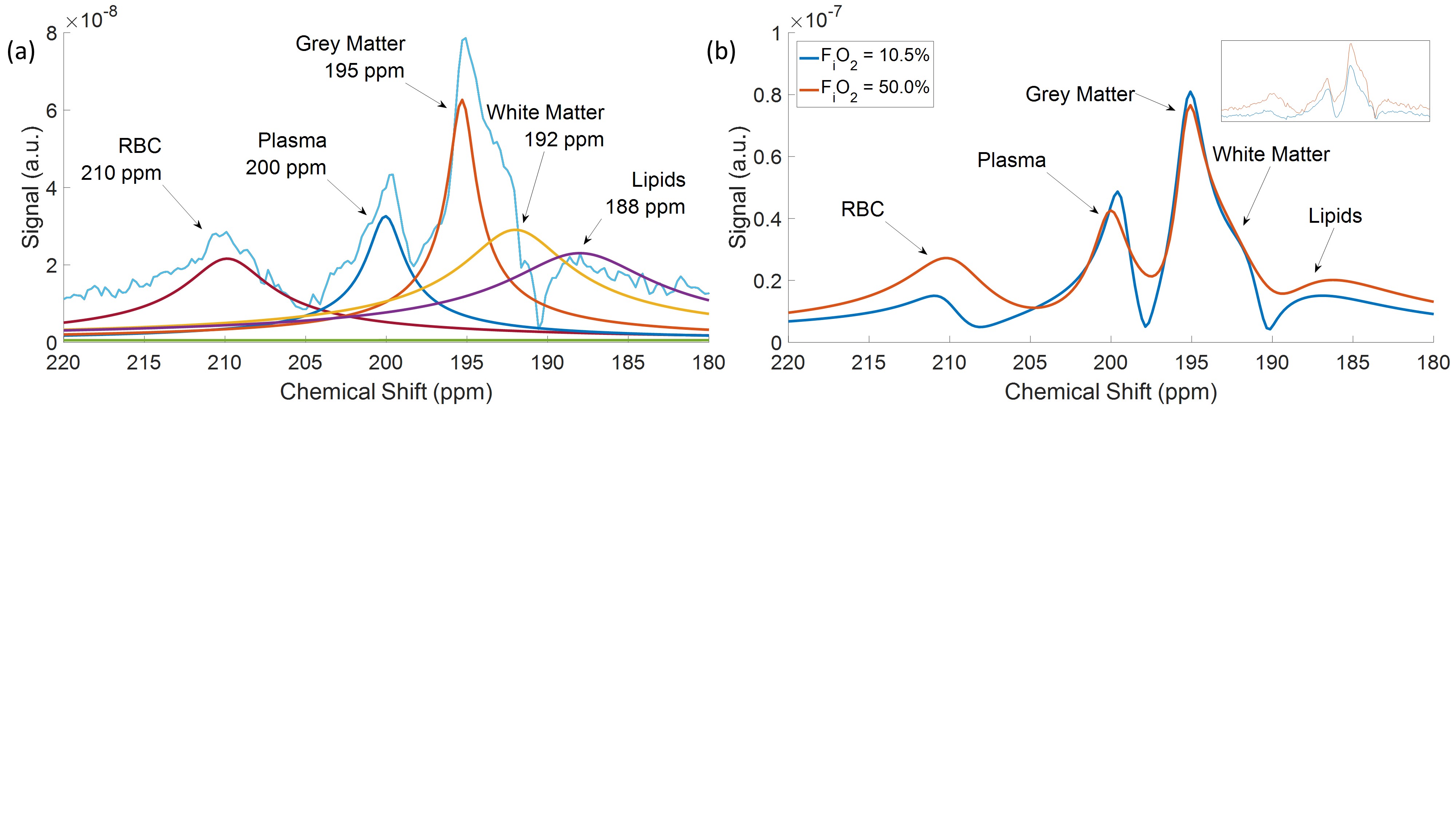
Alternating 3ml breaths of xenon (~86% 129Xe, ~15% polarization) and air or O2 were delivered to five mechanically-ventilated rats using four different breathing paradigms (Fig. 1) to vary the FiO2. Using a 3T MRI Scanner (Siemens, Germany) and quadrature transmit/receive birdcage coils (inner radii of 1.7cm or 6cm, Morris Instruments, Canada), 129Xe spectra were acquired following an excitation pulse delivered every 1s during ventilation (30° flip angle, 3-lobe sinc, pulse of length 1280μs, transmit frequency at 200ppm from gas, slice thickness of 4cm, centered on the rat brain). Once steady 129Xe signal was reached in the brain, the spectra were averaged and decomposed in the time domain into six additive exponential components (five dissolved components and one gas component) using spectral analysis code provided by the Duke Center for In Vivo Microscopy NIH/NIBIB (P41 EB015897)7. The signal of each dissolved component was calculated as the area under each Lorentzian curve in the spectral domain, normalized to the gas signal.
Results
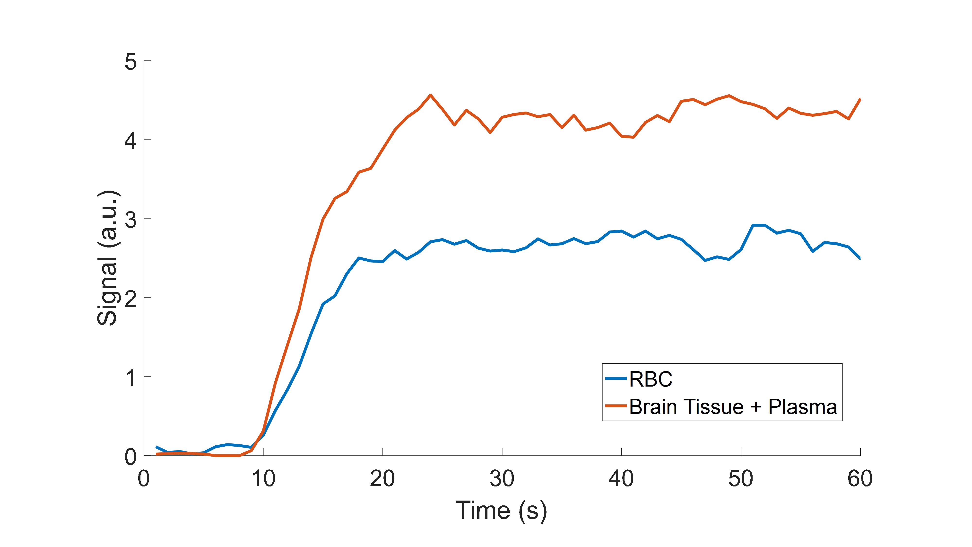
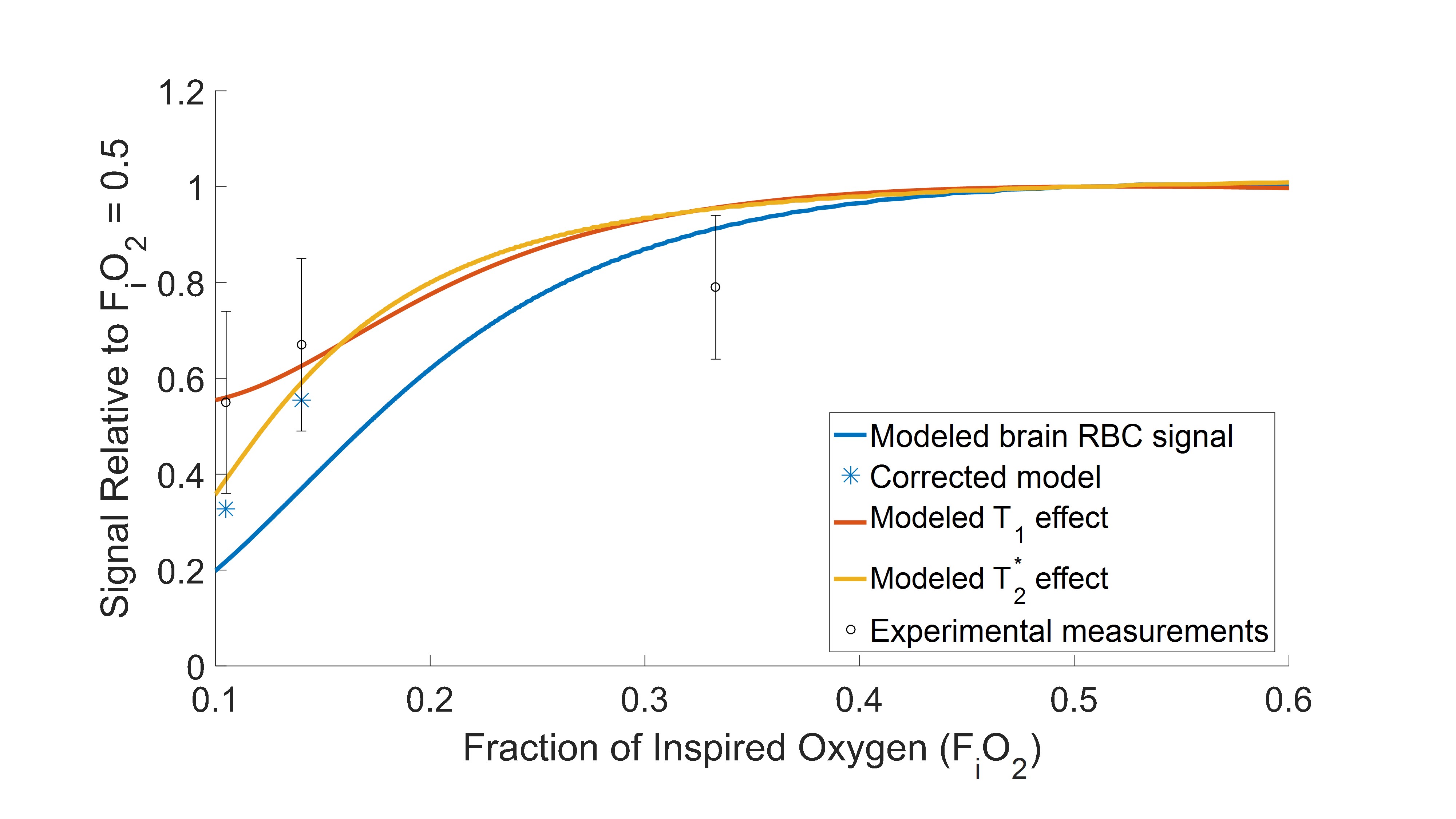
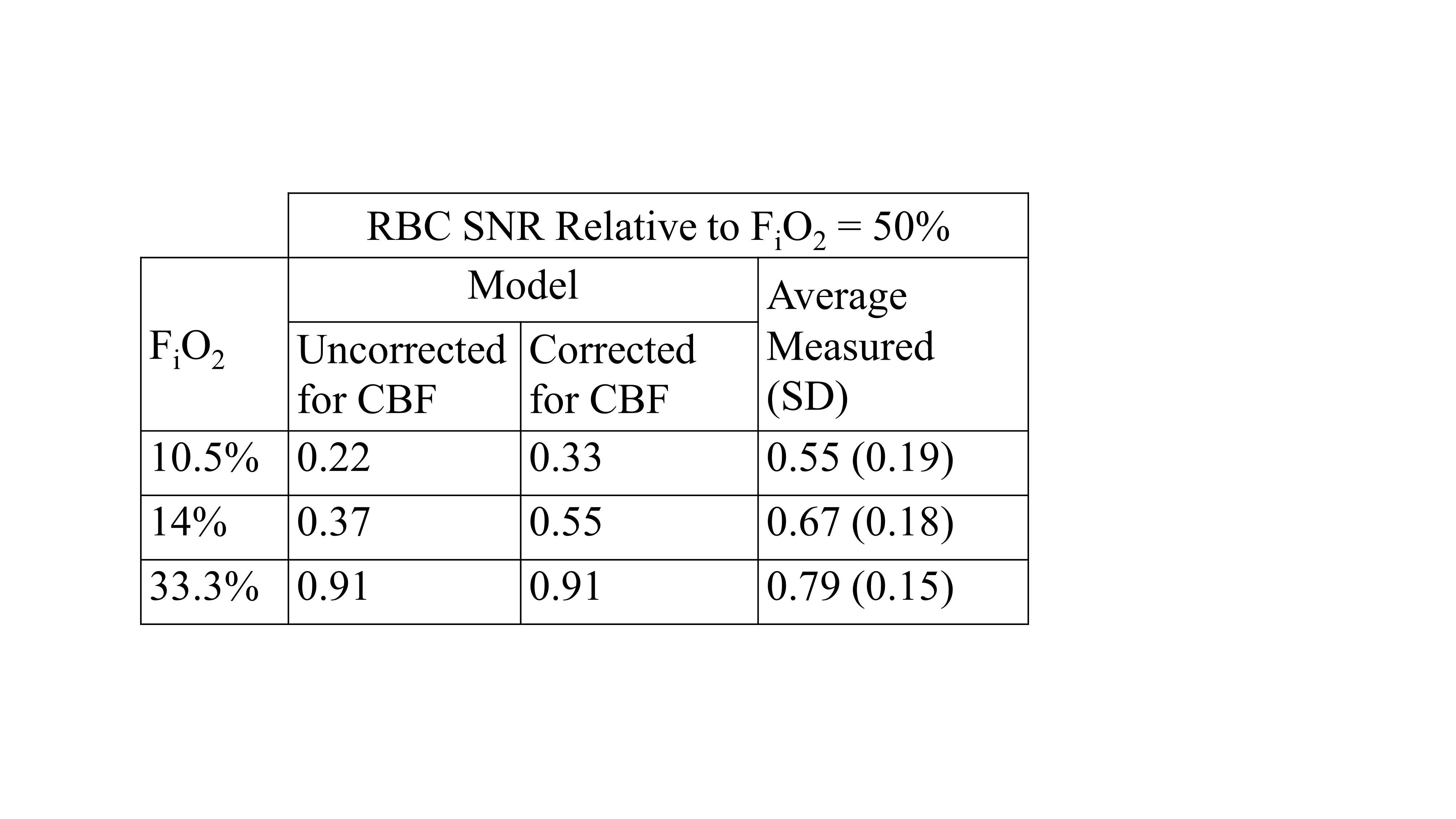
A representative 129Xe signal time-course is shown in Fig. 2 where a steady-state signal is reached at approximately 20s after initiation of the paradigms shown in Fig. 1. A representative 129Xe spectrum is shown in Fig. 3a. The chemical shift of each spectral component agrees with previously reported values8–10. Fig. 3b shows a comparison of two spectra acquired in the same rat but at different FiO2. The measured and predicted signal values as a function of FiO2 are shown in Fig. 4 and summarized in Table 1.Discussion
The 129Xe RBC signal measured in rat brain in vivo shows a strong dependence on FiO2 which agrees with the trends predicted by the theoretical model (Fig. 4). The measured 129Xe RBC signal reduction at FiO2 of 10.5% was 55%±19% relative to the signal at FiO2 of 50%. The drop in signal was statistically significant (t-test, P<0.0001). The maximum RBC signal, as predicted by the model, occurs at FiO2 = 61%. By fitting a model of 129Xe uptake in the brain5 to the time-course signal, the transit time for 129Xe to reach the brain from the lungs was measured to be 2.5s. Previous studies predicted a transit time of 4–5s in humans5,11. It is reasonable to expect the transit time in rats to be less than that in humans.
129Xe MRI of the brain has traditionally used the time-course of the 129Xe signal magnitude in the brain as a measure of CBF1,2,8,12–15. An important implication of this study is that the 129Xe signal depends on oxygen-induced relaxation effects, in addition to hemodynamic effects, thereby confounding measurements of CBF. For example, increased T1 relaxation in the blood during transit to the brain would reduce the 129Xe signal in the brain up to 40% by the end of a 30s breath hold16. 129Xe signal from RBCs in the brain would be further reduced by 20% due to T2* relaxation for the excitation pulse used in our study. This effect is further exemplified by the brain tissue spectra in Fig. 3b. An increased brain tissue signal is expected for the hypoxic paradigm due to an increase in CBF. However, the signal does not increase, suggesting a possible masking of the increased CBF by the increased relaxation rates.
Conclusion
A dependence of 129Xe RBC signal on FiO2 is observed in the rat brain, in qualitative agreement with a theoretical model based on changes in oxygen-induced T1 and T2* relaxation of 129Xe gas in the lung, dissolved 129Xe signal in the blood, and 129Xe uptake in the brain. These results underscore the need to optimize and control FiO2 when using 129Xe signals to probe brain function.Acknowledgements
Thanks to E. Stirrat, N. Kanhere, A. Lindenmaier, B. Zanette, and F. Morgado for their assistance with imaging experiments. This work was supported by grant funding from NSERC Discovery Grant (RGPIN-2015-03832). Y.F. was financially supported by a Queen Elizabeth II Graduate Scholarship in Science and Technology.References
1. Imai H, Kimura A, Akiyama K, Ota C, Okimoto K, Fujiwara H. Development of a fast method for quantitative measurement of hyperpolarized 129Xe dynamics in mouse brain. NMR Biomed. 2012;25(2):210-217. doi:10.1002/nbm.1733.
2. Rao M, Stewart NJ, Griffiths PD, Norquay G, Wild JM. Imaging Human Brain Perfusion with Inhaled Hyperpolarized 129Xe MR Imaging. Radiology. 2018;0(0):1-7.
3. Patz S, Hersman FW, Muradian I, et al. Hyperpolarized 129Xe MRI: A viable functional lung imaging modality? Eur J Radiol. 2007;64(3):335-344. doi:10.1016/j.ejrad.2007.08.008.
4. Norquay G, Leung G, Stewart NJ, Tozer GM, Wolber J, Wild JM. Relaxation and exchange dynamics of hyperpolarized 129 Xe in human blood. Magn Reson Med. 2015;74(2):303-311. doi:10.1002/mrm.25417.
5. Peled S, Jolesz FA, Tseng C-H, Nascimben L, Albert MS, Walsworth RL. Determinants of tissue delivery for 129Xe magnetic resonance in humans. Magn Reson Med. 1996;36(3):340-344. doi:10.1002/mrm.1910360303.
6. Gherase MR, Wallace JC, Cross AR, Santyr GE. Two-compartment radial diffusive exchange analysis of the NMR lineshape of 129Xe dissolved in a perfluorooctyl bromide emulsion. J Chem Phys. 2006;125(4):1-8. doi:10.1063/1.2217735.
7. Virgincar RS, Robertson SH, Nouls J, et al. Establishing an accurate gas phase reference frequency to quantify 129Xe chemical shifts in vivo. Magn Reson Med. 2016;77(4):1438-1445. doi:10.1002/mrm.26229.
8. Mazzanti ML, Walvick RP, Zhou X, et al. Distribution of Hyperpolarized Xenon in the Brain Following Sensory Stimulation: Preliminary MRI Findings. Brechbiel MW, ed. PLoS One. 2011;6(7):e21607. doi:10.1371/journal.pone.0021607.
9. Kershaw J, Nakamura K, Kondoh Y, Wakai A, Suzuki N, Kanno I. Confirming the existence of five peaks in 129Xe rat head spectra. Magn Reson Med. 2007;57(4):791-797. doi:10.1002/mrm.21186.
10. Rao M, Stewart NJ, Norquay G, Griffiths PD, Wild JM. High resolution spectroscopy and chemical shift imaging of hyperpolarized 129Xe dissolved in the human brain in vivo at 1.5 tesla. Magn Reson Med. 2016;75(6):2227-2234. doi:10.1002/mrm.26241.
11. Kilian W, Seifert F, Rinneberg H. Dynamic NMR spectroscopy of hyperpolarized (129)Xe in human brain analyzed by an uptake model. Magn Reson Med. 2004;51(4):843-847. doi:10.1002/mrm.10726.
12. Nakamura K, Kondoh Y, Wakai A, Kershaw J, Wright D, Kanno I. 129Xe spectra from the heads of rats with and without ligation of the external carotid and pterygopalatine arteries. Magn Reson Med. 2005;53(3):528-534. doi:10.1002/mrm.20399.
13. Duhamel G, Choquet P, Grillon E, et al. Global and Regional Cerebral Blood Flow Measurements Using NMR of Injected Hyperpolarized Xenon-129. Acad Radiol. 2002;9(2):S498-S500. doi:10.1016/S1076-6332(03)80275-1.
14. Kimura A, Imai H, Wakayama T, Fujiwara H. A simple method for quantitative measurement and analysis of hyperpolarized (129)Xe uptake dynamics in mouse brain under controlled flow. Magn Reson Med Sci. 2008;7(4):179-185. doi:10.2463/mrms.7.179.
15. Zhou X, Sun Y, Mazzanti M, et al. MRI of stroke using hyperpolarized 129Xe. NMR Biomed. 2011;24(2):170-175. doi:10.1002/nbm.1568.
16. Delahoche J, Delapille P, Lemaître F, Verin E, Tourny-Chollet C. Arterial Oxygen Saturation and Heart Rate Variation During Breath-Holding: Comparison between Breath-Hold Divers and Controls. Int J Sports Med. 2005;26(3):177-181. doi:10.1055/s-2004-820976.
17. Duong TQ, Iadecola C, Kim S-G. Effect of hyperoxia, hypercapnia, and hypoxia on cerebral interstitial oxygen tension and cerebral blood flow. Magn Reson Med. 2001;45(1):61-70. doi:10.1002/1522-2594(200101)45:1<61::AID-MRM1010>3.0.CO;2-8.
18. Bereczki D, Wei L, Otsuka T, et al. Hypoxia Increases Velocity of Blood Flow through Parenchymal Microvascular Systems in Rat Brain. J Cereb Blood Flow Metab. 1993;13(3):475-486. doi:10.1038/jcbfm.1993.62.
Figures