3999
In vivo $$$^{31}\hspace{0.1 mm}$$$P-MRS of triple negative breast cancer patients1Radiology, UMC Utrecht, Utrecht, Netherlands, 2Image Science Institute, UMC Utrecht, Utrecht, Netherlands, 3Medical Oncology, Academic Medical Centre Amsterdam, Amsterdam, Netherlands
Synopsis
31P-MR spectra of breast cancer tissue have shown very high GPC/PC ratios in xenografts for triple negative (TN) tumors. We set out to investigate if this is also visible in vivo in six TN breast cancer patients. In two of the patients the phosphocholine (PC) peak is missing or approaching zero. To the best of our knowledge, this is the first time that this phenomenon is shown in vivo in breast cancer patients, however, the relation of the presence or absence of PC and TN subtype remains to be investigated.
Introduction
Ex vivo 31P-MRS of breast cancer tissue and biopsies has been used to study the phospholipid metabolism in breast cancer and the relation between NMR characteristics with different gene profiles has been investigated (1). For example, a distinct 31P-MRS pattern could discriminate basal like and luminal like breast cancer where xenograft models of primary triple negative breast carcinomas had higher glycerophosphocholine (GPC)/ phosphocholine (PC) ratio than samples from ER+/PgR+ carcinomas (2,3). The introduction of high field human MRI systems (7 T) enabled 31P-MRS of the breast in vivo (4,5) with sufficient SNR and spectral resolution to discriminate between PE and PC in breast cancer lesions. However, to our knowledge, the specific 31P-NMR patterns described by Moestue et al. in triple negative breast cancer obtained ex-vivo have not been confirmed in vivo in triple negative breast cancer patients. Therefore, in this work we set out to investigate 31P NMR patterns in vivo in six triple negative breast cancer patients which are part of a larger study in which 31P-MRSI was acquired.Materials and methods
After signing informed consent, six breast cancer patients (5 patients recruited from an ongoing study and 1 patient recruited from the PROFILE study (6)) were scanned with a 2 channel unilateral 1H/31P dual-tuned coil (MR Coils, Drunen, Netherlands) (7) on a 7T MR system (Philips, Cleveland, USA). Image based B0 shimming was performed by a least square error optimization using a 3D B0 map followed by manual segmentation of the breasts and third order shimming (8). Before MRS measurements, a fat-suppressed 3D fast-field-gradient-echo sequence was used to image the breast tissue and locate the tumor (T1-weighted, selective water excitation, fat-suppressed, flip angle 10°, TR = 4.0 ms, TE = 2.0 ms, FOV = 160x160x160 mm3, voxelsize = 1x1x1 mm3, acquisition time = 51 sec).
31P-MRS was obtained using the AMESING sequence (9), in which 1 FID and 5 full echoes were acquired with TR = 6 s, ΔTE = 45 ms, FOV 160x160x160 mm3, 8x8x8 voxels, 2x2x2 cm3 nominal resolution, BW = 8200 Hz, sampling matrix size = 256, resulting in a total scan time of 25:36 min. All MRSI data were zero-filled and apodised (15 Hz Lorentzian) in the time domain and spatially Hamming filtered. The tumor spectra were analyzed by spectral fitting in jMRUI (10).
Results
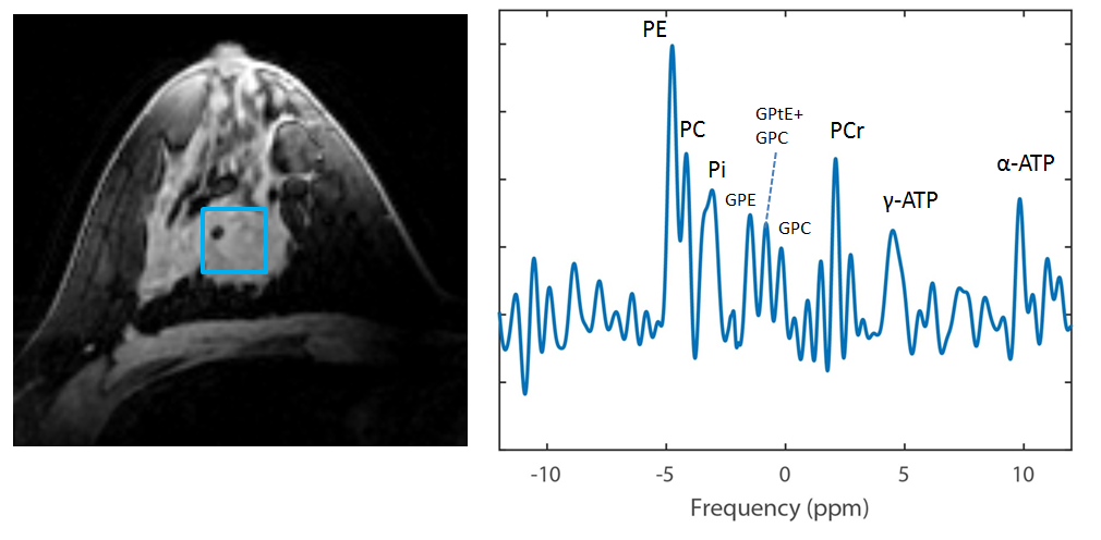
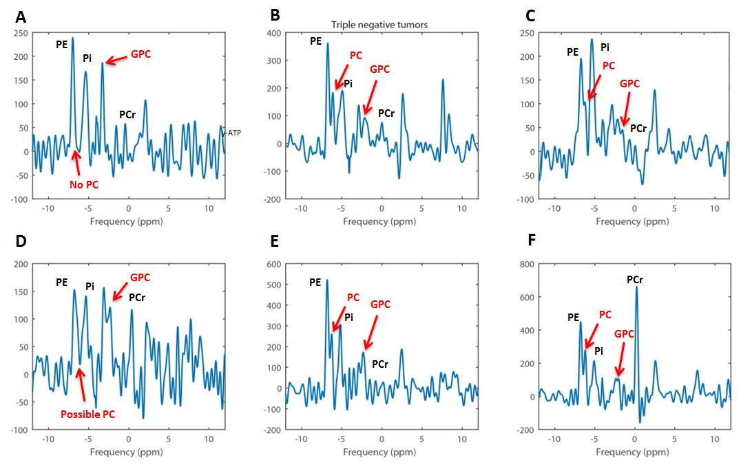
To illustrate what the 31P-spectrum looks like in a non-triple negative tumor, an example of an 31P –MR spectra of one of the non-triple negative patients from the ongoing study is shown in figure 1 (ER+, PR+ and Her2neu- in this case). Most of the 31P-spectra of the six triple negative tumors show a PC peak (Figure 2), yet the tumor in Figure 2A shows a clear absence of this PC peak. In only one case the B0 shimming was sub-optimal therefore unclear if PC was present or not.Discussion and conclusion
In the small set of triple negative breast cancer patients that we investigated, in at least one and at maximum two out of the six patients the GPC/PC ratio is high (or zero due to absence of PC peak). Earlier findings in ex vivo NMR of basal/luminal like breast cancer xenografts and ex vivo 31P NMR of biopsies from TN breast cancer patients show higher variation of this ratio in the basal-like samples compared to luminal-like samples. Also a majority showed higher GPC signal than PC signal (3), which we observe in three out of six TN breast cancer patients. When accumulating more 31P-MRS data of tumors of TN breast cancer patients the relation between the absence of the PC peak and tumor subtype can be investigated in more detail. To our knowledge this is the first time this phenomenon of the absent PC peak is shown in vivo in breast cancer patients and motivates further research on PC levels in TN breast cancer patients.Acknowledgements
No acknowledgement found.References
1. Podo F. Tumour phospholipid metabolism. NMR Biomed. 1999;12(7):413–439.
2. Moestue S, Borgan E, Huuse E. Distinct choline metabolic profiles are associated with differences in gene expression for basal-like and luminal-like breast cancer xenograft models. BMC Cancer. 2010;10(433):1–12http://www.biomedcentral.com/1471-2407/10/433/abstract%5Cnpapers3://publication/uuid/7BCE48D9-AAC8-4BD5-90DD-F39847E62F81.
3. Grinde MT, Skrbo N, Moestue SA, et al. Interplay of choline metabolites and genes in patient-derived breast cancer xenografts. Breast Cancer Res. 2014;16(1):R5http://breast-cancer-research.biomedcentral.com/articles/10.1186/bcr3597.
4. Klomp DWJ, van der Kemp WJM, Korteweg M, Wijnen JP, Bosch M Van De, Luijten PR. P MRS at 7T can be more sensitive and specific than 1 H MRS in monitoring breast cancer treatment . Proc Intl Soc Mag Reson Med. 2011;19:343.
5. van der Kemp WJM, Boer VO, Luijten PR, Stehouwer BL, Veldhuis WB, Klomp DWJ. Adiabatic multi-echo 31P spectroscopic imaging (AMESING) at 7 T for the measurement of transverse relaxation times and regaining of sensitivity in tissues with short T₂ values. NMR Biomed. 2013;26(April):1299–1307http://www.ncbi.nlm.nih.gov/pubmed/23553945.
6. Schmitz AMT, Veldhuis WB, Menke-Pluijmers MBE, et al. Preoperative indication for systemic therapy extended to patients with early-stage breast cancer using multiparametric 7-tesla breast MRI. PLoS One. 2017;12(9):1–14.
7. Klomp DWJ, Bank BL Van De, Raaijmakers A, et al. 31P MRSI and 1 H MRS at 7 T : initial results in human breast cancer. 2011;(January):1337–1342.
8. Hancu I, Govenkar A, Lenkinski RE, Lee SK. On shimming approaches in 3T breast MRI. Magn Reson Med. 2013;69(3):862–867.
9. van der Kemp WJM, Boer VO, Luijten PR, Stehouwer BL, Veldhuis WB, Klomp DWJ. Adiabatic multi-echo 31P spectroscopic imaging (AMESING) at 7 T for the measurement of transverse relaxation times and regaining of sensitivity in tissues with short T₂ values. NMR Biomed. 2013;26(April):1299–1307.
10. Vanhamme L, Van Den Boogaart A, Huffel S Van. Improved Method for Accurate and Efficient Quantification of MRS Data with Use of Prior Knowledge. J Magn Reson. 1997;129:35–43.
11. Lehmann BD, Pietenpol JA, Tan AR. Triple-negative breast cancer: molecular subtypes and new targets for therapy. Am Soc Clin Oncol Educ B. 2015;e31–e39http://meetinglibrary.asco.org/content/11500031-156.
Figures