3988
31P MRS of the frontal lobe of human brain at 7T1National Institutes of Health, Bethesda, MD, United States, 2Chongqing University of Posts and Telecommunications, Chongqing, China
Synopsis
The majority of neuropsychiatric disorders are thought to be originated in the frontal lobe. Increasing the SNR of frontal lobe 31P MRS using a surface coil can be very important because many MRS manifestations of psychiatric illnesses could be subtle. This study demonstrates the feasibility of high SNR 31P MRS of human frontal lobe using a surface coil and 3D CSI at 7 Tesla. Comparing to the occipital lobe spectra acquired from the same subject it was shown that the sensitivity and spectral resolution of 31P MRS spectra of the frontal lobe can be made comparable to that of the occipital lobe.
INTRODUCTION
The majority of neuropsychiatric disorders are thought to be originated in the frontal lobe. At 7 Tesla, 31P MRS of the occipital lobe using surface coil or half-volume coil has been demonstrated.1,2,5 Although the frontal lobe is accessible using a volume coil,3,4 increasing SNR of frontal lobe 31P MRS using a surface coil can be very important because many MRS manifestations of psychiatric illnesses could be subtle. The purpose of this study is to demonstrate the feasibility of high SNR 31P MRS of human frontal lobe using a surface coil and 3D CSI. For comparison, occipital lobe spectra from the same subject were also acquired in the same scan session.METHODS
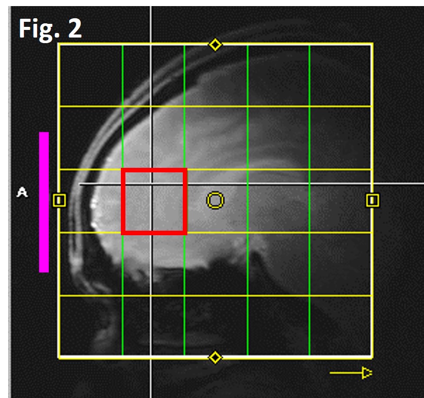
Hardware: All studies were performed on a Siemens 7T scanner with a home-built coil assembly (Fig 1). The proton coil was a shielded quadrature half-volume coil (two overlapping octagon loops). The 31P coil was a 7-cm surface coil. For each subject, the angle of the headrest was adjusted so that the surface of the forehead is parallel to B0 field. Fig 2 shows a gradient-echo image of the frontal lobe with the 31P coil (purple line) placed parallel to the B0 field. The similar coil assembly was also used to acquire 31P MRS from the occipital lobe.
SAR evaluation: RF power deposition for nuclear Overhauser enhancement (NOE) dominates the SAR. Thus, B1 field and SAR distribution in human frontal and occipital lobes were calculated using numerical simulation (Computer Simulation Technology) for the proton coil. An adult male model was used. RF coils were modeled with the same dimension as the actual coil assembly.
31P MRS: A 3x3x3 cm3 cubic voxel in the frontal lobe (the red box in Fig. 2) was shimmed using the FASTMAP method to adjust all 1st and 2nd order shims. The typical half-height water linewidth from the cube was 12~13 Hz. Localized 31P MRS was acquired using a 3D CSI method. FOV=15x15x15 cm3, CSI matrix=5x5x5, and voxel size=3x3x3 cm3 with the CSI voxel of interest coinciding with the shim box as shown in Fig 2. 31P hard pulse duration=250 ms, TR=2 s, NA=8, number of data points=1024, and SW=5 kHz. Elliptical phase encoding was applied to reduce acquisition time. The total scan time for the entire 3D CSI was 9 min. 31P flip angle was empirically optimized. Proton pulses for NOE were also applied to enhance 31P SNR.5 The average power for the scan was limited to 3.5 W. For comparison, the same procedure was repeated to acquire spectra from the occipital lobe of the same subject in a single scan session. Localized CSI data was zero-filled to 16 k and broadened with LB=10 Hz. No baseline correction was applied.
RESULTS
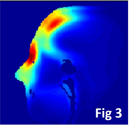
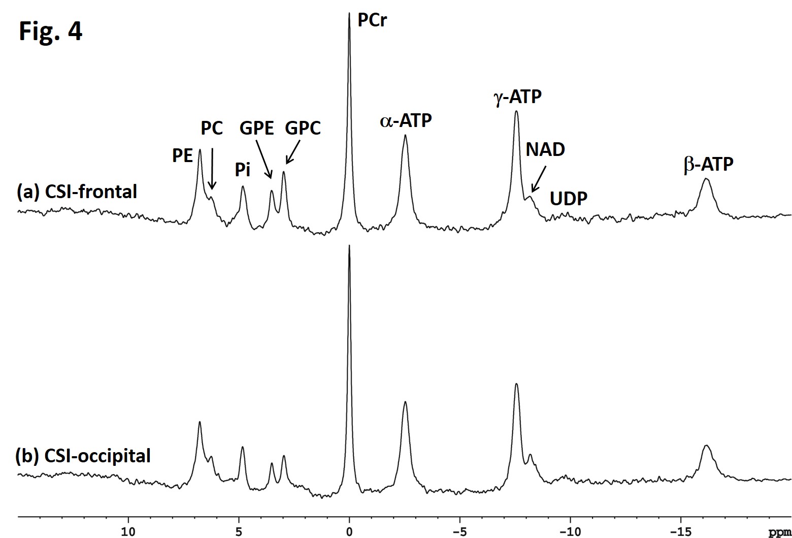
Fig 3 shows a sagittal view of calculated SAR distribution from the frontal lobe simulation where the maximum local SAR is at nasal bridge. Similar simulation was done on the occipital lobe as well. For one watt of input RF power at 297 MHz, the maximum 10-g averaged local SAR was 1.74 W/kg and 1.67 W/kg for the frontal and occipital lobe, respectively. For the limited power of 3.5 W, the maximum local SAR was 6.09 W/kg which is well below the safety guideline of 10 W/kg. Fig 4a shows localized frontal lobe spectrum from the CSI voxel of interest. The spectrum shows eleven phosphorus resonances: phosphoethanolamine (PE), phosphocholine (PC), inorganic phosphate (Pi), glycerophosphoethanolamine (GPE), glycerophophocholine (GPC), phosphocreatine (PCr), γ-, α-, β-adenosine triphosphate (ATP), nicotinamide (NAD), and uridine diphospho glucose (UDP). For comparison, a localized 31P MRS spectrum from the occipital lobe of the same subject acquired using a similar coil assembly and the same sequence parameters was shown in Fig 4b.DISCUSSION
The coil assembly shown in Fig. 1 was optimized to provide patient comfort and optimal SNR as it makes the major flux of the 31P coil perpendicular to the main B0 field. Our results showed that 31P MRS of the frontal lobe with NOE enhancement using a surface coil assembly can be safely performed. Due to close proximity to the nasal cavity, the linewidth of the frontal lobe voxel is broader than the corresponding occipital lobe as expected (20.1 vs 18.8 Hz for PCr and 47.5 Vs 45.6 Hz for α-ATP in Fig. 4). SNR of PCr in the frontal and the occipital lobe (Fig 4) was found to be 70.7 and 94.1, respectively. Our results demonstrate that high SNR spatially localized 31P MRS of the frontal lobe can be achieved using a surface coil assembly and 3D CSI at 7 T.Acknowledgements
This work was supported by the Intramural Research Program of the National Institute of Mental Health, National Institutes of Health.References
1. Lei H, Zhu XH, Zhang XL, et al. In vivo 31P magnetic resonance spectroscopy of human brain at 7 T: an initial experience. Magn Reson Med 2003;49:199-205.
2. Ren J, Sherry AD, Malloy CR. 31P-MRS of healthy human brain: ATP synthesis, metabolite concentrations, pH, and T1 relaxation times. NMRBiomed 2015;28:1455-1462.
3. Zhu XH, Qiao H, Du F, et al. Quantitative imaging of energy expenditure in human brain. NeuroImage 2012;60:2107-2117.
4. Lagemaat MW, van de Bank BL, Sati Pascal, et al. Repeatability of 31P MRSI in the human brain at 7T with and without the nuclear Overhauser effect. NMRBiomed 2016;29:256-263.
5. Li S, An L, Ferraris-Araneta M, et al. Broadband NOE for 7T 31-phosphorous MRS in human brain. ISMRM 2016, Singapore, Program # 4015.
Figures
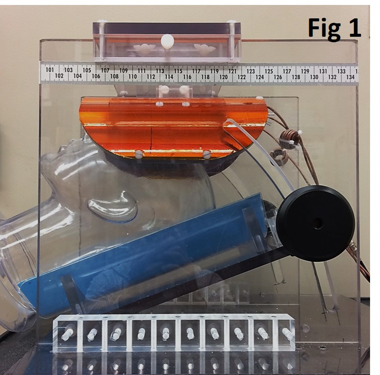
Fig 1. Blue: adjustable headrest; Amber: coil assembly