3964
Test-retest reproducibility of quantitative proton MRS using short-TE STEAM and semi-LASER sequences in young adult volunteer brains at 7T.1Kyoto University, Kyoto, Japan, 2Siemens Healthcare K.K., Tokyo, Japan, 3Siemens Healthcare, USA, Malvern, PA, United States
Synopsis
Recently, 7T-MR system has been approved for clinical use in Europe and USA; however, its clinical configuration is limited to single-channel transmit so B1+ shimming is not feasible. This study investigated reproducibility of single-voxel MRS using short-TE STEAM and semi-LASER using a single-transmit & 32-receiver head coil at 7T. Fifteen healthy young volunteers were scanned twice at the posterior cingulate. SNR was higher in semi-LASER, but coefficients of variation were comparable ranging mainly from 5-10% and better in short-TE STEAM in low-concentration J-coupled peaks. Even with clinical setups, 7T shows high reliability and will contribute to MRS investigation.
INTRODUCTION
Reliable assessment of brain metabolites is important to study a variety of neurological and neuropsychiatric disorders. High-quality, quantifiable MR spectra has been shown in various regions at 7T utilizing a 16-channel transceiver coil and B1+ shimming1, where an excellent test-retest reproducibility of single-voxel MRS was reported using a semi-localized adiabatic selective refocusing (semi-LASER) sequence even with low-concentration metabolites2. Glutathione (GSH) and γ-aminobutyric acid (GABA) were measured with Cramer-Rao lower bounds (CRLB) < 7% using a J-difference editing semi-LASER sequence at 7T with an 8-channel transceiver system3. Recently, 7T-MR system has been approved for clinical use in Europe and the USA; however, this configuration is limited to single-channel transmit, so B1+ shimming is not feasible. This may result in lower reliability and needs to be evaluated. There is another potentially useful sequence, Stimulated echo acquisition mode (STEAM) for 7T because of low RF power requirement and short-TE capability. STEAM must be validated as it has been compared with other sequences only for some selected metabolites3,4,5. This study investigated test-retest reproducibility of short-TE STEAM and semi-LASER MRS measurement using a single-transmit 7T system in view of clinical use of 7T MRS.METHODS
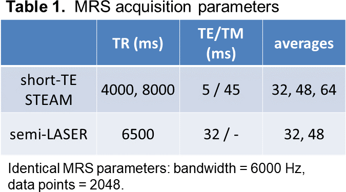
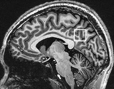
MRS scan was performed on a 7T whole-body scanner (Siemens, Erlangen, Germany) using a single-transmit volume coil and a 32-receiver head coil (Nova Medical, MA, USA). Fifteen healthy volunteers (9 males and 6 females, mean age 25 years, aged 20-38 years) were examined twice with off-magnet intermission under approval of IRB. An MRS voxel of 20 cubic mm was positioned at the posterior cingulate across the mid-sagittal plane on acquired T1-weighted images (Figure 1). Consistent voxel placement among brains was confirmed by the same neuroradiologist for all scans. FASTMAP shimming (prototype) and transmit amplitude adjustment were performed in the MRS voxel. Proton MR spectra were acquired using short-TE (5 ms) STEAM and semi-LASER (TE = 32 ms) pulse sequences (prototypes) with water (VAPOR5) and outer volume suppressions. MRS parameters are listed in Table 1. Water unsuppressed spectra of the same MRS voxel were also acquired. Spectral analysis was carried out using LCModel version 6.3-1L (LA Systems, Tokyo, Japan). Basis-set of STEAM was supplied as a standard option of the LCModel and that of semi-LASER was developed in-house. Eddy current correction and water-scaling for quantification were performed using the water unsuppressed spectra6. Signal-to-noise ratio (SNR), standard error estimates of CRLB for spectral fit, test-retest coefficients of variance (CoV) were compared between the sequences with different TRs and the number of signal averages.RESULTS
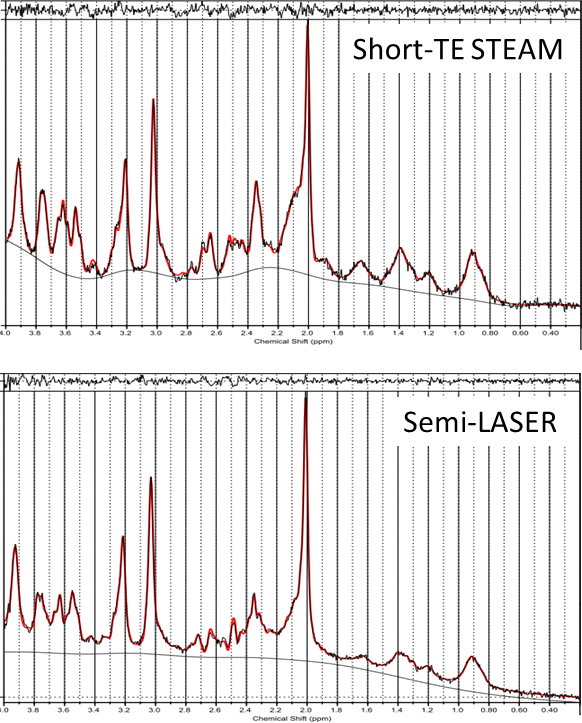
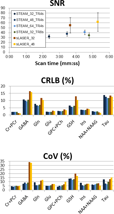
Representative MR spectra of short-TE STEAM and semi-LASER from the posterior cingulate in the same brain, corresponding LCModel fit curves and residuals are shown in Figure 2. Sensitivity of J-coupled peaks in 2.1-2.8 and 3.3-3.8 ppm compared to that of singlet peaks at 2.0, 3.0, 3.2, and 3.9 ppm is higher in short-TE STEAM than that in semi-LASER. Small fitting errors around J-coupled peak regions are observed in both residuals compared with those levels between 0.2-1.8 ppm. SNR, CRLB and CoV for metabolites with mean CRLB < 20 % obtained with different scan conditions are shown in Figure 3. Even though SNRs of semi-LASER spectra were higher than those of short-TE STEAM, CRLBs and CoVs of semi-LASER were worse than those of short-TE STEAM, especially for GABA, Gln and GSH. Reproducibility with CoV around 5 % were achieved using the short-TE STEAM sequence to detect Cr+PCr, Gln, Glu, GPC+PCh, Ins, NAA and Tau.DISCUSSION
Semi-LASER showed higher SNR compared with STEAM that has inherently lower SNR for using the stimulated echo. However, it did not necessarily result in lower CRLB or CoV values. One possible cause is that whole spectral SNR of semi-LASER didn’t show full advantage as a spin-echo sequence. Long TR, which was a minimum value under SAR limitation, reduced efficiency of signal averaging. Another possibility is T2 signal decay. Short-T2 metabolite peaks, e.g. 63-ms in GABA at PCC7, suffer less from signal decay in short-TE STEAM compared with semi-LASER. Even in semi-LASER, both CRLB and CoV were low for metabolites without singlet peaks, including Glu and Ins that have characteristic spectrum peaks. In addition to semi-LASER, short-TE STEAM and J-difference editing sequences3 would be reasonable choices for high reproducibility of low-concentration metabolites without singlet peaks like GABA, Glu and Ins.Acknowledgements
We acknowledge his technical contribution of Dr. Moran R Gerald, Siemens Healthcare Canada, and express our sincere gratitude to him.References
1. Marjanska M, Auerbach E, Valabregue R, et al. Localized 1H NMR spectroscopy in different regions of human brain in vivo at 7T: T2 relaxation times and concentrations of cerebral metabolites. NMR Biomed. 2012; 25: 332–339.
2. Terpstra M, Cheong I, Lyu T, et al. Test-Retest Reproducibility of Neurochemical Profiles with Short-Echo, Single-Voxel MR Spectroscopy at 3T and 7T. Magn Reson Med, 2016; 76:1083–1091.
3. Prinsen H, de Graaf RA, Mason GF, et al. Reproducibility Measurement of Glutathione, GABA, and Glutamate: Towards In Vivo Neurochemical Profiling of Multiple Sclerosis With MR Spectroscopy at 7T. J Magn Rson Imaging 2017; 45:187–198.
4. Marsman A, Boer VO, Luijten PR, et al. Detection of Glutamate Alterations in the Human Brain Using 1H-MRS: Comparison of STEAM and sLASER at 7T. Front Psych 2017;8:a60.
5. Oz G, and Tkac I. Short-echo, single-shot, full-intensity proton magnetic resonance spectroscopy for neurochemical profiling at 4T: Validation in the cerebellum and brainstem. Magn Reson Med 2011; 65:901-910.
6. Gasparovic C, Song T, Devier D, et al. Use of tissue water as a concentration reference for proton spectroscopic imaging. Magn. Reson. Med. 2006; 55(6): 1219–1226.
7. Intrapiromkul J, Zhu H, Cheng Y, et al. Determining the In Vivo Transverse Relaxation Time of GABA in the Human Brain at 7T. J Magn Rson Imaging 2013; 38:1224-9.
Figures