3951
Magnetic targeting and imaging of super-paramagnetic iron-oxide nanoparticles to subcutaneous tumour models1UCL, london, United Kingdom, 2Kings College, london, United Kingdom
Synopsis
Magnetic targeting of iron-oxide nanoparticles using
Introduction
Delivering a high concentration of drug to the tumour tissue whilst reducing systemic dosing and limiting off-target side effects is one of the main challenges in clinical chemotherapy. Magnetic nanoparticles, in combination with applied magnetic fields, enable therapeutic drugs1 and cells2 to be guided to specific sites within the body as well as acting as a MRI contrast agent, enabling non-invasive imaging of particle delivery at the site of injury.
The main impediment for clinical translation of magnetic targeting is the poor penetration depth of the magnetic field gradients resulting in sub-optimal targeting to deeper tissues in the human body. Using the magnetic field gradients inherent to all MRI systems, also known as magnetic resonance targeting3 (MRT), has the potential to provide a possible solution for this problem. To tailor such a method to targeting of magnetic nanoparticles, we employed a systematic approach to investigating different magnetic targeting parameters such as nanoparticle size, flow velocity, magnetic field gradient and magnet configuration together with the effect on T2 and T2* and spatial distribution of targeted nanoparticles to subcutaneous tumours using MRI.
Methods
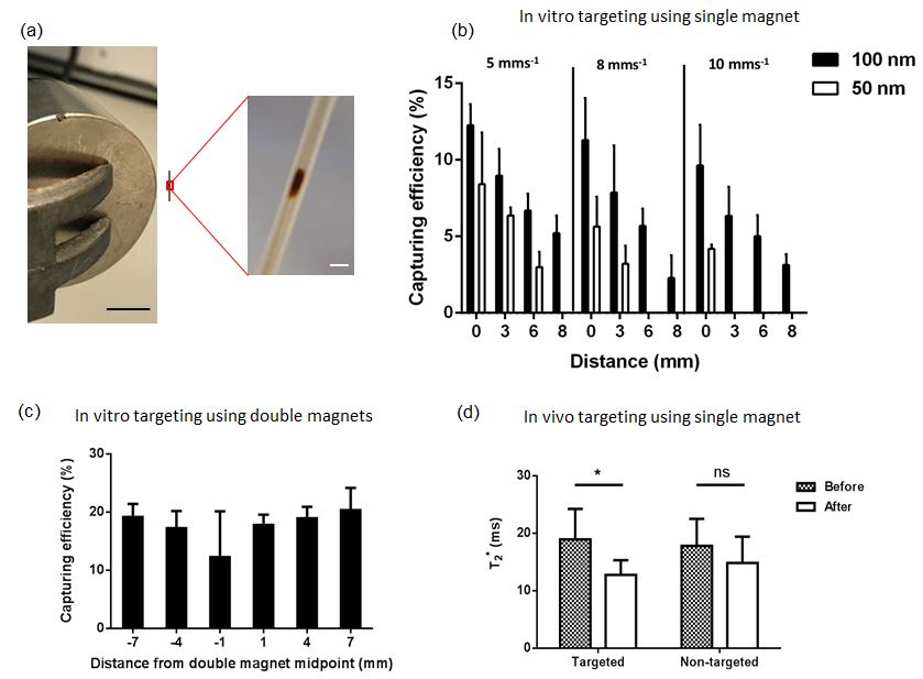
An in vitro flow model was used to simulate magnetic targeting of circulating nanoparticles within the blood vessels of the tumour in a simplified model. This was investigated with two sizes of nanoparticles (50, 100 nm), three different permanent magnet designs at different flow velocities and magnet distances from the capillary tube.
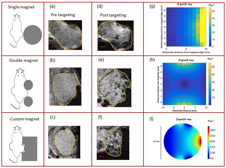
Super-paramagnetic iron-oxide nanoparticles, Fluid Mag-CT particles (Chemicell, 100 nm diameter) were intravenously injected into the mouse with the magnet positioned over the surface of the subcutaneous tumour for 20 minutes. This was explored with three different magnet systems (see fig 2): i) single magnet, ii) double magnet and iii) custom built high gradient magnet (Giamag Technologies Inc, Norway) with a high and homogenously distributed magnetic field gradient across the tumour (see fig 2 I). T2-weighted images and T2* maps were acquired before and after particle administration using respiratory-gated fast spin echo and multiple gradient echo sequences respectively using a 9.4T MRI scanner (Agilent Technologies Inc, USA) and a 1T compact MRI scanner (Bruker corporation, USA).
Results
In the flow phantom, a notable difference was observed between the capturing efficiency of 100 nm particles compared with 50 nm particles at all flow velocities and distances from the single magnet. We also observed a marked relationship between capturing efficiency and distance as well as flow; fewer particles were attached at greater magnet distances and faster flow velocities (Fig. 1 b). When the similar experiment was repeated with the double magnet, the capturing efficiency of 100 nm particles was almost doubled and the spatial distribution was improved (Fig. 1 c).
T2* maps indicated a significant reduction in T2* of tumours after injection of 100 nm nanoparticles and magnetic targeting using the single magnet (Fig. 1 d). No significant difference was observed in the contralateral control tumour experiencing no magnetic targeting. Notable hypo-intensities were observed in tumours after magnetic targeting using all three magnet designs due to the accumulation of iron oxide nanoparticles (Fig. 2 a-f). As expected, using a single magnet the hypo-intensities observed were more prevalent at locations close to the magnet (Fig. 2 d), as this was the location with the maximum magnitude of magnetic field gradient. In vivo targeting experiments using the double and custom designed magnet systems demonstrated increased hypo-intense regions deeper within the tissue (Fig. 2 e,f). The product of magnetic field strength and magnetic field gradient maps of the three magnets (Fig. 2 g-I) depict the deeper regional distribution of accumulated particles within tumours.
Discussion
Results presented here indicate that magnetic field gradients can enhance the delivery of intravenously injected 100 nm nanoparticles to a level detectable by quantitative MR imaging, and the regional distribution of nanoparticles can be improved with modified magnetic field gradient shape. Ongoing and future work involves manipulating a custom gradient set (Tesla engineering ltd, UK) on our 9.4T scanner (Agilent Technologies Inc, USA) to perform the magnetic targeting/imaging in real time.Conclusion
Magnetic targeting of iron oxide conjugated drugs shows great promise for enhancing delivery of chemotherapy to tumours. A key factor influencing efficient nanoparticle delivery is magnetic field gradient shape across the whole tumour.Acknowledgements
No acknowledgement found.References
(1) Vicky V. Mody Arthur Cox Samit Shah et al., Magnetic nanoparticle drug delivery systems for targeting tumor, Applied Nanoscience, 2014; 385–392
(2) Riegler J, Lau KD, Garcia-Prieto et al., Magnetic cell delivery for peripheral arterial disease: A theoretical framework, Med Phys, 2011; 38, 3932
(3) Munitta Muthana, Aneurin J. Kennerley, Russell Hughes et al., Directing cell therapy to anatomic target sites in vivo with magnetic resonance targeting, Nature Comms. 2015; 10.1038
Figures