3948
A 3D Spiral/Radial sequence for both anatomical and DCE imaging of pulmonary metastases in mice1CRMSB UMR5536, CNRS-Univ.Bordeaux, Bordeaux, France, 2LAMC U1029, INSERM, Talence, France
Synopsis
In order to detect and characterize pulmonary metastases in small animals, a 3D UTE sequence using a hybrid radial/spiral encoding, was employed while free breathing. Due to the high contrast and the high spatial resolution (125μm isotropic), early-growing metastases (representing less than 10 voxels) were detected in mice in only 12min. In parallel, through the acceleration of the acquisition by 4, the same sequence was used to perform, for the 1st time, DCE-MRI on pulmonary metastases in mice. The new pulmonary-dedicated sequence enables to obtain either anatomical or DCE-MRI information in mice.
INTRODUCTION
Many cancers develop metastases in the lungs. However, the lack of signal from the lungs on conventional MRI as well as respiratory and cardiac movements make this organ a technological challenge in biomedical imaging, especially in small animals where these rates are high. In addition, most of preclinical studies are performed at high magnetic fields where lungs have extremely short T2*, that hampers their detection. The few studies carried out on pulmonary metastases in small animals have used 2D sequences with large slice thickness1 preventing the detection of early-growing tumors and limiting the accuracy of volume measurements. A 3D radial sequence has then been developed to detect lung metastases in mice but lasted 1h due to respiration-triggering2. To further characterize metastases, the leakage of Gadolinium-based contrast agents (Gd-CA) through the vascular barrier in the tumor can be employed as an indicator of a success of chemotherapy3,4. Nevertheless it has not been employed on pulmonary metastases. Consequently, a rapid 3D MR sequence insensitive to respiration motion, that enables to obtain high metastasis contrast, is needed. This sequence should generate high spatially resolved images so as to detect early-growing pulmonary metastases. In addition, short acquisition times are necessary to follow the intake and clearance of Gd-CA. Our team recently developed a Ultra short TE (UTE) 3D sequence using a hybrid radial/spiral trajectories5. This exotic encoding decreases the sensitivity to respiratory motion. Moreover, acceleration of the image acquisition is possible through undersampling. Our objective was thus to optimize this MRI sequence in mice at 7T to obtain 1) anatomical information on early-growing pulmonary metastases without respiratory-synchronization and 2) DCE-MRI data on every metastases.MATERIALS and METHODS
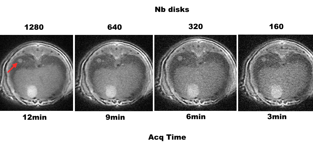
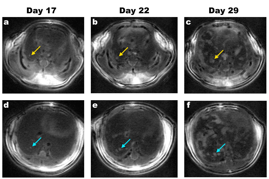
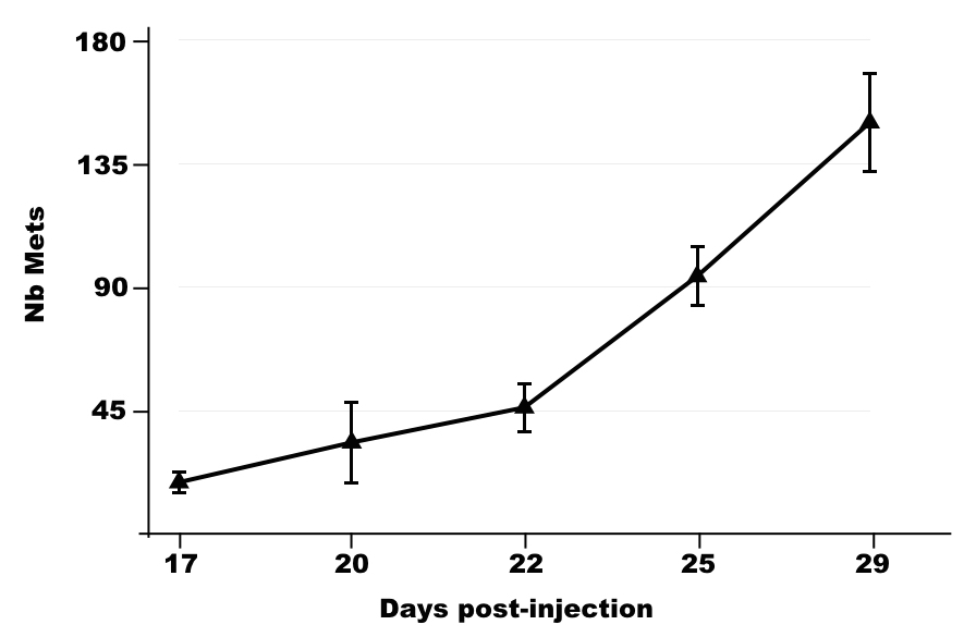
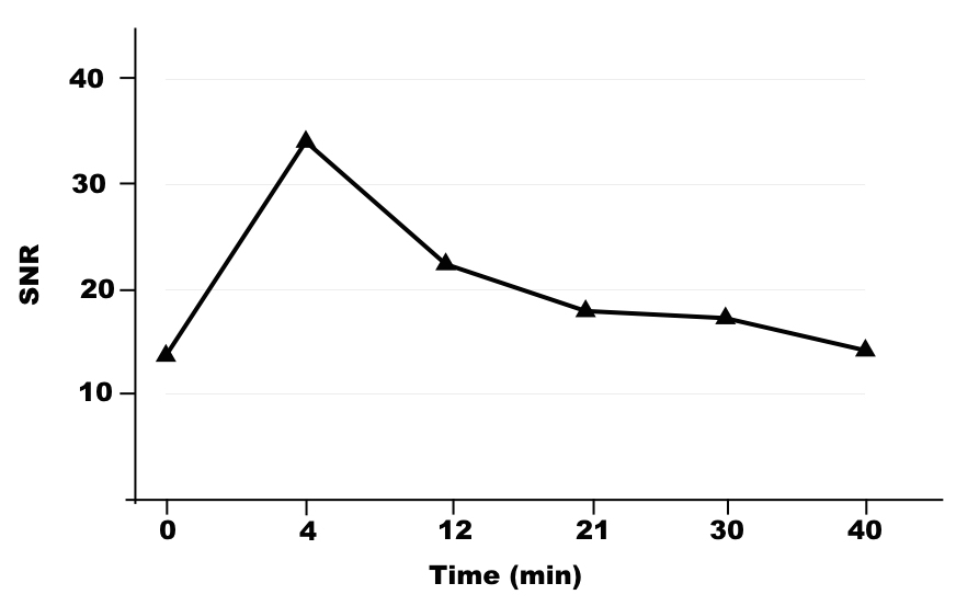
Experiments were performed on a preclinical 7T system (Bruker, Germany) equipped with a whole body excitation coil and a volume 4-channels receiver coil (Rapid Biomed, Germany). A 3D UTE sequence was used with a disk-like trajectory, in which each disk contained spirals. The 3D encoding was performed by tilting the disks with a golden angle. The amount of disks was increased from 160 to 1280 leading to acquisition time from 3min to 12min. To obtain anatomical images with high spatial resolution or to perform fast DCE-MRI, the following parameters were used: FOV=203; matrix= 1603; FA=10°; BW=400kHz. Four BalbC mice were intravenously injected with 1x10⁵ RENCA cells as a model of pulmonary metastases. Two weeks later, the anatomical follow-up started until day 29 post-injection. In addition, once a week, they were intravenously injected with Gd-DTPA (ProHance) and DCE-MRI was performed over 1 hour. Image reconstruction was performed on Matlab. Image analysis was performed using Matlab and ImageJ software.RESULTS
The optimized anatomical sequence made it possible to obtain 3D images of the lungs of the mouse, without motion artifact even though respiration triggering was not employed (Fig 1). Increasing the amount of disks increased both the SNR and the resolution/sharpness of the images. Using 1280 disks (12min), a very high spatial resolution (125μm isotropic) was reached. Pulmonary metastases were detected with high contrast (CNR between metastases and healthy lung tissue: 8.4±2.1). Very small metastases were detected: 0.012mm3 and 0.021mm3, representing 6 and 11 in-plane voxels at day 17 (Fig 2 yellow and blue arrows, respectively). The number of metastases counted over time was similar in each mouse, demonstrating a high reproducibility of the model (Fig 3). In parallel, the same sequence was performed with only 160 disks in order to decrease the acquisition time to 3min. This enabled to perform DCE-MRI in each metastasis. Gd-DTPA extravasated rapidly from the blood vessels and was cleared in 1h (Fig 4).DISCUSSION
The optimization of the 3D UTE Spiral sequence enabled to fully characterize pulmonary metastases in small animals, either through detection over time using high spatial resolution or DCE-MRI with fast acquisitions. The anatomical images were obtained 4-times faster than in the literature. In addition, this study is the first one to obtain 3D DCE-MRI information on pulmonary metastases in small animals.CONCLUSION
The advantages of this new pulmonary-dedicated MR sequence are the full coverage of the lungs, the 3D high spatial resolution and the rapidity, combined with the exemption of sacrificing multiple animals at every time points. This will be a tremendous asset for future studies evaluating the efficiency of new cancer therapies.Acknowledgements
No acknowledgement found.References
[1] Bianchi A et al. In vivo MRI for effective non-invasive detection and follow-up of an orthotopic mouse model of lung cancer. NMR Biomed. 2014; 27: 971–979
[2] Kobayashi N et al. SWIFT MRI Enhances Detection of Breast Cancer Metastasis to the Lung. Magn Reson Med 2015;73:1812–1819
[3] Weidensteiner C et al. Tumour T1 changes in vivo are highly predictive of response to chemotherapy and reflect the number of viable tumour cells – a preclinical MR study in mice. BMC Cancer 2014;14:88
[4] Ravoori MK et al. Tumor T1 Relaxation Time for Assessing Response to Bevacizumab Anti-Angiogenic Therapy in a Mouse Ovarian Cancer Model. PLoS ONE 2015;10(6): e0131095. doi:10.1371/ journal.pone.0131095
[5] Castets CR et al. Fast 3D Ultrashort Echo-Time Spiral Projection Imaging Using Golden-Angle: A Flexible Protocol for In Vivo Mouse Imaging at High Magnetic Field. Magn Reson Med 2016; DOI 10.1002/mrm.26263
Figures